


Introduction
Glomus tumor is a benign hamartomatous neoplasm originating from the glomus body, a specialized neuromyoarterial structure involved in thermoregulation through arteriovenous shunting [1, 2]. These tumors account for approximately 1–5% of all soft-tissue tumors of the hand and most commonly occur in the subungual region of the fingers and thumb [1, 2]. Despite their benign nature and small size, glomus tumors are clinically significant due to the intense pain that they produce, often leading to delayed diagnosis and prolonged patient suffering.
The glomus body consists of modified smooth muscle cells surrounding arteriovenous channels within the Sucquet-Hoyer canal. Hyperplasia or neoplastic proliferation of these structures results in formation of a glomus tumor. The tumor is highly vascular and richly innervated, which explains the classical symptom triad of severe localized pain, pinpoint tenderness, and cold hypersensitivity.
Subungual glomus tumors are the most common presentation, although extradigital locations including the forearm, foot, and visceral organs have also been described. The diagnosis is often delayed because the lesion may not be externally visible and radiographs can be normal in early stages. Patients are frequently treated conservatively for prolonged periods before definitive diagnosis is established. Magnetic resonance imaging (MRI) remains the imaging modality of choice due to its excellent soft-tissue resolution and ability to identify even very small lesions [3]. Surgical excision remains the gold standard treatment and provides immediate and dramatic symptomatic relief when complete tumor removal is achieved [2, 4].
This case highlights the importance of maintaining a high index of suspicion for glomus tumor in patients with chronic fingertip pain and demonstrates excellent clinical outcome following nail-sparing surgical excision.
Case Report
A 21-year-old male presented with complaints of severe pain over the left thumb for 6 months. The pain was insidious in onset and progressive in nature. The patient described excruciating localized pain near the nail bed, particularly aggravated during exposure to cold weather and while washing hands. Pain was partially relieved with rest and avoidance of cold exposure. The patient had difficulty performing activities of daily living including writing, gripping objects, and using mobile devices. There was no history of trauma, inflammatory arthritis, chronic steroid use, smoking, or alcohol consumption. There were no known comorbidities. The patient had previously been treated conservatively elsewhere with analgesics and local medications without significant improvement. Due to worsening symptoms and persistent functional limitation, he presented to our institution for further evaluation.
Clinical examination
On examination, there was no obvious swelling, nail deformity, discoloration, or local warmth. Marked pinpoint tenderness was elicited over the subungual region of the left thumb. Cold sensitivity test was positive, with immediate aggravation of pain on cold exposure. There was mild restriction of thumb movements secondary to pain. Love’s pin test reproduced severe localized pain over the lesion site. Hildreth’s test demonstrated temporary reduction in pain following proximal tourniquet application.
Investigations
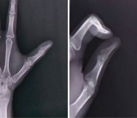
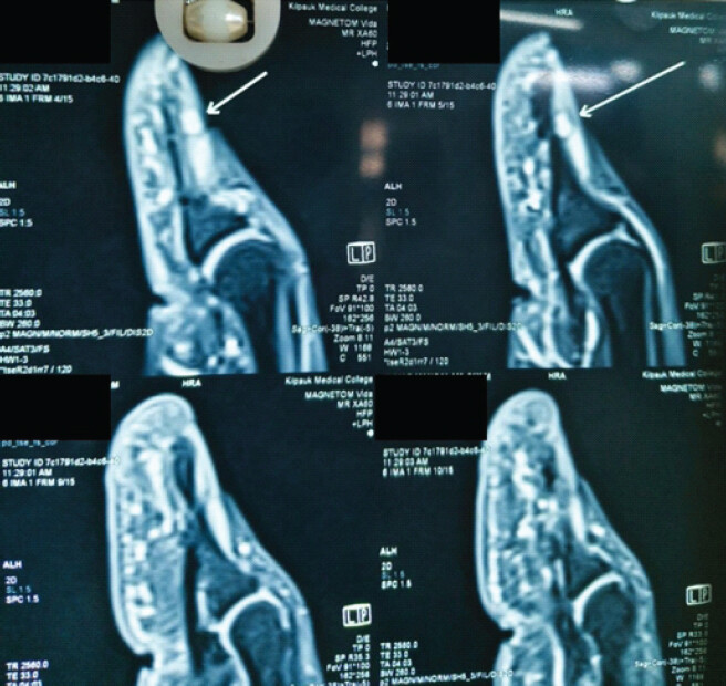
Plain radiographs of the left thumb demonstrated subtle cortical scalloping and mild erosion involving the dorsal aspect of the distal phalanx without evidence of fracture or aggressive bony destruction (Fig. 1). Computed tomography scan provided better delineation of the cortical erosion and confirmed the presence of a small subungual soft-tissue lesion with pressure effect over the distal phalanx. MRI of the thumb demonstrated a well-defined subungual lesion that appeared hypointense on T1-weighted images and hyperintense on T2-weighted and short tau inversion recovery sequences, with intense post-contrast enhancement (Fig. 2). These findings were highly suggestive of glomus tumor. Based on clinical examination and imaging findings, a diagnosis of subungual glomus tumor of the left thumb was established.
Surgical technique
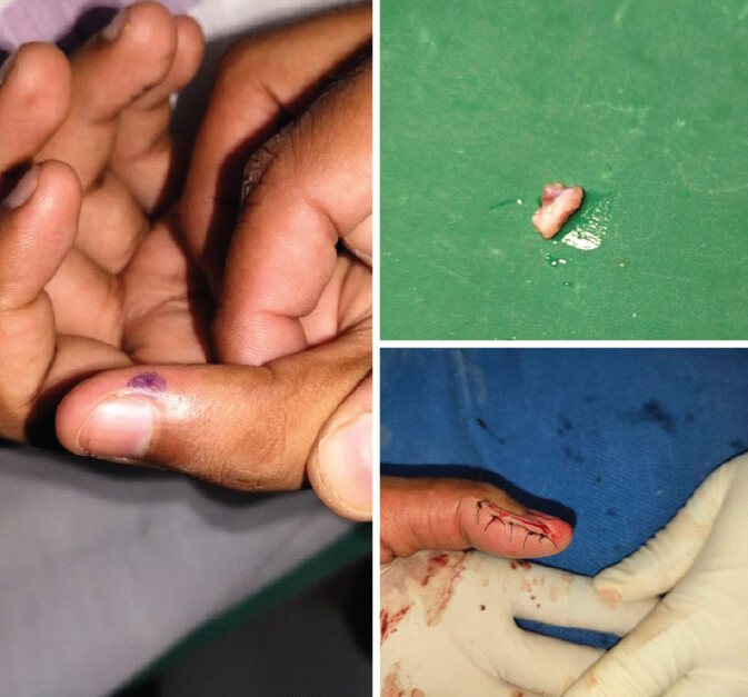
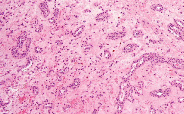
Under regional anesthesia and tourniquet control, the patient was positioned supine with the affected hand placed on a hand table. Before anesthesia preoperatively, the point of maximum tenderness was marked to center the surgical incision over the tumor (Fig. 3a). A nail-sparing paraungual approach was planned to preserve nail integrity and improve cosmetic outcome. A longitudinal paraungual incision was made along the lateral nail fold, followed by careful elevation of the soft tissues to expose the subungual region. The nail plate was preserved. A well-circumscribed bluish vascular lesion was identified beneath the nail bed. The tumor was meticulously dissected and completely excised in toto under magnification (Fig. 3b). Care was taken to avoid injury to the nail matrix and surrounding soft tissues. Curettage of the underlying cortical indentation was performed to reduce the risk of recurrence. Hemostasis was achieved following tourniquet release, and the wound was closed in layers (Fig 3c). Sterile dressing was applied. The excised specimen was sent for histopathological examination, which demonstrated nests of uniform round glomus cells surrounding vascular channels, confirming the diagnosis of glomus tumor (Fig. 4).
Follow-up
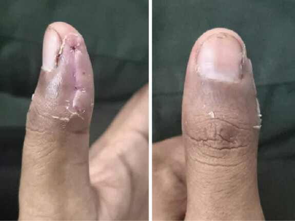
The patient was followed up at regular intervals of 2 weeks, 1 month, 2 months, and 3 months postoperatively. At 2 weeks, the surgical wound had healed well without infection or nail deformity. The patient reported marked reduction in pain. At 1 month follow-up, complete relief from resting pain and cold hypersensitivity was noted. The patient was able to resume most routine activities comfortably. At 2 months, thumb movements were painless with restoration of normal grip and pinch function. At 3 months follow-up, the patient remained symptom-free with full functional recovery and excellent cosmetic appearance (Fig. 5). At 1 year follow-up, there was no recurrence.
Functional outcome assessment
The patient demonstrated dramatic symptomatic improvement following complete excision of the glomus tumor. The visual analog scale score improved from 9/10 preoperatively to 3/10 at 1 month, 1/10 at 2 months, and complete pain relief (0/10) at 3 months follow-up. Cold hypersensitivity resolved completely by the second post-operative month, and the patient regained painless thumb function with return to normal daily activities. No recurrence was noted at the end of 1 year follow-up (Table 1).
Post-operative follow-up and functional outcome
| Parameter | Pre-operative | 1 Month | 2 months | 3 Months | 1 Year |
|---|---|---|---|---|---|
| Visual analog scale score | 09-Oct | 03-0ct | O1-Oct | 0/10 | 0/10 |
| Rest pain | Severe | Minimal | Absent | Absent | Absent |
| Cold hypersensitivity | Severe | Mild | Absent | Absent | Absent |
| Thumb movements | Painful restricted | Improved | Full painless | Full painless | Full painless |
| Daily activities | Significantly affected | Partial recovery | Near normal | Normal | Normal |
| Recurrence | – | Absent | Absent | Absent | Absent |
| Cosmetic outcome | – | Good | Good | Excellent | Excellent |
Discussion
Glomus tumor is a rare but important cause of chronic fingertip pain. The diagnosis is frequently delayed due to the tumor’s small size and absence of obvious external swelling. The average duration between onset of symptoms and diagnosis reported in literature ranges from several months to many years [2, 5]. The characteristic triad of severe localized pain, pinpoint tenderness, and cold hypersensitivity remains highly suggestive of glomus tumor [1, 5, 6]. Among these, cold sensitivity is considered particularly characteristic due to the tumor’s origin from thermoregulatory glomus bodies. Several clinical tests aid in diagnosis. Love’s pin test helps localize the lesion by reproducing focal tenderness, while Hildreth’s test demonstrates reduction in pain after temporary vascular occlusion [1, 4]. Although these tests are clinically useful, imaging plays a major role in confirming diagnosis.
Plain radiographs may appear normal in many cases, although chronic lesions may produce cortical scalloping or erosions of the distal phalanx due to pressure effects. MRI is considered the gold standard imaging modality due to its high sensitivity and specificity, particularly for detecting small lesions and evaluating suspected recurrence [3, 7]. Typical MRI findings include low-signal intensity on T1-weighted images and high-signal intensity on T2-weighted images with strong contrast enhancement. The differential diagnosis includes subungual exostosis, neuroma, hemangioma, mucous cyst, enchondroma, epidermoid inclusion cyst, and melanoma [4, 8].
Complete surgical excision remains the definitive treatment [2, 4]. Inadequate excision is the most common cause of recurrence. Several surgical approaches have been described including transungual, lateral subperiosteal, and paraungual techniques. The transungual approach provides excellent exposure but may result in postoperative nail deformity. The nail-sparing paraungual approach used in the present case allowed adequate tumor exposure while preserving nail aesthetics [4, 8, 9].
Published literature reports recurrence rates ranging from 5% to 10%, usually secondary to incomplete excision or presence of multiple lesions [2, 10]. Early recurrence within weeks may indicate incomplete removal, whereas late recurrence may represent development of a new lesion. The present case demonstrates classical clinical presentation with excellent postoperative outcome following complete excision. Early recognition and appropriate imaging are critical in preventing prolonged patient morbidity.
Conclusion
Glomus tumor, though rare, should always be considered in patients presenting with chronic fingertip pain associated with cold hypersensitivity and pinpoint tenderness. Delayed diagnosis is common due to subtle clinical findings and small lesion size. MRI plays a key role in accurate diagnosis and localization. Complete surgical excision remains curative in the majority of patients and provides dramatic symptomatic relief. The nail-sparing paraungual approach is an effective surgical technique that allows complete tumor removal while preserving cosmetic appearance and nail integrity.
Clinical Message
Persistent severe fingertip pain with cold hypersensitivity should never be ignored. Even a tiny subungual glomus tumor can cause disabling pain and significant functional impairment. Early MRI-based diagnosis and meticulous complete excision provide immediate pain relief and excellent functional recovery.
Conflict of Interest:
Nil
Source of Support:
Nil
Consent:
The authors confirm that informed consent was obtained from the patient for publication of this article
References
- Carroll RE, Berman AT. Glomus tumors of the hand: Review of the literature and report on twenty-eight cases. J Bone Joint Surg Am 1972;54:691-703. Google Scholar | PubMed
- Van Geertruyden J, Lorea P, Goldschmidt D, De Fontaine S, Schuind F, Kinnen L. Glomus tumours of the hand. A retrospective study of 51 cases. J Hand Surg Br Eur 1996;21:257-60. Google Scholar | PubMed
- Drapé JL, Idy-Peretti I, Goettmann S, Wolfram-Gabel R, Dion E, Grossin M. Subungual glomus tumors: Evaluation with MR imaging. Radiology 1995;195:507-15. Google Scholar | PubMed
- Tang CY, Tipoe T, Fung B. Where is the lesion? Glomus tumours of the hand. Arch Plast Surg 2013;40:492-5. Google Scholar | PubMed
- McDermott EM, Weiss AP. Glomus tumors. J Hand Surg Am 2006;31:1397-400. Google Scholar | PubMed
- Bhaskaranand K, Navadgi BC. Glomus tumour of the hand. J Hand Surg Br 2002;27:229-31. Google Scholar | PubMed
- Theumann NH, Goettmann S, Le Viet D, Resnick D, Chung CB, Bittoun J. Recurrent glomus tumors of fingertips: MR imaging evaluation. Radiology 2002;223:143-51. Google Scholar | PubMed
- Lee DW, Yang JH, Chang S, Won CH, Lee MW, Choi JH. Clinical and pathological characteristics of extradigital and digital glomus tumours: A retrospective comparative study. J Eur Acad Dermatol Venereol 2011;25:1392-7. Google Scholar | PubMed
- Vasisht B, Watson HK, Joseph E, Lionelli GT. Digital glomus tumors: A 29-year experience with a lateral subperiosteal approach. Plast Reconstr Surg 2004;114:1486-9. Google Scholar | PubMed
- Hazani R, Houle JM, Kasdan ML, Wilhelmi BJ. Glomus tumors of the hand. Eplasty 2008;8:e48. Google Scholar | PubMed
© 2026 Journal of Orthopaedic Case Reports - Published by Indian Orthopaedic Research Group
About the Authors
-

Dr. Prashanth Kempulraj
-

Dr. S Veerakumar
-

Dr. R Karu Shanmuga Karthikeyan
-

Dr. Vishnu Senthil
Related Articles in Journal of Orthopaedic Case Reports
 September 1, 2026 Glomus Tumor of the 4th Toe: A Case Report
 August 1, 2026 Intramuscular Glomus Tumor of Foot Presenting as Neuroma of Sural Nerve: A Case Report
 July 1, 2026 Extradigital Glomus Tumor of the Knee Mimicking Osteoarthritis: A Case Report
 January 1, 2026 Extradigital Glomus Tumor of the Forearm: Lessons from a Missed Diagnosis