


Introduction
Pathological fractures in the lumbosacral region secondary to vertebral hemangioma are exceedingly rare, occurring in <0.3% of all vertebral hemangioma cases [1]. While laminectomy and decompression remain widely employed, especially in the thoracic spine, the biomechanical demands of the lumbosacral junction are fundamentally different [2]. This transitional zone experiences higher shear forces, and vertebral body collapse here can exacerbate lumbosacral kyphosis, leading to progressive spinopelvic and global spinal imbalance [3].
Given the paucity of literature on managing such cases at the lumbosacral level, this report presents a novel, multi-modality approach integrating pre-operative embolization, low-pressure vertebral body stenting, and navigation-assisted spinopelvic fixation. This technique offers both mechanical restoration and enhanced safety in addressing the challenges of anterior column reconstruction in hypervascular spinal tumors.
Case Report
A 69-year-old male shopkeeper presented with severe low back pain (Visual Analog Scale [VAS] 9/10), accompanied by radiating pain to the left lower limb (VAS 8/10). This was a sudden worsening of diffuse, non-radiating low back pain that had persisted for the past six months. The pain was notably aggravated by positional changes and turning in bed, and it significantly impaired his ability to stand or walk. There were no bowel or bladder disturbances.
On examination, deep tenderness was elicited over the lumbosacral region. Neurologically, the patient exhibited motor weakness (grade 2/5) in the left extensor hallucis longus (EHL) and flexor hallucis longus (FHL), along with hypoesthesia in the L5 dermatome and an absent left ankle reflex.
Lateral radiographs revealed approximately 60% collapse of the L5 vertebral body, with a local Cobb angle of 28°. Magnetic resonance imaging demonstrated pan-vertebral involvement of L5 with posterior cortical bulging and thecal sac compression (Fig.1). Computed tomography (CT) imaging confirmed these findings, showing the classical “polka-dot” sign suggestive of a vertebral hemangioma.
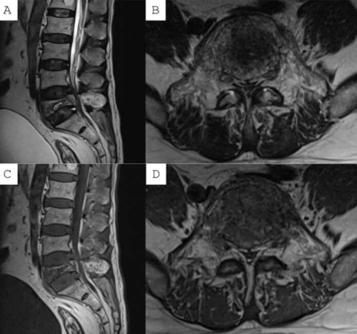
Given the combination of axial and radicular pain, vertebral collapse, and evolving neurological deficit, the patient was scheduled for navigation-guided L3–S2-alar-iliac (S2AI) spinopelvic fixation, left-sided L5 vertebral body stentoplasty, and left L5 hemilaminectomy with preservation of the posterior midline structures.
Operative procedure
-
Pre-operative percutaneous embolization was performed to reduce lesion vascularity
-
Under general anesthesia and prone positioning, a midline incision was made with bilateral exposure from L3 to S2AI
-
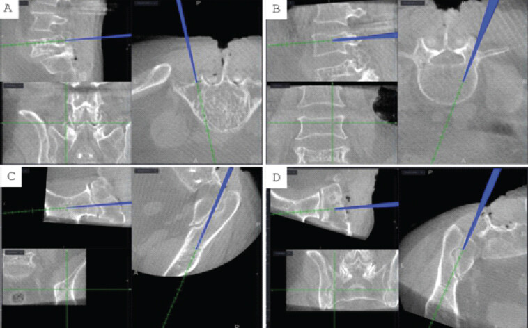
A reference frame was attached to the L3 spinous process, and an initial O-arm spin was acquired for navigation
-
Navigation-guided pedicle screws were inserted from L3 to S2AI, excluding L5 pedicles (Fig. 2)
-
Left-sided L5 hemilaminectomy was performed to decompress the thecal sac
-
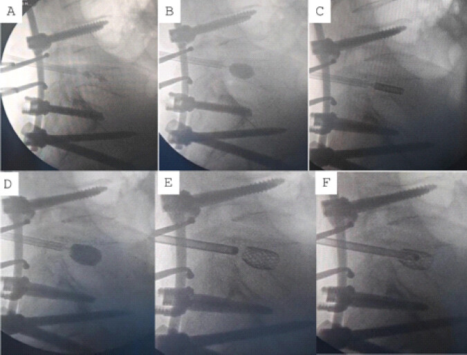
Under real-time fluoroscopic guidance, a medium-sized stentoplasty balloon was introduced and inflated to restore vertebral body height
-
Gel foam mixed with saline was injected into the cavity to embolize venous channels and reduce the risk of cement embolization [4]
-
A mesh stent was deployed and expanded to provide internal support and vertebral scaffolding (Fig. 3)
-
Low-pressure polymethyl methacrylate cement was injected into the stent to achieve anterior column reconstruction
-
Posterior rods were placed, and a final O-arm scan confirmed decompression, vertebral height restoration, cement containment, and accurate screw placement
-
The wound was closed in layers over a suction drain.
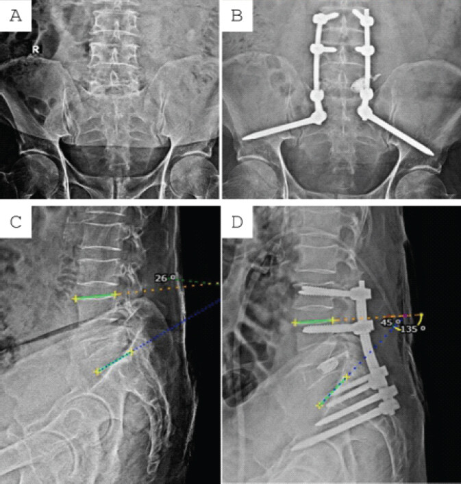
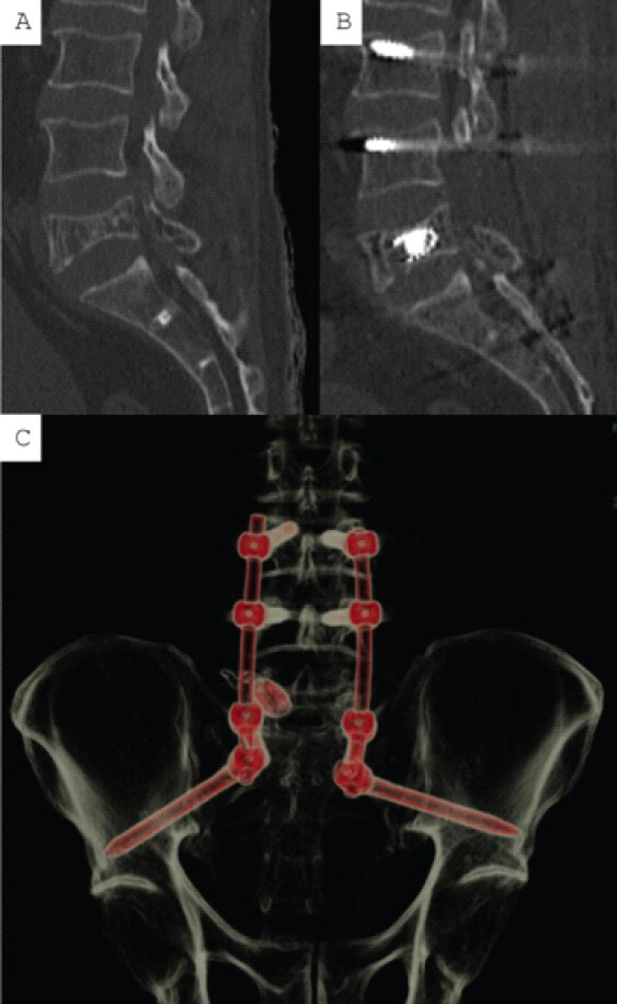
The post-operative course was uneventful, and the patient experienced immediate relief from radicular symptoms. He was mobilized on the 1st post-operative day. Post-operative radiographs confirmed successful anterior column reconstruction with correction of the lumbosacral angle from 26° to 45° (Fig. 4). The patient was discharged on the 3rd post-operative day, and sutures were removed at 2 weeks. Histopathology supported the diagnosis of hemangioma. At the 12-month follow-up, the patient reported sustained pain relief (VAS 1/10), with complete recovery of motor function in the EHL and FHL. 12-month follow-up CT imaging demonstrated maintained vertebral body height, proper spinal alignment, and complete clearance of the spinal canal (Fig. 5).
Discussion
Although literature on lumbosacral vertebral hemangiomas causing pathological fractures is limited, this region is biomechanically unstable and prone to non-union, especially in pathological fractures from other etiologies. Surgical stabilization often requires spinopelvic fixation, with options including posterior fixation to S1/S2, circumferential fusion, iliac screws with offset connectors, or S2AI screws for improved stability and alignment.
Vertebroplasty was originally developed in France in the 1980s to halt the progression of painful vertebral body hemangiomas and metastatic lesions, leveraging both its mechanical stabilization and thermal necrosis effects [5]. Even in contemporary practice, vertebroplasty demonstrates over 80% success in achieving significant or complete pain relief in symptomatic vertebral hemangiomas. However, the overall complication rate remains around 29%, with epidural cement leakage being the most common (14%) [6]. This is partly due to high-pressure cement injection and further compounded by the hypervascular nature of the lesion and poor bone quality, including cortical thinning, characteristic of vertebral hemangiomas.
In our case, L3 and L4 were selected as cranial anchor points, and S1 along with S2AI screws served as caudal anchors. The choice of S2AI screws was based on their in-line trajectory with L5 and S1 pedicles, eliminating the need for offset connectors while maintaining construct stability and reducing implant prominence [7]. Fixation limited to S1 or S2-alar screws alone was deemed inadequate due to poor sacral bone quality from osteoporosis and the high biomechanical demands at the lumbosacral junction [8].
We opted for stentoplasty over vertebroplasty to restore vertebral body height and facilitate internal scaffold formation via cancellous bone compaction with an expandable silicone balloon. The mesh stent provided additional structural integrity and supported the cement mass in maintaining the restored height [9]. Importantly, stenting allowed for low-pressure cement injection, thereby significantly reducing the risk of cement extravasation. Furthermore, gel foam embolization was performed before cement injection to occlude venous channels and trabecular spaces, further minimizing the risk of cement embolism.
Since vertebral hemangiomas primarily involve the anterior column, the role of the posterior midline complex in maintaining spinal biomechanics is critical and cannot be overstated [10]. Accordingly, we preserved the posterior midline structures in our case to maintain native stability. Intraoperative O-arm imaging was employed to ensure accurate placement of pedicle and S2AI screws, to evaluate the extent of decompression, and to detect any inadvertent cement leakage into the spinal canal or anteriorly.
Conclusion
This case highlights a rare but clinically significant presentation of a symptomatic lumbosacral vertebral hemangioma complicated by pathological fracture and neurological deficit. Successful management was achieved through a multimodal approach combining pre-operative embolization, vertebral body stenting, and navigation-guided spinopelvic fixation using S2AI screws. This strategy allowed safe anterior column reconstruction, effective decompression, and sustained neurological recovery, while minimizing cement-related complications.
Clinical Message
In rare cases of pathological L5 fractures due to vertebral hemangioma, combining vertebral body stenting with cement augmentation and spinopelvic fixation provides effective decompression and stabilization. A concept-driven strategy that respects lumbosacral biomechanics, minimizes cement-related risks, and preserves native anatomy can lead to excellent pain relief, functional recovery, and alignment maintenance.
Conflict of Interest:
Nil
Source of Support:
Nil
Consent:
The authors confirm that informed consent was obtained from the patient for publication of this article
References
- Li C, Zhang HB, Zhang H, Li Q, Zhang J, Wang J. Severe pathological fractures caused by vertebral hemangiomas with posterior decompression, bone cement augmentation and internal fixation. Orthop Traumatol Surg Res 2016;102:489-94. Google Scholar | PubMed
- Chopra H, Hoffman H, Richardson TE, Galgano MA. Surgical management of symptomatic vertebral hemangiomas: A case report and literature review. Surg Neurol Int 2021;12:56. Google Scholar | PubMed
- Oxland TR. Biomechanics of the L5/S1 Junction and the Effect of Spondylolisthesis and SpondyloptosisHarms J, Stürz H. Severe Spondylolisthesis. Heidelberg: Steinkopff; 2002. 53-66. Available from: https://link.springer.com/10.1007/978-3-642-57525-9_5 [Last accessed on 2025 Jun 23]. Google Scholar | PubMed | CrossRef
- Bhatia C, Barzilay Y, Krishna M, Friesem T, Pollock R. Cement leakage in percutaneous vertebroplasty: Effect of preinjection gelfoam embolization. Spine (Phila Pa 1976) 2006;31:915-9. Google Scholar | PubMed
- Galibert P, Déramond H. Percutaneous acrylic vertebroplasty as a treatment of vertebral angioma as well as painful and debilitating diseases. Chirurgie 1990;116:326-34; discussion 335. Google Scholar | PubMed
- Gupta AK, Phukan P, Bodhey N. Percutaneous vertebroplasty for the treatment of symptomatic vertebral hemangioma with long-term follow-up. Interdiscip Neurosurg 2021;23:100968. Google Scholar | PubMed
- Von Glinski A, Pierre C, Frieler S, Mahoney JM, Harris JA, Amin DB. Fixation strength of modified iliac screw trajectory compared to traditional iliac and s2 alar-iliac trajectories: A cadaveric study. World Neurosurg 2021;154:e481-7. Google Scholar | PubMed
- Yamada K, Abe Y, Satoh S. Safe insertion of S-2 alar iliac screws: Radiological comparison between 2 insertion points using computed tomography and 3D analysis software. J Neurosurg Spine 2018;28:536-42. Google Scholar | PubMed
- Moura DL, Isidoro D, Lourenço P, Jardim C. Stent-armed kyphoplasty in osteoporotic thoracolumbar fractures-clinical and functional results and a center experience over 10 years. J Spine Surg 2022;8:462-76. Google Scholar | PubMed
- Li J, Huang S, Tang Y, Wang X, Pan T. Biomechanical analysis of the posterior bony column of the lumbar spine. J Orthop Surg Res 2017;12:132. Google Scholar | PubMed
© 2026 Journal of Orthopaedic Case Reports - Published by Indian Orthopaedic Research Group
About the Authors
-

Dr. Shailesh Hadgaonkar
-

Dr. Sameer Nagpal
-

Dr. Parag Sancheti
Related Articles in Journal of Orthopaedic Case Reports
 July 1, 2026 Brown’s Tumor Presenting as a Pathological Humerus Fracture: A Rare Diagnostic Pitfall
 June 1, 2026 Intralesional Triamcinolone versus Oral Bisphosphonate for Benign Cystic Bone Lesions: A Prospective Comparative Study of Radiological and Pain Outcomes
 May 1, 2026 Aneurysmal Bone Cyst of Lateral Cuneiform: Challenge in Diagnosis and Management
 April 1, 2026 A Rare Case of Left Femoral Neck Fracture in a 6-Year-Old Boy with Osteopetrosis