


Introduction
Sutures are foreign biomaterials implanted into tissue, and their interaction with host immune mechanisms may lead to inflammation, infection, or foreign body reactions. Different suture materials – natural or synthetic, absorbable or non-absorbable – demonstrate variable tissue reactivity, with braided sutures generally producing greater inflammatory responses than monofilament sutures [1]. Although surgical site infection (SSI) following total knee arthroplasty (TKA) is relatively uncommon, occurring in approximately 1% of procedures [2], post-operative wound complications remain a significant concern for surgeons. In addition to microbial infection, host factors such as immune status, comorbidities, and the local tissue environment may influence the likelihood of adverse tissue reactions to sutures [1].
Occasionally, post-operative wound inflammation and discharge may clinically resemble SSI but represent a distinct non-infective entity known as suture-related pseudo-infection (SRPI), also described as Vicryl reaction or suture granuloma. SRPI is a rare complication following TKA in which a foreign body reaction to suture material mimics post-operative infection, often leading to diagnostic uncertainty [3]. The pathophysiology of this reaction involves a foreign body immune response characterized by protein adsorption, inflammatory cell infiltration, macrophage activation, and formation of foreign body giant cells around the suture material [4].
Vicryl reaction typically presents as a delayed post-operative inflammatory response, most commonly occurring 6–9 weeks after surgery, which corresponds to the absorption phase of Vicryl sutures (approximately 56–70 days) [5]. This timing suggests that the reaction is related to degradation of the absorbable suture material and the resulting foreign body response. Clinically, patients may present with pain, erythema, swelling, and wound discharge, occasionally accompanied by mild fever features that closely mimic prosthetic joint infection (PJI) and often raise diagnostic concern [6]. In some cases, patients develop mild pain with a discharging sinus along the surgical scar, further increasing suspicion of periprosthetic joint infection [3]. Such presentations can make differentiation from true SSI challenging.
Recognizing this entity is important because it can closely mimic infection yet requires different management. In this report, we present a case series of four patients who developed Vicryl-related reactions following TKA, highlighting the clinical presentation, diagnostic challenges, and management considerations.
Case Series
We report a case series of four patients who developed signs of infection following TKA. All patients initially had an uneventful post-operative recovery and subsequently presented with delayed wound-related symptoms along the surgical incision line. The most common clinical presentation was a non-healing wound over the suture line, frequently associated with sero-sanguineous, serous, or occasionally sero-purulent discharge. The infra-patellar region, particularly around the tibial tuberosity, was the most commonly affected site in all cases.
On clinical examination, patients demonstrated localized tenderness over the suture site with surrounding erythema. However, an important distinguishing feature was that the range of motion (ROM) of the knee joint remained full, painless, and unrestricted in all patients. None of the patients exhibited signs suggestive of deep joint involvement, and no pus was found in the knee joint aspirate.
Microbiological assessment showed negative culture results in three of the four patients, supporting a non-infective etiology. One patient in the series had a history of rheumatoid arthritis, which may have contributed to an altered and exaggerated immune response. The clinical presentation in these patients closely resembled post-operative infection, leading to diagnostic uncertainty.
Case Descriptions
Case 1
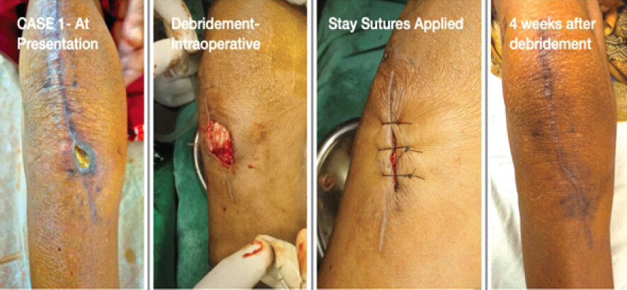
Sixty four years old woman who underwent TKA presented approximately 40 days after surgery with complaints of a non-healing wound over the infra-patellar region along the surgical suture line. Slough was visible over the wound site, but there was no active pus discharge and no associated fever. Clinical examination revealed that the ROM of the knee was full and painless. Knee aspiration yielded sero-sanguineous fluid, with no evidence of intra-articular pus (Fig. 1).
Case 2
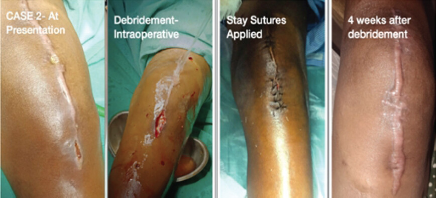
A 43-year-old woman who was a known case of rheumatoid arthritis underwent bilateral TKA and presented about 55 days after TKA with persistent wound discharge over the surgical suture line, predominantly in the infra-patellar region. The patient initially noticed watery discharge on and off soon after suture removal, which gradually progressed to serous discharge. There was no history of fever, and the knee joint demonstrated full, painless ROM. Knee aspirate was serous in nature (Fig. 2).
Case 3
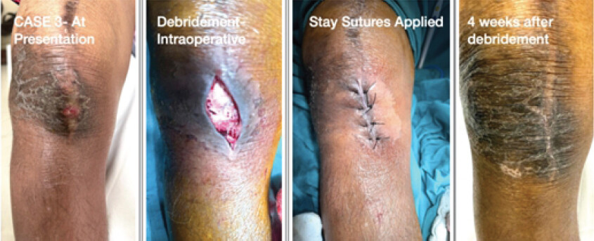
A 70-year-old patient presented approximately 134 days after TKA with localized swelling over the infra-patellar region along the surgical suture line for 10–12 days. The patient reported scanty thick discharge from the infra-patellar region for 2 days. On examination, there was expressed pus discharge from the site. The patient had a history of fever for 1 week, raising suspicion of infection. However, clinical examination demonstrated full and painless knee ROM, and knee aspiration revealed serous fluid, indicating absence of deep joint infection (Fig. 3).
Case 4
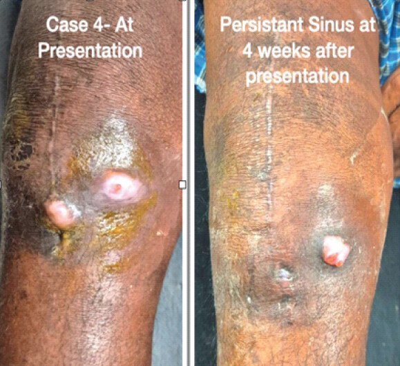
A 60-year-old gentleman presented approximately 28 days after TKA with complaints of watery discharge from the infra-patellar suture line, which began 3–4 days after suture removal. There was no associated fever, and examination showed painless knee ROM which was terminally restricted. Knee aspiration revealed sero-sanguineous fluid, with no evidence of intra-articular purulence (Fig. 4).
All four cases were operated on by the same surgeon in the same setup. All standard protocols for prevention of infection were followed. Vicryl or Braided Coated Polyglactin 910 (2-0 size, continuous locking pattern) was used to close the arthrotomy in all the patients. Across all cases, pain and the discharge were localized to the infra-patellar suture line with local rise of temperature and erythema limited to the immediate vicinity. These symptoms pointed toward probable PJI.
However, on taking detailed history and performing further investigations, we found that fever was absent in 3 out of 4 cases, with only one patient (Case 3) presenting with fever, indicating that systemic signs were largely absent despite local wound findings. Total leukocyte count (TLC) values were within normal limits or only mildly elevated across all cases (range: ~5.0–11.9 ×10³/mm3), again suggesting a lack of significant systemic infection. C-reactive protein (CRP) was elevated in 3 cases (Cases 1, 2, and 3), with notably higher values in Cases 2 and 3, while Case 4 showed near-normal levels. Erythrocyte sedimentation rate (ESR) was elevated in all cases, particularly in Case 2 and Case 3, reflecting a chronic inflammatory response rather than definitive infection. X-ray of the knee showed normal radiological findings in all cases, with no evidence of prosthesis-related complications or loosening, supporting the absence of deep infection. Furthermore, preserved and painless knee mobility and absence of pus in the joint aspirate created a dilemma supporting an alternative diagnosis rather than true PJI.
Therefore, we decided to investigate further. We approached the patients as follows.
-
The patients were examined clinically and the ROM of the knee was checked. Swab from the discharge and knee aspirate were sent for Gram stain and culture and sensitivity.
-
All relevant blood investigations including TLC, CRP, and ESR were done.
-
As none of the patients had signs of sepsis or presence of an abscess in the knee joint, we did not start antibiotics for the patients. Rather, we decided to locally debride the non-healing wound first. The patient described in Case 4 refused the debridement procedure, and hence, prophylactic oral antibiotics were prescribed right after sending his wound discharge and knee aspirate sample for microbiological examination.
-
Intra-operative debrided tissue was sent for histopathological examination, and discharge was sent for Gram stain and culture and sensitivity for the first 3 cases.
-
The wound was closed with only stay sutures for a second look after 5 days.
-
Empirical broad-spectrum antibiotic was started for the first 3 cases after the debridement procedure, namely injectable piperacillin-tazobactam.
-
Wound was opened for the 2nd time 5 days after the first procedure for a probable presence of slough or infection, and definite closure was performed with Nylon sutures only.
-
Sutures were removed after 12–14 days.
Intra-operative findings (Fig. 1, 2, 3)
-
None of the cases had any evidence of deep-seated infection.
-
Cases 1 and 2 had remnants of Vicryl suture visible intra-operatively. This finding prompted us to ponder in the line of a probable “Vicryl reaction,” and hence, we sent debrided tissue for histopathological examination.
-
Definitive closure was performed using only Nylon Sutures.
The results of histopathological examination (available in 3 cases) consistently showed features of foreign body reaction, including macrophage infiltration, foreign body giant cell formation, and granulomatous inflammation/non-specific inflammatory changes.
The results of clinical, microbiological, histopathological, radiological, and blood investigations for all four cases are summarized in Table 1.
Investigation Findings
| Case 1 | Case 2 | Case 3 | Case 4 | |
|---|---|---|---|---|
| Age/Sex | 64/F | 43/F | 70/F | 60/M |
| Suture Material used | Braided Coated Polyglactin 910 | Braided Coated Polyglactin 910 | Braided Coated Polyglactin 910 | Braided Coated Polyglactin 910 |
| Time Of Presentation | 40 Days | 55 days | 139 days | 28 days |
| Fever | Absent | Absent | Present | Absent |
| TLC | 5.04 X 10³/ mm3 | 10.3 X 10³/ mm3 | 11.97 X 10³/ mm3 | 6 X 10³/ mm3 |
| CRP | 6 mg/L | 43 mg/dL | 38 mg/L | 3 mg/dL |
| ESR | 15 mm/hr | 76 mm/hr | 42 mm/hr | 29 mm/hr |
| Abscess | Frank Pus Absent | Frank Pus Absent | Frank pus expressed | Frank pus Absent |
| Slough present | Sero-sanguinous discharge | Serous discharge | ||
| Wound Swab C/S | No growth | E. Coli | No growth | No growth |
| Nature of Knee Aspirate | Sero-sanguinous | Serous | Serous | Sero-sanguinous |
| Knee Aspirate C/S | No growth | No growth | No growth | No growth |
| Intra-operative Sample C/S | No growth | Enterobacter Cloacae | No growth | —————- |
| Vicryl remnant | Present | Present | Absent | —————- |
| Histopathological examination | Macrophage | Non-specific inflammation | Macrophage | —————- |
| Giant cell formation | Foreign body reaction with occasional giant cells | Lymphocytic inflammatory infiltrare | ||
| Granuloma | Presence of foreign body giant cells | |||
| Sinus | Absent | Healed after surgery | Healed after surgery | Persistant |
| Knee Radiology | Normal | Normal | Normal | Normal |
| No obvious signs of loosening | No obvious signs of loosening | No obvious signs of loosening | No obvious signs of loosening | |
| Knee ROM | Preserved and Painless | Preserved and Painless | Preserved and Painless | Terminally restricted and Painless |
Results (Table 1)
Post-operative/4 weeks follow-up
-
Cases 1, 2, and 3 who underwent local debridement were followed up, and sutures were removed after 12–14 days
-
Wounds were healed completely and showed no signs of inflammation after 4 weeks for Cases 1, 2, and 3 (Fig. 1, 2, 3).
-
Case 4 who had refused debridement was followed up, and persistent sinus was found at 4 weeks after presentation (Fig. 4).
Discussion
Vicryl (polyglactin 910) is a commonly used absorbable braided suture material in orthopedic surgery because of its favorable handling characteristics and predictable absorption profile. However, as with any implanted biomaterial, it may occasionally provoke foreign body reactions that mimic post-operative infection. The first evidence of Vicryl-related hypersensitivity reactions available in the literature was probably described in 1977 in a case of strabismus surgery, where the authors highlighted that although such reactions are rare, surgeons should consider suture hypersensitivity when unexplained post-operative inflammation occurs to avoid misdiagnosis and inappropriate treatment [7].
The clinical presentation of Vicryl reaction or SRPI can closely resemble PJI. Patients may present with localized wound inflammation, discharge, and occasionally mild systemic symptoms, making differentiation from infection difficult. Previous studies have reported that laboratory investigations may show mildly elevated inflammatory markers such as ESR or CRP while white blood cell counts remain normal or only slightly elevated [6]. Similar findings were observed in our series, where TLCs were within normal limits or only marginally elevated, while CRP was raised in three of the four patients and ESR was raised in two of the four cases. The exceptionally high CRP and ESR in Case 2 of our series could be due to the acute exacerbation of inflammation due to rheumatoid arthritis.
Imaging modalities such as ultrasound may reveal small subcutaneous or fascial collections without intra-articular involvement, which further supports the diagnosis of superficial pathology rather than deep joint infection [3,5]. In addition, microbiological cultures are often negative, and histopathology typically demonstrates a foreign body granuloma with inflammatory infiltrates and giant cells, confirming a suture-related reaction rather than true infection [3].
In our series, the clinical presentation was largely consistent with the literature. All patients demonstrated localized wound complications over the infra-patellar suture line while maintaining painless and full knee ROM, suggesting absence of intra-articular involvement. The reason why the infra-patellar region over the tibial tuberosity was affected in all four cases needs explanation. One of the speculated reasons could be the lack of ample soft tissue coverage and the presence of less fatty tissue. Literature largely lacks an explanation regarding the involvement of this typical location.
Importantly, none of the patients had pus within the knee joint, and all were operated by the same surgeon in the same setup following strict standard infection prevention protocols, reducing the likelihood of true deep infection. Three patients underwent surgical debridement with removal of the affected tissue along with 2–3 mm margin of healthy tissue and subsequently recovered without the need for arthrotomy or opening of the joint, which is consistent with previous reports showing rapid clinical improvement following removal of the offending suture material and the affected tissue [6]. Conversely, the fourth patient who declined debridement continued to experience persistent watery discharge and sinus despite prolonged empirical antibiotic therapy, highlighting that removal of the causative suture material is crucial for resolution.
Microbiological evaluation in such cases may occasionally produce confusing results. In our series, three patients had negative cultures, while one patient showed bacterial growth in both pre-operative swab and intraoperative samples; however, the organisms isolated (namely, Escherichia coli and Enterobacter Cloacae) were different and are known skin contaminants [8,9]. Histopathology in this patient revealed foreign body giant cell reaction, supporting the diagnosis of Vicryl reaction. It is possible that the culture positivity represented sample contamination or a superimposed superficial infection overlying a suture-induced inflammatory reaction.
Braided sutures such as Vicryl have been shown to provoke higher tissue reaction, whereas monofilament sutures like Monocryl produce less inflammatory response [10]. Although modifications such as triclosan-coated sutures have been proposed to reduce SSIs, clinical trials have shown no significant reduction in SSI rates following hip or knee arthroplasty, questioning their routine use [2]. However, the literature does not report an increase or decrease in the rate of hypersensitivity reactions with their use.
Management of post-operative wound complications following TKA requires careful evaluation, as delayed or inappropriate treatment may lead to progression from superficial pathology to deep peri-prosthetic infection [10]. Literature suggests that surgical irrigation and debridement often reveal sterile abscesses with or without retained Vicryl sutures, and removal of the suture material along with surrounding tissue can lead to resolution of symptoms [5, 10]. In some cases, recurrence of inflammatory symptoms has been reported when the same absorbable suture material was reused during subsequent procedures, further supporting the theory of suture-induced reaction [5]. Histopathological examination demonstrating foreign body granuloma with giant cells remains an important diagnostic tool for confirming the condition and avoiding unnecessary aggressive procedures such as prosthesis removal [6].
The findings from our case series reinforce the concept that Vicryl-related reactions can closely mimic infection yet represent a distinct clinical entity requiring different management. Early recognition is essential to prevent prolonged antibiotic therapy, repeated investigations, or unnecessary revision surgery.
Preventive strategies may include the preferential use of monofilament sutures instead of braided absorbable sutures such as Vicryl in selected cases, particularly in patients with suspected suture hypersensitivity, and avoiding the re-use of absorbable sutures during wound closure following debridement to minimize the risk of recurrent foreign body reaction.
Although well-defined diagnostic criteria for Vicryl reaction are lacking in the literature, definitive diagnosis relies on histopathological examination demonstrating a foreign body granulomatous reaction. However, as Vicryl reaction and infection may coexist, the presence of a foreign body reaction should not lead to exclusion of infection, particularly when wound discharge, knee aspirate, or intra-operative cultures yield pathogenic organisms. Therefore, microbiological findings should always be interpreted in conjunction with clinical, laboratory, and histopathological evidence.
Surgeons should therefore maintain a high index of suspicion for suture-related reactions when patients present with localized wound complications several weeks after arthroplasty, particularly in the absence of joint involvement.
Conclusion
This case series highlights Vicryl reaction or SRPI as an important and often under-recognized cause of delayed wound complications following TKA. The findings emphasize that not all post-operative wound discharge represents PJI, and careful clinical assessment is crucial to avoid misdiagnosis.
A key takeaway from this study is that preserved, painless ROM with absence of intra-articular pus strongly suggests a superficial pathology rather than deep periprosthetic infection. In all our cases, the pathology was localized to the infra-patellar suture line, and none required arthrotomy or prosthesis removal. This reinforces the importance of correlating clinical findings with investigations rather than relying solely on wound appearance or isolated culture results. Secondly, the study demonstrates that laboratory parameters such as CRP may be elevated despite absence of true infection, and microbiological cultures can be negative or misleading. Therefore, histopathological confirmation of foreign body granuloma remains a valuable diagnostic tool in differentiating Vicryl reaction from infection.
Timely surgical debridement with removal of offending suture material and inflamed tissue is both diagnostic and therapeutic, leading to complete resolution in most cases. In contrast, delayed or conservative management, as seen in one patient, may result in persistent sinus and prolonged morbidity.
Finally, this series underscores the need for preventive strategies, including the preferential use of monofilament sutures over braided materials like Vicryl in selected cases, and avoiding re-use of absorbable sutures during closure after debridement. Overall, increased awareness of this entity can help surgeons avoid unnecessary investigations, prolonged antibiotic therapy, and radical procedures such as two-stage revision arthroplasty, thereby improving patient outcomes and optimizing resource utilization.
Conflict of Interest:
Nil
Source of Support:
Nil
Consent:
The authors confirm that informed consent was obtained from the patient for publication of this article
References
- Holzheimer RG. Adverse events of sutures: Possible interactions of biomaterials? Eur J Med Res 2005;10:521-6. Google Scholar | PubMed
- Sprowson AP, Jensen C, Parsons N, Partington P, Emmerson K, Carluke I. The effect of triclosan-coated sutures on the rate of surgical site infection after hip and knee arthroplasty: A double-blind randomized controlled trial of 2546 patients. Bone Joint J 2018;100-B:296-302. Google Scholar | PubMed
- Durrani S, Dua A, Mathur M, Sharma N. An exceptional case of suture related pseudo-infection (SRPI) / Granuloma following total knee replacement: Case report. Int J Orthop Sci 2024;10:252-4. Google Scholar | PubMed
- Berkay AF, Buyukkuscu MO. Vicryl reaction following arthroscopic surgery: A case report. Cureus 2024;16:e65255. Google Scholar | PubMed
- Pierannunzii L, Fossali A, De Lucia O, Guarino A. Suture-related pseudoinfection after total hip arthroplasty. J Orthop Traumatol 2015;16:59-65. Google Scholar | PubMed
- Sayegh S, Bernard L, Stern R, Pache JC, Szalay I, Hoffmeyer P. Suture granuloma mimicking infection following total hip arthroplasty. A report of three cases. J Bone Joint Surg 2009;85:2006-9. Google Scholar | PubMed
- Martin-Casals A, Scott WE. Reaction to polyglactin 910 (Vicryl): A case report. J Pediatr Ophthalmold Strabismus 1977;14:178-9. Google Scholar | PubMed
- Pearson ML, Pegues DA, Carson LA, O'Donnell R, Berger RH, Anderson RL. Cluster of enterobacter cloacae pseudobacteremias associated with use of an agar slant blood culturing system. J Clin Microbiol 1993;31:2599-603. Google Scholar | PubMed
- Weinstein MP. Blood culture contamination: Persisting problems and partial progress. J Clin Microbiol 2003;41:2275-8. Google Scholar | PubMed
- Ihsan MR, Eu WC, Lim MY, Zairul A. Stitch abscess post total knee arthroplasty. Google Scholar | PubMed
© 2026 Journal of Orthopaedic Case Reports - Published by Indian Orthopaedic Research Group
About the Authors
-

Dr. Alok Sahu
-

Dr. Rajesh Senapati
-

Dr. Hemanth Raj
-

Dr. Debashish Meher
-

Dr. Hemant Sharma
-

Dr. Nikita Jajodia
Related Articles in Journal of Orthopaedic Case Reports
 March 1, 2026 Sequential Bilateral Prosthetic Knee Infections Managed with Divergent Surgical Strategies: A Case Report
 May 10, 2024 Uncommon Pathogen: Achromobacter xylosoxidans Infection Following Total Knee Arthroplasty
 September 1, 2026 A Case of Catastrophic Failure of Tibial Component with Extensive Metallosis Following Total Knee Arthroplasty: A Case Report
 September 1, 2026 A Prospective Study of Functional Outcome of Total Knee Arthroplasty in Kellgren–Lawrence Grade IV Osteoarthritis