


Introduction
Gunshot wounds represent a subset of high-energy trauma capable of causing extensive soft-tissue destruction in combination with comminuted fractures. Such injuries require a multidisciplinary approach addressing both skeletal stabilization and soft-tissue reconstruction. Gustilo-Anderson Type IIIB open tibial fractures are limb-threatening injuries with a reported mean union time of 28–52 weeks and infection rates ranging from 6% to 50% [1,2]. Optimal management typically involves close collaboration between orthopedic and plastic surgery teams.
The “fix and flap” strategy – comprising aggressive debridement, skeletal stabilization, and early vascularized soft-tissue coverage – has long been advocated as the standard of care [3]. For large soft-tissue defects, free tissue transfer is generally recommended within the classic reconstructive ladder algorithm. However, free flap procedures carry substantial risks, including donor site morbidity, anastomotic complications, partial or total flap loss, and the need for multiple operative interventions [4]. Practical constraints such as prolonged operative time, requirement for post-operative intensive care monitoring, and high resource utilization further limit the universal applicability of microsurgical reconstruction [5].
Contemporary advances in external fixation and Ilizarov-based principles have introduced viable alternatives for managing complex extremity injuries. Circular external fixator systems such as the Ilizarov frame and the Taylor Spatial Frame (TSF) permit simultaneous management of bone and soft tissue, even in the most challenging scenarios [6,7]. Intentional acute shortening and deformation – collectively termed “Artificial Deformity Creating” (ADCr) – has been described as a method to achieve primary closure of large soft-tissue defects without adjunctive flap procedures [8,9].
In 2022, Plotnikovs et al. published a comprehensive systematic review proposing the unified terminology of ADCr to encompass shortening, angulation, and rotation techniques, and demonstrated that this approach significantly reduces the need for microsurgery while enabling accurate multiplanar deformity correction with computer-controlled hexapod frames [10]. Pierrie and Beltran subsequently outlined updated indications in their 2023 review, identifying contraindications to free tissue transfer (chronic peripheral vascular disease, non-reconstructable vascular injury, and severe comorbidities) and absence of a skilled microsurgeon as appropriate settings for ADCr [11].
In this report, we present a case of Gustilo-Anderson Type IIIB distal tibial pilon fracture resulting from a gunshot wound, managed successfully with intentional acute deformation and gradual correction using TSF, without the need for free tissue transfer. The case is discussed in the context of the current literature, addressing indications, technical considerations, complications, and areas requiring further research.
Case Report
Presentation and initial assessment
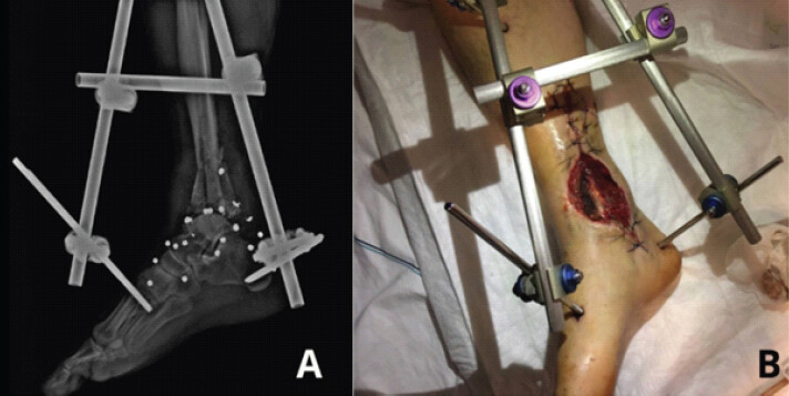
A 32-year-old male was brought to the emergency department following a gunshot wound to the right lower leg. Physical examination and radiological evaluation revealed a Gustilo-Anderson Type IIIB open fracture of the distal tibia and fibula (Fig. 1). The patient was hemodynamically stable, with no evidence of compartment syndrome or neurovascular deficit on initial assessment. Emergency irrigation with copious normal saline and wide-field debridement were performed. Provisional skeletal stabilization was achieved using a temporary unilateral external fixator.
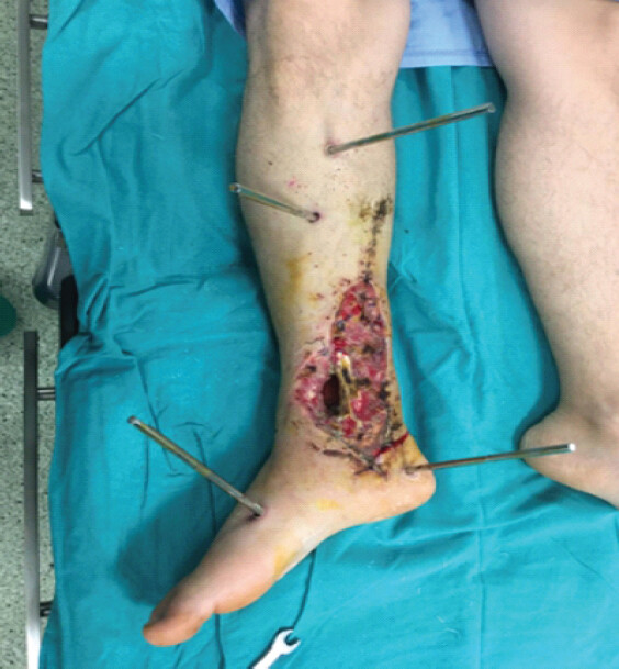
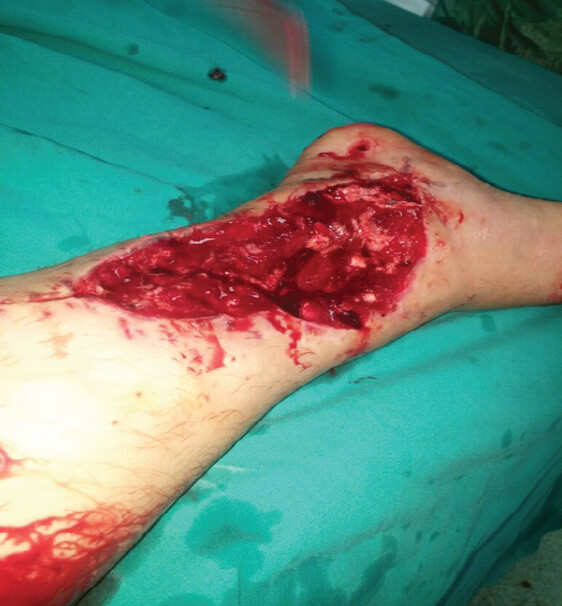
Forty-eight hours after the initial intervention, a split-thickness skin graft was applied to address the soft-tissue defect; however, the graft became infected and subsequently necrotic, resulting in enlargement of the soft-tissue defect (Fig. 2). The patient received intravenous broad-spectrum antibiotics and underwent a second-look surgical debridement (Fig. 3), followed by two additional debridement sessions.
Surgical planning and TSF application
Conventional soft-tissue coverage with split-thickness skin grafting had already been attempted; however, infection led to graft necrosis and enlargement of the wound. Therefore, repeat grafting was not considered a reliable option, and acute deformation with primary tension-free closure was selected. Given the persistent soft-tissue defect and failure of conventional wound closure methods, TSF application was planned approximately 10 days after the initial injury. Pre-operative planning included assessment of wound dimensions, fracture geometry, and vascular anatomy. Debridement of the necrotic and infected bone fragments caused by the gunshot injury at the distal tibia resulted in an approximately 5-cm tibial bone defect. A separate 5-cm segment of the distal fibula was excised to prevent the fibula from protruding through the soft tissues and mechanically obstructing creation of the acute deformity. Thus, the fibular resection facilitated the deformation maneuver and was not the cause of the 5-cm limb shortening.
An intentional acute varus deformity of 110° was then created at the fracture site (Fig. 4), enabling the wound edges to be approximated and closed primarily without tension. The limb was secured in this deformed position with the TSF (Fig. 5). Vascular integrity was carefully monitored throughout the procedure; pulsatile flow in the dorsalis pedis and posterior tibial arteries was confirmed both by clinical palpation and Doppler ultrasonography, and the absence of arterial kinking was verified.
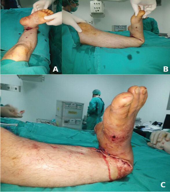
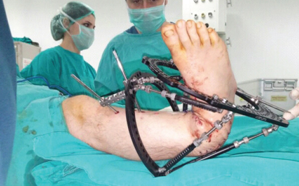
Because the fracture was located immediately adjacent to the ankle joint and the soft-tissue injury extended to the ankle capsule, preservation of ankle motion was not considered a primary objective of limb salvage. During the treatment course, the severely injured ankle developed post-traumatic arthrosis and progressed to fusion. This loss of ankle motion was accepted as a necessary trade-off for achieving a stable, plantigrade, infection-free limb; no separate formal ankle arthrodesis procedure was performed.
The limb was maintained in the deformed position for 15 days to allow complete soft-tissue healing. Throughout this period, no wound dehiscence, tension-related complications, or circulatory compromise were observed.
Deformity correction and limb lengthening
Following confirmed soft-tissue healing, the deformity and mounting parameters were entered into the computer-assisted TSF software (Direct Scheduler Utility module). The wound closure line was designated as the reference zone (Fig. 6), and correction was performed according to soft-tissue tolerance rather than at a fixed daily rate. Whenever soft-tissue tension increased and the skin became taut and shiny, correction was temporarily paused and resumed only after the tension had subsided. Using this individualized, tissue-guided protocol, the 110° varus deformity was corrected over 14 days until the soft tissues had been gradually distracted and the ankle-foot relationship had been restored to a clinically acceptable position.
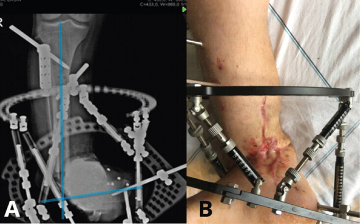
The approximately 5-cm limb-length discrepancy reflected the distal tibial bone defect created by excision of the necrotic and infected fragments and subsequent compression at the fracture site, rather than the length of the fibular resection. On day 14, after completion of the initial gradual correction and adequate soft-tissue distraction, the patient was returned to the operating room. A proximal tibial corticotomy was performed for limb lengthening, and the TSF at the distal tibia/ankle level was revised (Fig. 7). Because the severe distal tibial and ankle injury made conventional radiographic anatomical landmarks unreliable, the residual deformity was managed using clinically determined TSF residual deformity parameters, with a plantigrade foot during weight bearing as the correction target. This residual ankle-foot correction was then continued simultaneously with proximal tibial lengthening. Distraction osteogenesis at the proximal tibial corticotomy was performed at a rate of 1 mm/day from day 7 onward, progressively restoring limb length to match the contralateral side.
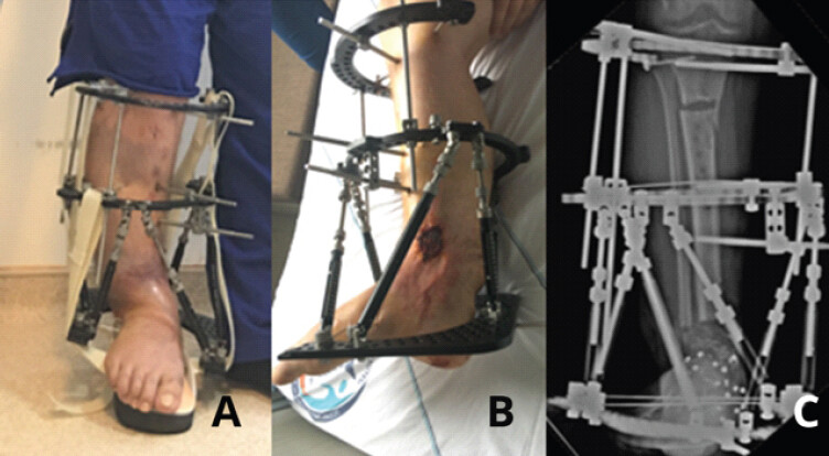
Clinical outcome
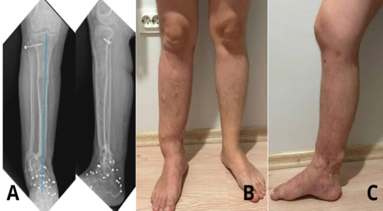
After approximately 7 months of TSF treatment, adequate callus formation at the lengthening site and solid post-traumatic ankle fusion, which had developed during the treatment course, were confirmed radiologically, and the fixator was removed. Clinically, the tibial fracture had healed without any signs of infection (Fig. 8). At final follow-up, the mechanical axis of the limb was anatomically restored and knee range of motion was preserved. The ankle was in a plantigrade position and the patient was able to bear full weight without assistive devices. No neurovascular complications occurred throughout the treatment course, and complete wound healing was achieved without any additional soft-tissue surgical procedure.
Discussion
The management of Type IIIB open tibial fractures secondary to gunshot wounds classically requires combined orthopedic and reconstructive surgical expertise. This case demonstrates the success of intentional limb deformation and shortening – as an alternative strategy when standard flap procedures are unavailable or have failed. In this technique, the fracture fragments and overlying soft tissues are transiently brought into an abnormal position to achieve wound closure, after which bone alignment and length are gradually restored using advanced external fixation systems [8,12,13].
Plotnikovs et al., in their 2022 systematic review, proposed the unifying term “Artificial Deformity Creating (ADCr)” to encompass all techniques involving shortening, angulation, and rotation – individually or in combination. This review demonstrated that the ADCr method is applicable in both acute trauma and chronic complication scenarios (infected non-union and pseudoarthrosis) and requires considerably fewer resources compared to microsurgical reconstruction [10].
Hernandez-Irizarry et al. reported a multicenter retrospective study of 19 patients with Gustilo-Anderson Type IIIB or IIIC open tibial fractures treated with intentional temporary limb deformation and hexapod frame correction. All limbs were shortened by an average of 25.1 mm; after skeletal correction, the mean residual limb length discrepancy was 5.7 mm, and no patient required amputation [5].
Pierrie and Beltran, in their 2023 updated review, defined relative indications for these techniques in detail: Contraindications to free tissue transfer (chronic peripheral vascular disease, non-reconstructable vascular injury, and severe systemic disease), absence of an experienced microsurgeon, and low-resource healthcare settings. The authors also discussed the complication profile, noting that minor complications (pin site infection, delayed wound healing, and pain) and major complications can be managed with vigilant patient monitoring [11].
The use of TSF enabled accurate correction of angular and rotational deformities even after wound closure [7,14]. Unlike conventional uniplanar external fixators, the TSF is a computer-controlled hexapod system that permits simultaneous three-dimensional correction through its six adjustable struts. Accurate calculation of mounting parameters has been shown to enhance correction precision [15], allowing us to gradually and reliably correct even an extreme 110° varus deformity in this case.
Lahoti et al. demonstrated in a series of seven patients that the Direct Scheduler Utility module of the TSF web-based software enables complete deformity correction, closing soft-tissue defects ranging from 3 to 10 cm without additional plastic surgical intervention [16]. This observation is consistent with our findings, where all required corrections were made throughout the treatment course without the need to reassemble or alter the fundamental frame configuration.
A critical aspect of this technique is the real-time assessment of limb perfusion during and after acute deformity creation. Nho et al. emphasized meticulous monitoring of arterial circulation throughout the shortening and deformation maneuver, recommending a transition to gradual incremental correction if pulses are lost [8]. In our case, continuous vascular monitoring using both clinical palpation and Doppler ultrasonography ensured the absence of arterial kinking and excessive tension throughout the deformation and subsequent distraction phases.
Similar approaches have been successfully applied in the pediatric population. Halsey et al. reported a case of a child with a Gustilo IIIB open leg injury in whom acute deformation enabled wound closure, followed by gradual correction with infection-free functional recovery [17]. Ugaji et al. further reported the efficacy of temporary shortening and deformation in three cases with massive bone and soft-tissue defects [18].
The technique has also been applied in combat casualty care under resource-limited conditions. Hsu and Beltran successfully managed a series of war wounds involving various anatomical sites using shortening and angulation to achieve soft-tissue coverage at a forward combat support hospital [19]. These data suggest that the ADCr technique is not restricted to advanced referral centers and holds broad applicability within a multidisciplinary treatment framework. The Ilizarov method has also been used successfully for simultaneous management of tibial bone and soft-tissue defects [20].
An important unanswered question in the literature concerns the maximum size of soft-tissue defect that can be closed using ADCr without adjunctive microsurgery. Plotnikovs et al. noted in their systematic review that no author has defined a specific threshold, and that the largest defects reported differ considerably across studies [10]. Similarly, no universally accepted threshold has been established for the maximum amount of acute shortening that can be safely performed. Although approximately 3 cm has frequently been cited as a practical reference for isolated acute shortening, the acceptable magnitude depends on whether shortening, angulation, or a combination of both is used, as well as on the size and location of the soft-tissue defect and the neurovascular tolerance of the limb. Greater composite defects may therefore be managed through combined shortening and angulation followed by gradual, tissue-guided correction rather than by acute shortening alone [10].
Given that the majority of existing evidence consists of case reports and small case series, prospective randomized controlled trials evaluating long-term functional outcomes and health-related quality of life following ADCr are warranted. Pierrie and Beltran have highlighted that robust large-scale evidence supporting these powerful techniques remains limited, and that careful patient selection and surgeon experience play a decisive role in determining outcomes[11].
Conclusion
In high-energy open extremity fractures, the strategy of acute shortening and deformation using TSF (ADCr) represents an effective and reliable option for limb salvage in appropriately selected patients. When performed in experienced centers with a multidisciplinary approach, this technique enables closure of large soft-tissue defects and restoration of skeletal integrity. Current systematic reviews and multicenter studies support ADCr as a method capable of eliminating the need for free tissue transfer – particularly when microsurgical reconstruction is contraindicated or unavailable.
The present case demonstrates that acute deformity as extreme as 110° can be safely applied with meticulous vascular monitoring and precise use of the TSF computer-assisted software, followed by successful anatomical restoration. Nevertheless, prospective studies are needed to clarify the indications, define maximum deformity parameters, and evaluate long-term functional outcomes of this technique.
Clinical Message
In carefully selected patients with severe open tibial fractures and soft-tissue loss, intentional temporary angulation or shortening may avoid free-flap reconstruction by enabling tension-free wound closure. This strategy requires meticulous debridement, continuous neurovascular monitoring, precise hexapod-frame planning, and staged correction by an experienced multidisciplinary limb-reconstruction team.
Conflict of Interest:
Nil
Source of Support:
Nil
Consent:
The authors confirm that informed consent was obtained from the patient for publication of this article
How to Cite this Article
Karakoyun Ö, Olçar AH. Soft-Tissue Closure through Acute Deformation and Gradual Correction Using Taylor Spatial Frame in a Gustilo-Anderson Type IIIB Open Tibial Pilon Fracture: A Case Report. Journal of Orthopaedic Case Reports 2026 October;16(10): 16-22.
References
- Giannoudis PV, Papakostidis C, Roberts C. A review of the management of open fractures of the tibia and femur. J Bone Joint Surg Br 2006;88-B:281-9. [Google Scholar] | [PubMed]
- Sharma H, Nunn T. Conversion of open tibial IIIB to IIIA fractures using intentional temporary deformation and the taylor spatial frame. Strategies Trauma Limb Reconstr 2013;8:133-40. [Google Scholar] | [PubMed]
- Gopal S, Majumder S, Batchelor AG, Knight SL, De Boer P, Smith RM. Fix and flap: The radical orthopaedic and plastic treatment of severe open fractures of the tibia. J Bone Joint Surg Br 2000;82:959-66. [Google Scholar] | [PubMed]
- Choudry U, Moran S, Karacor Z. Soft-tissue coverage and outcome of Gustilo grade IIIB midshaft tibia fractures: A 15-year experience. Plast Reconstr Surg 2008;122:479-85. [Google Scholar] | [PubMed]
- Hernandez-Irizarry R, Quinnan SM, Reid JS, Toney CB, Rozbruch SR, Lezak B. Intentional temporary limb deformation for closure of soft-tissue defects in open tibial fractures. J Orthop Trauma 2021;35:e189-94. [Google Scholar] | [PubMed]
- Ilizarov GA. The tension-stress effect on the genesis and growth of tissues: Part II. The influence of the rate and frequency of distraction. Clin Orthop Relat Res 1989;239:263-85. [Google Scholar] | [PubMed]
- Küçükkaya M, Karakoyun O, Armağan R, Kuzgun U. Correction of complex lower extremity deformities with the use of the Ilizarov-Taylor spatial frame. Acta Orthop Traumatol Turc 2009;43:1-6. [Google Scholar] | [PubMed]
- Nho SJ, Helfet DL, Rozbruch SR. Temporary intentional leg shortening and deformation to facilitate wound closure using the Ilizarov/Taylor spatial frame. J Orthop Trauma 2006;20:419-24. [Google Scholar] | [PubMed]
- Pierrie SN, Hsu JR. Shortening and angulation strategies to address composite bone and soft tissue defects. J Orthop Trauma 2017;31 Suppl 5S32-5. [Google Scholar] | [PubMed]
- Plotnikovs K, Movcans J, Solomin L. Acute shortening for open tibial fractures with bone and soft tissue defects: Systematic review of literature. Strategies Trauma Limb Reconstr 2022;17:44-54. [Google Scholar] | [PubMed]
- Pierrie SN, Beltran MJ. Acute shortening and angulation for complex open fractures: An updated perspective. OTA Int 2023;6;Suppl 4:e245. [Google Scholar] | [PubMed]
- Sen C, Kocaoglu M, Eralp L, Gulsen M, Cinar M. Bifocal compression-distraction in the acute treatment of grade III open tibia fractures with bone and soft-tissue loss: A report of 24 cases. J Orthop Trauma 2004;18:150-7. [Google Scholar] | [PubMed]
- El-Rosasy MA. Acute shortening and re-lengthening in the management of bone and soft-tissue loss in complicated fractures of the tibia. J Bone Joint Surg Br 2007;89:80-8. [Google Scholar] | [PubMed]
- Sökücü S, Karakoyun O, Arıkan Y, Küçükkaya M, Kabukçuoğlu Y. Efficacy of the Taylor spatial frame in the treatment of deformities around the knee. Acta Orthop Traumatol Turc 2013;47:86-90. [Google Scholar] | [PubMed]
- Küçükkaya M, Karakoyun O, Armağan R, Kuzgun U. Calculating the mounting parameters for Taylor spatial frame correction using computed tomography. J Orthop Trauma 2011;25:449-52. [Google Scholar] | [PubMed]
- Lahoti O, Findlay I, Shetty S, Abhishetty N. Intentional deformation and closure of soft tissue defect in open tibial fractures with a Taylor spatial frame -- a simple technique. J Orthop Trauma 2013;27:451-6. [Google Scholar] | [PubMed]
- Halsey JN, Iobst CA, Pearson GD. Acute intentional deformation of open tibial fractures for complex soft tissue closure in the pediatric patient. EPlasty 2022;22:e35. [Google Scholar] | [PubMed]
- Ugaji S, Matsubara H, Aikawa T, Tsuchiya H. Efficacy of temporary intentional leg shortening and deformation for treatment of massive bone and soft tissue defects in three patients. Strategies Trauma Limb Reconstr 2022;17:195-201. [Google Scholar] | [PubMed]
- Hsu JR, Beltran MJ. Skeletal Trauma Research Consortium. Shortening and angulation for soft-tissue reconstruction of extremity wounds in a combat support hospital. Mil Med 2009;174:838-42. [Google Scholar] | [PubMed]
- Rozbruch SR, Weitzman AM, Watson JT, Freudigman P, Katz HV, Ilizarov S. Simultaneous treatment of tibial bone and soft-tissue defects with the Ilizarov method. J Orthop Trauma 2006;20:197-205. [Google Scholar] | [PubMed]
© 2026 Journal of Orthopaedic Case Reports - Published by Indian Orthopaedic Research Group
About the Authors
-

Özgür Karakoyun
-

Ahmet Hamdi Olçar
Related Articles in Journal of Orthopaedic Case Reports
 October 1, 2026 Development of a Non-ossifying Fibroma in the Same Femur 6 Years after Limb-Salvage Surgery for Osteosarcoma: A Case Report
 October 1, 2026 Early Surgical Escalation and Structured Multidisciplinary Pathways Improve Limb-Salvage Outcomes in Necrotizing Fasciitis: A Two-Cycle Quality Improvement Project
 October 1, 2026 Management of Severe Knee Flexion Contracture of 140° with Fibrous Ankylosis in a Resource-Limited Setting: A Case Report
 September 1, 2026 Limb Salvage for Entire Tibial Ewing Sarcoma in a 4-year-Old Child Using Extracorporeal Irradiation and Free Flap Reconstruction: A Case Report and Surgical Technique