


Introduction
Giant cell tumor (GCT) of bone is a benign but locally aggressive primary osseous neoplasm, comprising approximately 5–6% of all primary bone tumors and nearly 20% of benign bone tumors [1]. It most commonly arises in the epiphyseal–metaphyseal region of long bones in skeletally mature individuals, particularly around the knee and distal radius [1,2]. Involvement of the bones of the foot is distinctly uncommon, constituting less than 1% of reported cases, and phalangeal involvement is sporadic [3].
GCTs occurring in the small bones of the hand and foot are known to demonstrate more aggressive biological behavior compared to those arising in long bones. These tumors tend to present at a younger age, show rapid cortical destruction, and have a higher propensity for local recurrence [4]. Among these, involvement of the distal phalanx of the great toe is exceedingly rare, with only isolated case reports documented in the literature [3].
Given the rarity of this presentation, there is no consensus on optimal management. While intralesional curettage may be adequate for less aggressive lesions, recurrence rates in small-bone GCTs are significantly higher, often requiring wide excision or amputation to achieve appropriate local control [5].
We report a rare case of GCT of the distal phalanx of the great toe, initially managed with biopsy and distal phalanx resection, which subsequently recurred and required amputation of the great toe.
Case Report
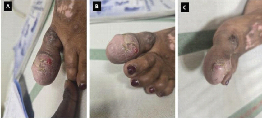
A young adult female patient in her early twenties presented with progressive pain and swelling involving the distal aspect of the great toe. The symptoms had gradually progressed over several months, without any antecedent trauma or infection. Clinical examination revealed a firm, tender swelling of the distal great toe, with stretched overlying skin, without local inflammatory signs or neurovascular deficits (Fig. 1).
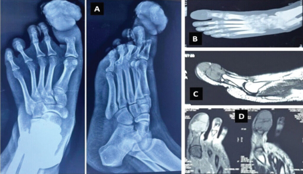
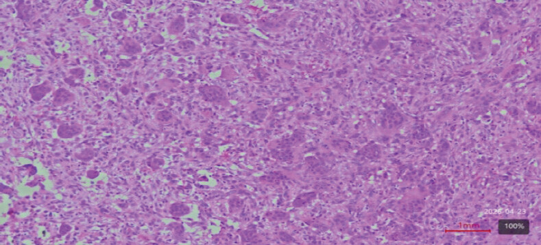
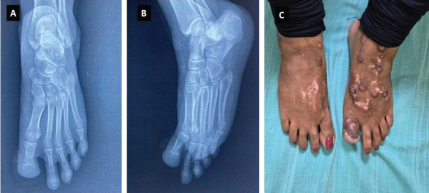
Plain radiographs demonstrated an expansile osteolytic lesion involving the distal phalanx of the great toe, with cortical thinning and destruction, and no periosteal reaction or matrix calcification (Fig. 2a). Computed tomography scan also confirmed similar findings (Fig. 2b). An magnetic resonance imaging of the foot showed a lobulated lesion involving the distal phalanx that is hypointense to intermediate on T1-weighted images and heterogeneous intermediate-to-high signal on fluid-sensitive sequences, with areas of relatively low signal. There extends up to the articular surface with preserved joint cortex, without neurovascular encasement and also to subungual region and soft tissue involvement, consistent with a locally aggressive bone tumor (Fig. 2c and d). Based on the radiological appearance, a giant cell–rich lesion was suspected. A biopsy was performed, which revealed sheets of mononuclear stromal cells interspersed with numerous osteoclast-like multinucleated giant cells, consistent with GCT of bone (Fig. 3). The patient took three doses of denosumab. Following histopathological confirmation, surgical resection of the distal phalanx was undertaken. The immediate postoperative period was uneventful. Besides, the skin lesions on the dorsum of foot were “Lichen simplex chronicus” and she received intralesional steroids and tarolimus ointments besides antihistaminic in dermatological department. Postoperative follow-up was performed for 1 year, and no further recurrence was observed; the patient remains symptom-free (Fig. 4).
Discussion
GCT of bone is a benign but locally aggressive neoplasm that mostly affects the epiphyseal–metaphyseal regions of long bones in skeletally mature individuals [1,2]. Involvement of the foot is very uncommon, accounting for <1% of cases, and phalangeal involvement is among the rarest presentations [6,7]. Among these, occurrence in the distal phalanx of the great toe is extremely rare, with only isolated case reports available in the literature [3,8].
Several studies have demonstrated that GCT bones (GCTB) arising in the small bones of the hand and foot behave more aggressively than those in long bones, despite identical histological features [4,7]. These tumors tend to present at a younger age, show rapid cortical destruction, early soft-tissue extension, and have a significantly higher rate of local recurrence following intralesional procedures [4,5,7]. The limited bone stock of the phalanges, proximity to joints and the nail bed, and early cortical breach contribute to the difficulty of achieving adequate margins with conservative surgery.
The differential diagnosis of an expansile lytic lesion in the distal phalanx includes aneurysmal bone cyst, giant cell reparative granuloma, brown tumor of hyperparathyroidism, infection, enchondroma, and chondroblastoma [9]. Due to this overlap, histopathological confirmation through biopsy remains essential before definitive management. In the present case, the diagnosis was established on biopsy, followed by distal phalanx resection.
Recurrence rates following curettage for GCTB of the small bones have been reported to range from 50% to 80%, significantly higher than those observed in long-bone lesions [4,5,10]. Wide excision or amputation offers superior local control in recurrent or aggressive cases and is often recommended when tumor margins are compromised or soft-tissue extension is present [5,10]. In the present case, amputation was done for definitive disease control [3,8]. Although uncommon, malignant transformation of GCT is well-documented, especially in recurrent or poorly treated lesions [11]. In cases of locally aggressive tumors, resection or amputation of the affected digits offers the best results by preventing any such changes [12].
The great toe plays a critical role in normal gait, functioning as the principal load-bearing and propulsive unit during terminal stance and push-off. Biomechanical studies have demonstrated that the first metatarsophalangeal joint and hallux complex transmit approximately 40–60% of body weight during normal walking, with substantially higher forces – up to 2–3 times body weight – during athletic or high-demand activities [13,14,15]. At toe-off, the hallux stabilizes the medial column of the foot, facilitates efficient forward propulsion, and maintains plantar pressure distribution across the forefoot [16]. Loss of the great toe, therefore, predictably alters gait mechanics, resulting in lateral transfer of load to the lesser metatarsal heads and reduced push-off efficiency. Nevertheless, gait analysis and clinical outcome studies have shown that patients adapt well following hallux amputation through compensatory mechanisms, particularly when pain-free local control is achieved, and adjacent joints are preserved [6,16]. In the setting of GCT of the distal phalanx, the oncological benefit of definitive excision outweighs the anticipated biomechanical compromise, making great toe amputation a rational and functionally acceptable treatment option.
Conclusion
GCT of the distal phalanx of the great toe is a rare and locally aggressive entity. Despite its benign histology, it carries a high risk of recurrence following limited surgical procedures. Early diagnosis, appropriate biopsy, and definitive surgical intervention are essential for achieving appropriate local control. Amputation remains a reliable treatment option in recurrent cases.
Clinical Message
GCT of the distal phalanx of the great toe is rare, tends to be more aggressive with higher recurrence, and therefore, amputation is a definitive treatment with acceptable functional outcomes.
Conflict of Interest:
Nil
Source of Support:
Nil
Consent:
The authors confirm that informed consent was obtained from the patient for publication of this article
How to Cite this Article
Sarma EA, Jain M, Mishra H, Aabid AMZ, Khan S, Panda J. Giant Cell Tumor of the Distal Phalanx of the Great Toe: Case Report. Journal of Orthopaedic Case Reports 2026 October;16(10): 52-56.
References
- Campanacci M, Baldini N, Boriani S, Sudanese A. Giant-cell tumor of bone. J Bone Joint Surg Am 1987;69:106-14. [Google Scholar] | [PubMed]
- Goldenberg RR, Campbell CJ, Bonfiglio M. Giant-cell tumor of bone. An analysis of two hundred and eighteen cases. J Bone Joint Surg Am 1970;52:619-64. [Google Scholar] | [PubMed]
- Bachhal V, Rangdal S, Saini U, Sament R. Giant cell tumor of distal phalanx of great toe. A case report. Foot (Edinb) 2011;21:198-200. [Google Scholar] | [PubMed]
- Szendröi M. Giant cell tumor of bone. J Bone Joint Surg Br 2004;86:5-12. [Google Scholar] | [PubMed]
- Turcotte RE. Giant cell tumor of bone. Orthop Clin North Am 2006;37:35-51. [Google Scholar] | [PubMed]
- O'Keefe RJ, O'Donnell RJ, Temple HT, Scully SP, Mankin HJ. Giant cell tumor of bone in the foot and ankle. Foot Ankle Int 1995;16:617-23. [Google Scholar] | [PubMed]
- Biscaglia R, Bacchini P, Bertoni F. Giant cell tumor of the bones of the hand and foot. Cancer 2000;88:2022-32. [Google Scholar] | [PubMed]
- Yokouchi M, Arishima Y, Nagano S, Shimada H, Nakamura S, Setoguchi T. Giant cell tumor of the distal phalanx of the fourth toe: A case report. J Foot Ankle Surg 2016;55:306-9. [Google Scholar] | [PubMed]
- Lang S. Differential diagnosis of giant cell-rich lesions of bone. Pathologe 2008;29 Suppl 2:245-9. [Google Scholar] | [PubMed]
- Balke M, Schremper L, Gebert C, Ahrens H, Streitbuerger A, Koehler G. Giant cell tumor of bone: Treatment and outcome of 214 cases. J Cancer Res Clin Oncol 2008;134:969-78. [Google Scholar] | [PubMed]
- Palmerini E, Picci P, Reichardt P, Downey G. Malignancy in giant cell tumor of bone: A review of the literature. Technol Cancer Res Treat 2019;18:1533033819840000. [Google Scholar] | [PubMed]
- Hashimoto K, Nishimura S, Ito T, Akagi M. Malignant giant cell tumor of toe: A case report and review of literature. Medicine (Baltimore) 2022;101:e29471. [Google Scholar] | [PubMed]
- McCormick JJ, Anderson RB. Turf toe: Anatomy, diagnosis, and treatment. Sports Health 2010;2:487-94. [Google Scholar] | [PubMed]
- Najefi AA, Jeyaseelan L, Welck M. Turf toe: A clinical update. EFORT Open Rev 2018;3:501-6. [Google Scholar] | [PubMed]
- Frimenko RE, Lievers W, Coughlin MJ, Anderson RB, Crandall JR, Kent RW. Etiology and biomechanics of first metatarsophalangeal joint sprains (turf toe) in athletes. Crit Rev Biomed Eng 2012;40:43-61. [Google Scholar] | [PubMed]
- Embaby OM, Elalfy MM. First metatarsophalangeal joint: Embryology, anatomy and biomechanics. World J Orthop 2025;16:102506. [Google Scholar] | [PubMed]
© 2026 Journal of Orthopaedic Case Reports - Published by Indian Orthopaedic Research Group
About the Authors
-

Evani Anirudh Sarma
-

Mantu Jain
-

Harshit Mishra
-

A Muhammad Zainul Aabid
-

Shahnawaz Khan
-

Jagatjyoti Panda
Related Articles in Journal of Orthopaedic Case Reports
 September 1, 2026 “A Giant Shoulder”: Proximal Humerus Giant Cell Tumor Managed With Extended Curettage and Proximal Humerus Internal Locking System Plating with Fibular Strut Graft – A Case Report
 September 1, 2026 Proximal Tibial Giant Cell Tumor in a Skeletally Immature Child: A Diagnostic Dilemma and Review of the Literature
 September 1, 2026 Unnoticed Giant Cell Tumor of the Left Distal Tibia in a 52-Year-Old Male: A Rare Case Report
 September 1, 2026 Acetabular Giant Cell Tumor Presenting with Protrusio Acetabuli: A Rare Case Report