


Introduction
Myositis ossificans (MO) is a self-limiting, benign, pseudo-inflammatory condition characterized by heterotropic ossification occurring in extra-skeletal soft tissues [1]. Heterotopic ossification can clinically be seen in two forms. The first presents as peri-articular heterotopic ossification with co-existing severe joint stiffness and is commonly encountered in clinical practice at the elbow in burns patients or at the hip following total hip arthroplasty. The second form, however, is seldom encountered clinically and is known as MO, which presents as a well-defined mass within the muscle or the surrounding tissues. MO is most frequently encountered in the thigh muscles of active athletes (following intramuscular hematoma formation) in 2nd to 3rd decade of life and usually resolves with rest and non-steroidal anti-inflammatory drugs [1]. MO of rotator cuff muscles is an extremely rare entity with very few cases reported till date [2]. Though the clinical presentation is variable and dependent on the stage of the disease, classical clinical symptoms in cases of MO include localized pain, swelling, and joint stiffness following blunt soft-tissue trauma [3]. MO has characteristic radiological and histological features which change according to the stage of its progression. These features can be best appreciated in the mature forms of this pseudo-tumor [4], which can easily be mistaken for a malignancy. The few papers on MO of the rotator cuff are themselves single cases. We report this patient as both subscapularis and infraspinatus were involved, and as competing radiology reports delayed the correct label for some years. It is highlighted that incidence and treatment cannot be fixed from one case.
Case Report
We report an interesting case of a 23-year-old man who presented to our center, after reviewing at multiple hospitals, with complaints of chronic pain in his right shoulder, associated with increased stiffness and restricted mobility. The patient had sustained blunt trauma to his right shoulder on November 21, 2021, while playing contact sports (Kabaddi). The individual took rest and self-medicated with over-the-counter analgesics for a few days, which did not lead to relief from his pain and associated symptoms. Following this, he was admitted to a zonal hospital in Meerut. The patient continued to be symptomatic, after which he was transferred to a tertiary care facility in Delhi.
On investigation, a radiograph of the right shoulder was reported as normal, but a magnetic resonance imaging (MRI) was suggestive of the possibility of synovial chondromatosis and a sprain of the subscapularis muscle. The patient was offered surgery in the form of arthroscopic evaluation and biopsy but refused to undergo the procedure. The individual was managed conservatively, immobilized in an arm sling pouch for three weeks, and sent home for rest and recuperation.
After reporting back, the patient continued to have pain and restriction of motion in the right shoulder joint and was re-evaluated. Contrast-enhanced MRI of the right shoulder on January 21, 2022, reported an irregular lobulated lesion involving meta-diaphysis of the proximal humerus with associated periosteal thickening in the metaphysis and proximal diaphysis, with 2 lesions, one measuring 2.1 × 3.9 × 3.7mm and another measuring 8.7 × 7.2 × 17 mm. Positron emission tomography PET-computed tomography (CT) showed periosteal thickening along with non-fluorodeoxyglucose (FDG) and exophytic growth in the metadiaphyseal region of the right humerus, with mildly increased FDG uptake in surrounding soft tissue. CT-guided biopsy was reported to be non-representative and inadequate for opinion as it demonstrated skeletal muscle only. Therefore, despite best efforts, the diagnosis remained inconclusive, and the patient was asked to review after 3 months and sent back to his place of work.
The individual noticed some relief in his symptoms over the next 3 months and did not follow up for a consultation for the next 3 years until October-November 2024, when he started experiencing increased pain, paresthesias, and severe restriction of movement in his right shoulder. The patient was evaluated at a different hospital, where basic hematological and biochemical tests, including tests for metabolic bone health, were carried out and found to be normal. An MRI was done there on October 18, 2024, which revealed the presence of an ill-defined exophytic lesion arising from the proximal metadiaphyseal region of the right humerus, measuring 3.8 × 2.7 × 3.1 cm, with foci of calcifications. The lesion was indenting the adjacent muscles and causing secondary cortical thickening of the adjacent shaft of the humerus. The report was suggestive of osteochondroma with suspicion of malignant transformation.
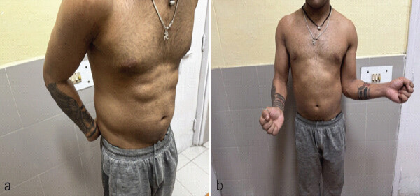
The patient was transferred to our hospital on December 20, 2024, and was symptomatic, complaining of severe restriction of mobility and pain in the right upper limb. Clinically, the shoulder had a normal contour with no wasting. However, there was tenderness on palpation, and movements were severely restricted, with a flexion-extension arc of 130°, abduction restricted to 90°, and severe restriction of rotation, with internal rotation limited to 20° and no external rotation (Fig. 1a and b). A CT scan showed eccentric cortical thickening in the lateral aspect of the meta-diaphyseal region of the right humerus, along with an adjacent dense solid, irregular periosteal reaction. In addition, a hypodense line separating the extra-humeral bony proliferation from the subjacent humeral bone was noted at certain places. The report suggested post-traumatic MO (Fig. 2a, Fig. 2b, c, d, e, f).
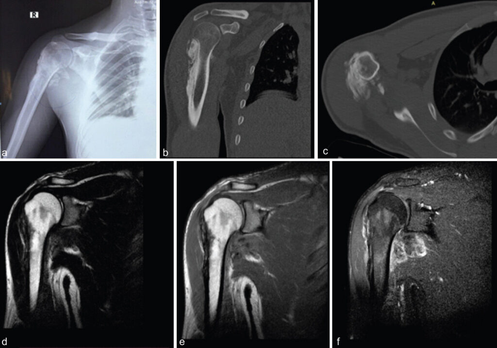
A CT-guided biopsy was repeated on January 16, 2025, but it was again reported as inadequate for any opinion, showing only skeletal muscle tissue. Therefore, an open biopsy of the lesion was performed on January 28, 2025, and samples from both bone and soft tissues were sent for histopathology. The report was consistent with MO in the subscapularis, infraspinatus, and proximal humerus (Fig. 3a and b).
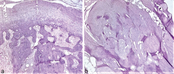
Due to the diffuse nature of the pathology, it was decided to manage it conservatively, focusing on physiotherapy, rest, and symptomatic treatment. Any surgical intervention would have required extensive tissue dissection, with a high risk of recurrence and increased severity. The risks and benefits of the procedure were explained to the patient, and a mutual decision was made to pursue conservative management. After the open biopsy, he was treated with rest, analgesics and physiotherapy. At later review, he described less pain and did not wish for excision. Movement of the shoulder had improved from the December 2024 examination, although rotation was still limited. Constant-Murley, Disabilities of the Arm, Shoulder, and Hand, and University of California, Los Angeles scores were not recorded at any visit. The patient had a recent follow-up and reports overall improvement of his symptoms by adherence to physiotherapy.
Discussion
Introduction
The term “Myositis Ossificans” is a misnomer when used to describe heterotropic ossification, as the condition does not exclusively affect the muscles and is also devoid of any primary muscle inflammation in the early stages [5]. MO is a rare, benign, self-limiting ossifying lesion that mimics inflammation, characterized by abnormal benign bone formation within soft tissue [6]. It frequently occurs at trauma-prone sites, such as large muscles of the extremities (i.e., the buttocks, thighs and elbow), but can anywhere in the body [7], including nerves, tendons and subcutaneous tissues. However, only five cases have been reported of MO involving the rotator cuff muscles till date, out of these three cases involved the supraspinatus [7], one involving the subscapularis [8], and one affecting the infraspinatus muscle [6]. Our case is the first to be reported, to the best of our knowledge, where MO involved both the subscapularis and infraspinatus muscles together. The exact causes of MO remain poorly understood, and in many cases, no specific contributing factors can be identified. However, various factors have been reported in the literature to play a significant role in its development, including soft-tissue trauma, typically occurring 4–12 weeks after injury, as well as bone fractures, dislocations, burns, or minor repetitive trauma, the latter being documented in up to 65% of cases. In addition, non-traumatic causes can arise from neurogenic conditions such as brain tumors, spinal cord injuries, strokes, and other neurological disorders [6].
Types
Noble’s classification identifies three types of MO: (i) Progressive MO, (ii) Traumatic MO circumscripta, and (iii) Non-traumatic MO circumscripta. Traumatic MO is the commonest subtype. Progressive MO, also known as progressive fibrodysplasia ossificans, is characterized by debilitating fibrosis and ossification of muscles caused by a rare, usually fatal, autosomal dominant inherited disorder that involves the bone morphogenetic protein receptor 1. Non-traumatic circumscripta MO is another rare form in which no prior trauma is involved [4].
Stages
MO progresses through three stages: Early (<4 weeks), intermediate (4–8 weeks), and mature (>8 weeks). As it progresses, it passes through various phases of ossification such that during the early stage, calcifications are typically not visible, but by the intermediate stage, amorphous calcifications begin to form, resulting in a densely calcified peripheral rim with a lucent center. The mature stage is classically marked by a peripheral calcified rim resembling cortical bone [9]. However, it is important to note that ossification attached to bone does not rule out the diagnosis of MO, as was the case with our patient, where the proximal humerus was involved as well.
Pathology
MO essentially consists of highly vascularized granulation tissue that undergoes ossification. The speed at which calcification appears and matures in MO largely depends on the patient’s age, much like callus formation. Just as callus develops and matures faster in younger individuals, the same principle applies to MO. Due to this age-related variability, the typical timelines assigned to different stages may not always be precise [9]. Although the precise pathophysiology of MO is not yet fully understood, its pathogenesis appears to involve a complex interplay of neurological, traumatic, and genetic factors [10]. Proposed mechanisms contributing to its development include individual predisposition, detached periosteal flaps within muscle tissue leading to ossification, metaplasia of intramuscular connective tissue cells, osteoblast proliferation due to periosteal rupture, and transformation of muscle hematoma into bone [11]. MO progresses through three distinct pathological phases, ultimately resulting in the characteristic “zone phenomenon” observed histologically. This zonal architecture consists of: (1) A peripheral rim of well-organized mature lamellar bone, (2) An intermediate osteoid region, and (3) A central focus of immature, non-ossified fibroblastic tissue. The complete transition through these phases typically occurs over approximately 3 months [12].
Clinical presentation
The clinical presentation of MO is highly variable. The classical clinical history is pain, swelling, and joint stiffness following blunt soft-tissue trauma; however, cases of atraumatic MO have also been reported in the literature [4]. While it may be discovered incidentally on imaging in some cases, other patients present with more acute symptoms – typically a rapidly growing, painful mass at the site of prior trauma, which may or may not be associated with restricted movement. Importantly, the clinical features of MO can closely resemble those of aggressive neoplastic processes (e.g., sarcomas), necessitating a high index of suspicion to avoid misdiagnosis [13,14]. The natural history of MO is generally self-limiting, with most lesions showing gradual regression in size over 1–2 years [15].
Diagnosis
The diagnosis of MO relies heavily on a thorough patient history (particularly identifying a preceding traumatic event), clinical evaluation, and supportive imaging and histological findings. The various modalities available to diagnose MO include radiological investigations and histopathological examination. The zoning pattern of peripheral maturation is the most important diagnostic feature [7]. During the early stage of MO, plain Radiographs are generally inconclusive or may lead to a misdiagnosis of calcification in the rotator cuff because calcification and ossification cast a similar shadow [2]. MO exhibits distinct imaging features across its maturation stages such that on CT and radiographs, early lesions may appear normal or show faint soft-tissue swelling, with calcifications becoming visible only after 2 weeks as peripheral rim-like ossification surrounding a radiolucent center. The classic “string sign” (a radiolucent cleft separating the lesion from adjacent bone) helps distinguish MO from malignancies [16]. MRI reveals temporal heterogeneity: Early stages display extensive perilesional edema and hemorrhage, mimicking aggressive tumors, while late stages develop a zonal pattern with peripheral low signal intensity (mature lamellar bone) and central fatty marrow signal. CT best demonstrates the diagnostic “zonal maturation” (peripheral calcification progressing inward), whereas MRI’s non-specific early findings (e.g., fluid-fluid levels, enhancement) often overlap with sarcomas [9, 17].
Differential diagnoses vary by the stage of disease, where early MO must be differentiated from extraskeletal osteosarcoma, synovial sarcoma, or abscess, as emphasized by Schultzel et al., who highlighted the need to exclude periosteal osteosarcoma and synovial sarcoma early on [18]. Synovial sarcomas are highly aggressive soft-tissue lesions that can mimic MO, as calcifications of those lesions are also concentrated at the periphery. However, Synovial sarcomas are known to cause bony erosion and destruction, a feature that is unheard of in cases of MO [19]. Intermediate MO may resemble parosteal osteosarcoma or chondrosarcoma, though the presence of a zonal pattern and string sign should direct orthopedician to bear in mind the possibility of MO. In some cases, parosteal osteosarcoma may also display the classical “string sign” but the calcifications are more centrally located, in contrast to peripheral calcifications seen in MO [19]. In the late stages (mature ossification), MO can mimic extra-skeletal osteosarcoma, chondrosarcoma, or metastatic carcinoma, but clinical history and lack of progression in the form of local invasion help to differentiate from MO [17,20]. Other conditions which can be considered as part of the differential diagnosis include dystrophic calcification and para-articular osteochondroma [21,22]. Biopsy is risky in early stages, as immature osteoid may be misdiagnosed as sarcoma; follow-up imaging is advocated. Our patient was diagnosed as a case of Synovial Chondromatosis and a sprain of the subscapularis muscle on initial presentation and was radiologically suspected as osteochondroma with suspicion of malignant transformation in the late stages. The patient refused to undergo biopsy in the early stage of the disease due to the fear of surgical intervention and opted to tolerate the pain. However, on worsening of symptoms over the next 3 years, the patient did consent for an open biopsy and histopathological examination, which ultimately confirmed the correct diagnosis. This case highlights that accurate diagnosis depends not only on recognizing the temporal evolution of imaging features such as zonal maturation on CT and edema resolution on MRI but also on correlating these findings with stage-specific histopathology. The diagnosis of MO should be considered ahead of malignancies in all cases where bone erosion and invasive features are missing, even at atypical locations like the rotator cuff.
Management
On confirmation of MO, the decision for surgical intervention depends on the severity of symptoms and the outcome expected post-operative. In our patient, any surgical intervention would have required extensive tissue dissection, with a high risk of recurrence and increased severity of symptoms. Thus, keeping the patient at the center of decision-making, it was decided to adopt a conservative approach to manage his symptoms. This included a combination of analgesia, in the form of non-steroidal anti-inflammatory drugs, rest, and physiotherapy.
In cases where surgical excision is considered appropriate, it should be followed by short doses of radiotherapy, which has proven to be beneficial in improving mobility and pain management [23,24]. Other non-conventional treatment therapies include iontophoresis treatment, acetic acid therapy, etidronate disodium, and magnesium therapy [6].
Limitations
As our case report describes only one patient, who presented late, after more than one hospital had already imaged the shoulder and offered a label, the findings cannot be stretched to all rotator cuff MO. Almost every earlier report is also a single case, so a comparison with a series is not available. The first 3 years of the patient’s history comes from notes of other units and from what the patient could recall. The first radiograph was called normal, and a full set of early films was not available with us. The paper therefore deals with mature disease and not with the week-to-week change of an immature lesion. The two CT-guided biopsies showed skeletal muscle only. There is a known sampling difficulty in this region, and it is not a complete histological map. Open biopsy later showed the usual zonal pattern. No validated shoulder score was used. Operative and non-operative care were not compared, as the patient was not willing for surgery. Genetic or molecular tests were not done; fibrodysplasia ossificans progressiva was not the clinical picture. The investigation reports from different centers did not agree with each other or with the final diagnosis, and that almost certainly added to the delay. The fact that the case was referred to a tertiary service may also mean that the presentation is more peculiar than average. These are ordinary limits of a rare referred case. For that reason, we offer a diagnostic caution and not an algorithm.
Conclusion
MO are benign lesions that can involve any extra-skeletal compartment. We present a rare case of MO affecting the subscapularis and infraspinatus muscles, with extension to the proximal humerus. Initially misdiagnosed as a malignant lesion due to its atypical presentation and location, the condition was subsequently accurately identified at our institution.
Thus, it is highlighted that MO may involve the rotator cuff and the neighboring proximal humerus. When it presents late, it can be read as synovial chondromatosis, osteochondroma, or even sarcoma. In this patient, the useful points were a clear episode of blunt trauma, no destruction of bone, and zonal ossification on CT. The useful caution was that needle biopsy failed twice. A diffuse mature lesion was left in place. The prolonged clinical course and diagnostic challenges encountered in this case underscore its significance for clinical reporting.
Clinical Message
This case report aims to highlight the importance of keeping a high index of suspicion to accurately diagnose cases of Myositis Ossificans masquerading as a malignancy, especially at atypical locations. After trauma to the shoulder, a mineralized mass in soft tissue that does not destroy bone should bring myositis ossificans to mind before malignancy, even when the rotator cuff is the site. A needle biopsy that shows only muscle is not the same as a negative diagnosis.
Conflict of Interest:
Nil
Source of Support:
Nil
Consent:
The authors confirm that informed consent was obtained from the patient for publication of this article
How to Cite this Article
Yasin S, Rajput M, Leelakanth K. Myositis Ossificans Affecting the Rotator Cuff Muscles and Proximal Humerus: A Rare Clinical Entity. Journal of Orthopaedic Case Reports 2026 October;16(10): 73-79.
References
- Al-Qattan MM, Al-Boukai AA, AlShehri Y, AlSaadi YA, Elaaqip HM, Alassiri AH. Idiopathic myositis ossificans of the deltoid muscle: A case report with unique presentation and MRI findings. Int J Surg Case Rep 2020;68:162-5. [Google Scholar] | [PubMed]
- Ichiseki T, Ueda S, Kaneuji A, Kawahara N. Case report massive tear of an ossified supraspinatus tendon: A case report and review of the literature. Int J Clin Exp Med 2017;10:1429-33. [Google Scholar] | [PubMed]
- Walczak BE, Johnson CN, Howe BM. Myositis ossificans. J Am Acad Orthop Surg 2015;23:612-22. [Google Scholar] | [PubMed]
- Rehman N, Sadashiva H, Madakshira MG, Raman DK. Non-traumatic myositis ossificans. Autopsy Case Rep 2021;11:e2021316. [Google Scholar] | [PubMed]
- Meyers C, Lisiecki J, Miller S, Levin A, Fayad L, Ding C . Heterotopic ossification: A comprehensive review. JBMR Plus 2019;3:e10172. [Google Scholar] | [PubMed]
- Saad A, Azzopardi C, Patel A, Davies AM, Botchu R. Myositis ossificans revisited - the largest reported case series. J Clin Orthop Trauma 2021;17:123-7. [Google Scholar] | [PubMed]
- Hatta T, Sano H, Watanuki M, Kishimoto KN, Hosaka M, Itoi E . Myositis ossificans of the rotator cuff muscle. J Orthop Sci 2013;18:675-8. [Google Scholar] | [PubMed]
- Martin DK, Zadow SP, Pandit S, Falworth MS. Myositis ossificans of the subscapularis: An unusual case of restricted internal rotation. J Shoulder Elbow Surg 2008;17:e21-4. [Google Scholar] | [PubMed]
- Kwee RM, Kwee TC. Calcified or ossified benign soft tissue lesions that may simulate malignancy. Skeletal Radiol 2019;48:1875-90. [Google Scholar] | [PubMed]
- Galanis N, Stavraka C, Valavani E, Kirkos J. Unsupervised exercise-induced myositis ossificans in the brachialis muscle of a young healthy male: A case report. Orthop J Sports Med 2017;5:2325967117718780. [Google Scholar] | [PubMed]
- Neupane B, Panthi S, Shrestha R, Sherchan RM, Sharma N, Shrestha S . Myositis ossificans in the shoulder -a case report. J Rapti Acad Health Sci 2024;1:72-4. [Google Scholar] | [PubMed]
- Palmer W, Bancroft L, Bonar F, Choi JA, Cotten A, Griffith JF . Glossary of terms for musculoskeletal radiology. Skeletal Radiol 2020;49:1-33. [Google Scholar] | [PubMed]
- Aneiros-Fernandez J, Caba-Molina M, Arias-Santiago S, O'Valle F, Hernandez-Cortes P, Aneiros-Cachaza J. Myositis ossificans circumscripta without history of trauma. J Clin Med Res 2010;2:142-4. [Google Scholar] | [PubMed]
- Rosenberg AE. Pseudosarcomas of soft tissue. Arch Pathol Lab Med 2008;132:579-86. [Google Scholar] | [PubMed]
- Kim SW, Choi JH. Myositis ossificans in psoas muscle after lumbar spine fracture. Spine 2009;34:E367-70. [Google Scholar] | [PubMed]
- Kransdorf MJ, Meis JM, Jelinek JS. Myositis ossificans: MR appearance with radiologic-pathologic correlation. Am J Roentgenol 1991;157:1243-8. [Google Scholar] | [PubMed]
- Parikh J, Hyare H, Saifuddin A. The imaging features of post-traumatic myositis ossificans, with emphasis on MRI. Clin Radiol 2002;57:1058-66. [Google Scholar] | [PubMed]
- Schultzel MM, Johnson MH, Rosenthal HG. Bilateral deltoid myositis ossificans in a weightlifter using anabolic steroids. Orthopedics 2014;37:e844-7. [Google Scholar] | [PubMed]
- Łuczyńska E, Kasperkiewicz H, Domalik A, Cwierz A, Bobek-Billewicz B. Myositis ossificans mimicking sarcoma, the importance of diagnostic imaging - case report. Pol J Radiol 2014;79:228-32. [Google Scholar] | [PubMed]
- Nishio J, Nabeshima K, Iwasaki H, Naito M. Non-traumatic myositis ossificans mimicking a malignant neoplasm in an 83-year-old woman: A case report. J Med Case Rep 2010;4:270. [Google Scholar] | [PubMed]
- Tong KA, Christiansen EL, Heisler W, Hinshaw DB Jr., Hasso AN. Asymptomatic myositis ossificans of the medial pterygoid muscles: A case report. J Orofac Pain 1994;8:223-6. [Google Scholar] | [PubMed]
- Ratcliff JR, Naqvi A, De La Roza G, Strauss JA, Damron TA. Soft tissue osteochondroma: Case report and immunohistochemistry for parathyroid hormone-related protein. Ann Diagn Pathol 2006;10:222-9. [Google Scholar] | [PubMed]
- Gokkus K, Sagtas E, Suslu FE, Aydin AT. Myositis ossificans circumscripta, secondary to high-velocity gunshot and fragment wound that causes sciatica. BMJ Case Rep 2013;2013 bcr2013201362.. [Google Scholar] | [PubMed]
- Pakos EE, Pitouli EJ, Tsekeris PG, Papathanasopoulou V, Stafilas K, Xenakis TH. Prevention of heterotopic ossification in high-risk patients with total hip arthroplasty: The experience of a combined therapeutic protocol. Int Orthop 2006;30:79-83. [Google Scholar] | [PubMed]
© 2026 Journal of Orthopaedic Case Reports - Published by Indian Orthopaedic Research Group
About the Authors
-

Saqib Yasin
-

Mayank Rajput
-

Kasukurthi Leelakanth
Related Articles in Journal of Orthopaedic Case Reports
 June 1, 2025 Rare Case of Subscapularis Myositis Ossificans with Intra-articular Extension
 December 10, 2023 Osteonecrosis of the Humeral Head Following Arthroscopic Repair of Rotator Cuff Tears: A Case Report
 June 10, 2023 Effective Management of Bilateral Massive Irreparable Rotator Cuff Tears through Implantation of Subacromial Balloon Spacers
 October 1, 2026 Myositis Ossificans of the Thenar Eminence mimicking a Fracture of an Accessory Trapezium: A Case Report