


Introduction
Distal radius fractures are a common injury in adults. While in the emergency department, displaced fractures are often closed reduced and splinted, which can be definitive treatment for certain patients. However, surgical treatment may be indicated and frequently consists of open reduction internal fixation (ORIF) using a volar plate and screws; the volar plate is not removed a vast majority of the time [1].
Extensor pollicis longus (EPL) injuries are well-documented complication of distal radius ORIF with volar plating, especially when dorsal comminution is present. EPL ruptures and tenosynovitis occurs 1.5% and 4.5% of the time after distal radius ORIF with volar plate, respectively [2]. Injury is associated with screw prominence into the third extensor compartment or with dorsal gapping with imperfect reduction [3]. The shape of the fracture involving Lister’s tubercle has also shown a role in EPL injury [4].
Heterotopic ossification (HO) is ectopic bone formation, which can cause significant pain and functional disability. Although it can have genetic etiologies, trauma is a common cause and most frequently occurs in the hip, knee, shoulder, and elbow and is less common in the wrist. For high-risk patients, prophylaxis using non-steroidal anti-inflammatory drugs may be used. However, once HO is formed, surgical excision of the ectopic bone is the only effective treatment.
No prior report has demonstrated HO formation leading to an encasement of the EPL tendon and subsequent tenosynovitis. Here, we provide a case of ectopic bone creating a narrow tunnel for the EPL tunnel; once excised, the patient had full resolution of his functional pain and limited range of motion.
Case Report
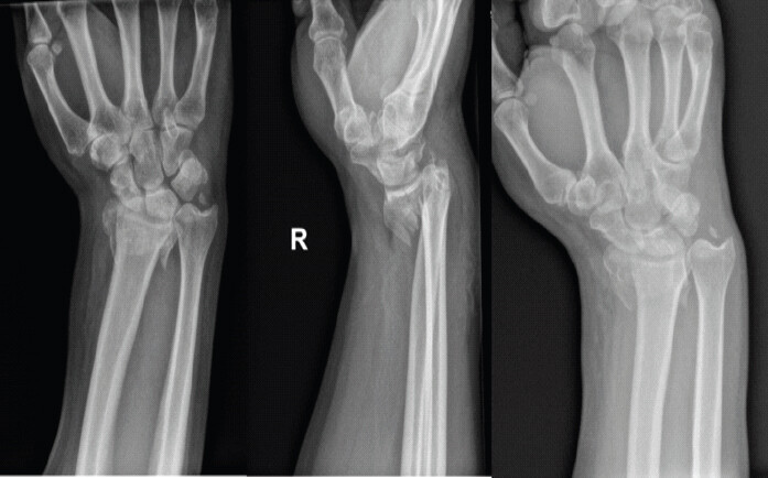
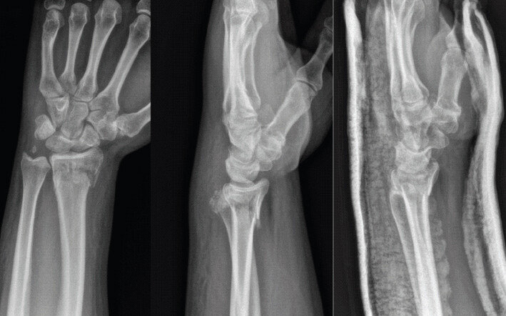
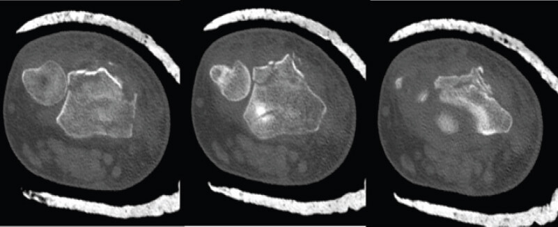
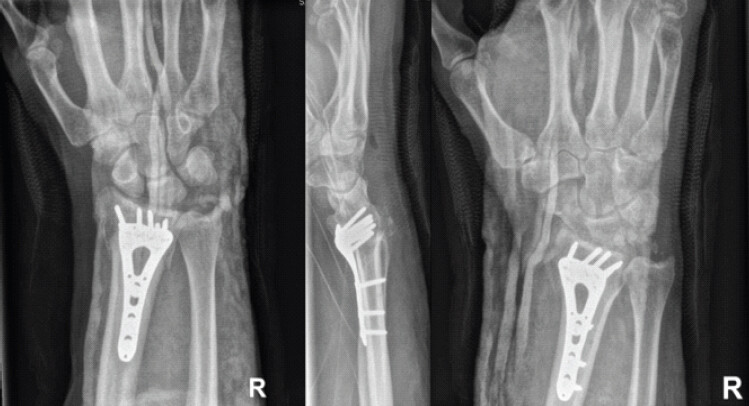
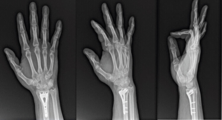
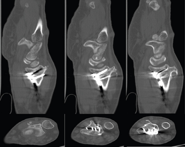
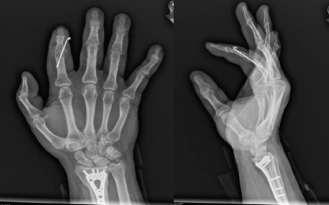
A 57-year-old healthy right-hand dominant male presents 2 years status post right distal radius ORIF with volar plate and screws and closed reduction of distal ulna for intra-articular distal radius and ulnar styloid fractures. Initial injury (Fig. 1), subsequent closed reduction (Fig. 2) radiographs, as well as representative axial computed tomography (CT) images (Fig. 3) are shown. He underwent volar plate ORIF (Fig. 4), and his initial post-operative course was uncomplicated. Twenty-two months postoperatively, the patient began having atraumatic persistent pain at the base of the thumb and had sudden inability to fully extend the thumb at the interphalangeal joint. Radiographs (Fig. 5) obtained in the clinic and CT (Fig. 6) demonstrated HO of the dorsal wrist. He followed up 1 month later; at that time, he reported a recent popping sensation of the thumb, which led to increased active range of motion for thumb extension. He continued to have persistent but less severe thumb pain after he felt the popping sensation.
Physical examination
Physical examination 23 months postoperatively demonstrated a well-healed previous surgical incision over the volar aspect of the wrist. The patient was non-tender to palpation over the right thumb CMC joint or into the dorsal aspect of the wrist, but there was a bony mass palpable over Lister’s tubercle. The patient was able to make a composite fist without issue. He had limited range of motion and weakness with thumb extension on the right hand compared to contralateral side and was unable to hold thumb fully extended when passively moved to full extension. Negative Finkelstein maneuver, negative right CMC grind test. Sensation intact distally to all fingers.
One month later, the patient reported a recent popping sensation and subsequent increased range of motion. Physical examination at this encounter demonstrated new findings of increased range of motion from prior but with continued weakness and pain with thumb extension on the right compared to contralateral side.
Surgical intervention
The patient underwent removal of HO of Lister’s Tubercle as well as removal of one screw from the volar distal radius plate that had been prominent, protruding from the dorsal cortex.
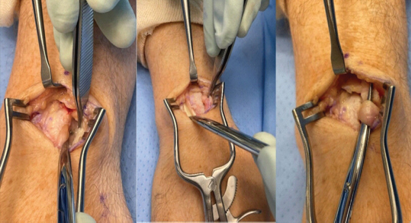
The patient underwent general anesthesia. Standard sterile prepping and draping was performed. A tourniquet was placed. First, a 3 cm dorsal wrist incision was used overlaying Lister’s tubercle and the bony mass. The extensor retinaculum was opened in a step-cut fashion, leading to identification of the 3rd and 4th dorsal compartment. Overlying the 3rd dorsal compartment, a significant amount of HO was found that had formed a bridging tunnel of bone (Fig. 7). A rongeur was used to remove the HO piece-by-piece. The bridging tunnel was strangulating the EPL. The EPL tendon had extensive tenosynovium surrounding the tendon, which was debrided; no significant fraying was present (Fig. 7). Dissection continued to underneath the 2nd dorsal compartment, and there was a screw with 3–4 mm of prominence with some fraying of the 2nd dorsal compartment. The decision was made to remove the screw (Fig. 8).
To remove the screw, a volar incision was centered over the distal half of the healed volar scar. A modified volar Henry approach was used in standard fashion. The prominent screw was removed without any other hardware removal without consequence. The patient wanted to return to full activity as soon as possible and preferred; through shared decision making, the plate was left to reduce risk of fracture
Once confirmation that the correct screw had been removed, the EPL was transposed superficial to the extensor retinaculum, which had been repaired with 0 Vicryl (Ethicon, Somerville, NJ) suture. Tourniquet was let down, irrigation was placed. The subcutaneous layers of both volar and dorsal incision were closed with 3-0 Vicryl (Ethicon, Somerville, NJ) followed by 4-0 nylon (Ethicon, Somerville, NJ) in a horizontal mattress fashion. The patient was placed into a sterile dressing and volar splint. He awoke from anesthesia without complication.
Post-operative course
His initial post-operative period was uncomplicated for the surgical site. On evaluation 10 days postoperatively, the patient had non-painful full range of motion of the thumb. The patient did have superficial thrombophlebitis on the contralateral upper extremity secondary to IV placement for surgery; he was treated with oral antibiotics. Sutures and splint were removed on post-operative day 10 without complication.
Two-months postoperatively, the patient continued to have excellent thumb range of motion; he also had no pain, well-healed surgical incision and was cleared for full activities. The patient unfortunately had a ipsilateral phalanx fracture and contralateral distal radius fracture 6 months later; at that time, the right wrist had progressed well and he had returned to full activity without limitation or pain. He has been seen in office over 1.5 years post-operative from this case presentation for other injuries, and he has continued to have no pain or limitation to the surgical wrist with full activity.
Discussion
This case demonstrates a novel complication of distal radius ORIF: HO over Lister’s tubercle creating a bony tunnel that entrapped the EPL tendon, causing tenosynovitis and functional impairment.
The patient had initially recovered well from distal radius ORIF but had subsequent surgery to excise heterotopic bone and remove a protruding screw. Both volar and dorsal incisions were made. The dorsal incision allowed for visualization of the tenosynovium of the EPL tendon as well as the excision of the heterotopic bone by rongeur; the volar incision was over the existing scar to remove the protruding screw. At the time of surgery, the EPL tendon was debrided and then transposed superficially to the extensor retinaculum due to the significant tenosynovitis seen intraoperatively for further protection. The patient wanted to a quick return to activity, so the decision was made to keep the majority of the volar hardware, as there was no other prominence or tendon fraying while in the operating room. At 2-month follow-up, the patient had no pain and had full range of motion of the thumb and continued to progress without difficulty beyond 1 year.
HO refers to ectopic bone formation through endochondral ossification, typically occurring after trauma through inflammatory cytokine activation [5]. While both HO and callus formation produce new bone through endochondral ossification, they differ fundamentally: Callus forms as part of normal fracture healing at the injury site, whereas HO represents pathologic bone formation in soft tissues away from the fracture. HO commonly affects the hips, knees, shoulders, and elbows [6, 7] but is rare in the wrist. One prior case reported HO of the volar ulnar-sided wrist causing ulnar nerve compression after minor trauma [8]. To the best of our knowledge, no prior publications have described HO extending from the dorsal radius over Lister’s tubercle with EPL tendon involvement or entrapment. The patient’s initial presentation of pain and weakness with thumb extension, followed by a “popping” sensation with improved range of motion 1 month later, likely represented intermittent mechanical adherence with locking and subsequent unlocking of the tendon within the ectopic bone tunnel.
EPL tendon injury is a recognized complication of distal radius ORIF with multiple etiologies. Attritional rupture commonly results from insufficient reduction and protruding volar screws [3,9] In addition, Cha et al. describe the “island-shape” fractures of of Lister’s tubercle narrowing the EPL groove, which may also lead to delayed rupture of the EPL [4]. Rivlin et al. reported EPL ruptures after corrective osteotomy for radius malunion caused by dorsal callus, prominent osteotomy edges, and osteophytes [10]. Recent evidence suggests dorsal comminution with EPL entrapment contributes to tendon injury even without prominent hardware [11]. In our case, both heterotopic bone and a protruding dorsal screw contributed to EPL tenosynovitis, though the ectopic bone tunnel represented the primary pathology.
Surgical excision of symptomatic HO is typically delayed 12–18 months to minimize recurrence [12]. Given the patient’s significant tenosynovitis and functional limitation, we proceeded with excision of the heterotopic bone and hardware removal almost 2 years after initial ORIF. The EPL tendon was transposed superficial to the extensor retinaculum for additional protection from residual bony irritation. The patient achieved complete symptom resolution and full thumb range of motion at 2-month follow-up and continued to have excellent outcomes over 1 year postoperatively.
This case highlights HO as a rare but important cause of EPL tenosynovitis after distal radius ORIF, which required surgical debridement to prevent additional injury or even rupture of the tendon. Surgeons should consider ectopic bone formation in addition to other causes of EPL injury after distal radius fracture.
Conclusion
This is a case of a healthy right-hand dominant male 2 years status post right distal radius ORIF with significant HO over Lister’s Tubercle creating a tunnel and causing tenosynovitis of the EPL tendon, which transiently led to inability to flex or extend the thumb. Radiographic evaluation demonstrated the ectopic bone formation over Lister’s tubercle as well as one protruding screw; surgical intervention consisted of screw removal, ectopic bone excision, and transposition of the EPL tendon superficial to the extensor retinaculum, resulting in decreased pain and restoration of thumb range of motion.
Clinical Message
Although rare, HO may be present after distal radius fractures treated with ORIF and can lead to irritation and tenosynovitis of the intact EPL tendon, leading to inability to flex or extend the thumb. Removal of the ectopic bone with transposition of the tendon can treat the pain and restore thumb motion.
Conflict of Interest:
Nil
Source of Support:
Nil
Consent:
The authors confirm that informed consent was obtained from the patient for publication of this article
How to Cite this Article
Barclay A, Greco V. Bridge over Troubled Tendon? A Novel Case of Heterotopic Ossification of Lister’s Tubercle Causing Extensor Pollicis Longus Tenosynovitis. Journal of Orthopaedic Case Reports 2026 October;16(10): 80-85.
References
- Palola V, Ponkilainen V, Huttunen T, Launonen A, Mattila VM. Incidence for volar locking plate removal following distal radius fracture surgery. Arch Orthop Trauma Surg 2021;141:1297-302. [Google Scholar] | [PubMed]
- Azzi AJ, Aldekhayel S, Boehm KS, Zadeh T. Tendon rupture and tenosynovitis following internal fixation of distal radius fractures: A systematic review. Plast Reconstr Surg 2017;139:717e-24. [Google Scholar] | [PubMed]
- Benson EC, DeCarvalho A, Mikola EA, Veitch JM, Moneim MS. Two potential causes of EPL rupture after distal radius volar plate fixation. Clin Orthop Relat Res 2006;451:218-22. [Google Scholar] | [PubMed]
- Cha SM, Shin HD, Lee SH. “Island-shape” Fractures of Lister's tubercle have an increased risk of delayed extensor pollicis longus rupture in distal radial fractures: After surgical treatment by volar locking plate. Injury 2018;49:1816-21. [Google Scholar] | [PubMed]
- Mujtaba B, Taher A, Fiala MJ, Nassar S, Madewell JE, Hanafy AK. Heterotopic ossification: Radiological and pathological review. Radiol Oncol 2019;53:275-84. [Google Scholar] | [PubMed]
- Shehab D, Elgazzar AH, Collier BD. Heterotopic ossification. J Nucl Med 2002;43:346-53. [Google Scholar] | [PubMed]
- Agarwal S, Loder S, Levi B. Heterotopic ossification following upper extremity injury. Hand Clin 2017;33:363-73. [Google Scholar] | [PubMed]
- Taha M, Gowda CS, Paniker J. Ulnar nerve compression at the wrist secondary to heterotopic ossification following a minor trauma: A case report. J Orthop Case Rep 2025;15:126-30. [Google Scholar] | [PubMed]
- Zenke Y, Sakai A, Oshige T, Moritani S, Menuki K, Yamanaka Y. Extensor pollicis longus tendon ruptures after the use of volar locking plates for distal radius fractures. Hand Surg 2013;18:169-73. [Google Scholar] | [PubMed]
- Rivlin M, Fernández DL, Nagy L, Graña GL, Jupiter J. Extensor pollicis longus ruptures following distal radius osteotomy through a volar approach. J Hand Surg Am 2016;41:395-8. [Google Scholar] | [PubMed]
- Laane CL, Husseini JS, Huang AJ, Balza Romero R, Stam M, Chen NC. Extensor pollicis longus entrapment on ct in acute distal radius fracture may be a predictor of tendon rupture. Hand (N Y) 2025;21:630-6. [Google Scholar] | [PubMed]
- Mavrogenis AF, Soucacos PN, Papagelopoulos PJ. Heterotopic ossification revisited. Orthopedics 2011;34:177. [Google Scholar] | [PubMed]
© 2026 Journal of Orthopaedic Case Reports - Published by Indian Orthopaedic Research Group
About the Authors
-

Anna Barclay
-

Victor Greco
Related Articles in Journal of Orthopaedic Case Reports
 October 1, 2026 Single-Stage Fixation of Bilateral Open Medial Hoffa Fractures Associated with Bilateral Patella Fracture and Bilateral Distal Radius Fractures: A Case Report
 September 1, 2026 Proximal Row Carpectomy for Established Post-Traumatic Radiocarpal Arthritis Following Distal Radius Volar Rim and Scaphoid Fractures: A Case Report
 September 1, 2026 Concomitant Distal Radius Fracture with Scaphoid Fracture-Dislocation and Carpal Instability: A Case Report
 September 1, 2026 Extensive Post-Traumatic Heterotopic Ossification with Multi-joint Involvement: A Case Report