


Introduction
A Hoffa fracture is a coronal-plane fracture of the femoral condyle and most commonly follows high-energy trauma. Because the fracture line may be obscured by overlapping structures on standard anteroposterior radiographs, these injuries can be missed during the initial assessment of a patient with multiple injuries [1,2]. These fractures most commonly involve the lateral femoral condyle; medial condyle involvement is less frequent, and bilateral Hoffa fractures are exceptionally rare, with only a limited number of cases reported in the literature [2].
Hoffa fractures are unstable intra-articular injuries and may be accompanied by periarticular damage including patellar fractures, disruption of the extensor mechanism, and ligamentous injury [3,4]. Open injuries add the risks of contamination, infection, soft-tissue loss, and difficulties in selecting the timing and method of fixation [5]. Simultaneous bilateral lower-limb and upper-limb injuries further complicate surgical prioritization and post-operative mobilization [6,7].
We report an unusual multi-extremity injury pattern comprising bilateral open medial Hoffa fractures, bilateral patellar fractures, posterior dislocation of the right knee with popliteal artery compression, and bilateral distal radius fractures. The case highlights emergency vascular assessment, fracture-specific fixation, the rationale for single-stage definitive surgery after physiological stabilization, and the challenges of rehabilitation when both wrists and both knees are injured.
Case Report
A 23-year-old man was brought to a level-1 tertiary trauma centre after a two-wheeler road traffic accident associated with a brief loss of consciousness. On arrival, the patient was in hypovolemic shock and underwent immediate resuscitation according to Advanced Trauma Life Support principles. Initial arterial blood gas analysis demonstrated a pH of 7.384, pCO2 of 47.2 mmHg, and pO2 of 69.5 mmHg. Resuscitation included oxygen supplementation, intravenous crystalloids and plasma expanders, together with transfusion of 2 units of packed red blood cells. As per the institutional open-fracture protocol, intravenous piperacillin-tazobactam was initiated immediately on arrival and tetanus prophylaxis was administered. The patient had sustained multiple injuries involving both lower limbs and both wrists.
The right knee had a Gustilo-Anderson grade IIIB open injury, with an anterior laceration measuring approximately 7 × 3 × 3 cm and an additional distal thigh wound measuring 10 × 4 × 4 cm. Both wounds were communicated with the underlying injury. The left knee had a Gustilo-Anderson grade IIIB open injury, with an anterior wound measuring approximately 10 × 3 × 3 cm, exposed soft tissue with contamination (Fig. 1).
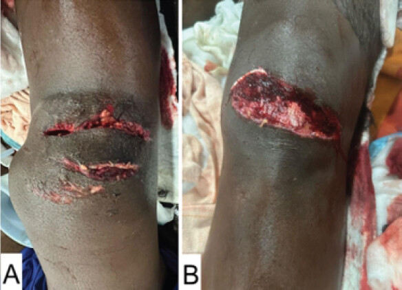
The right wrist had a closed injury with deformity. The left wrist had a puncture wound over the volar aspect of the distal radius communicating with the fracture, along with a deep dermal abrasion. Distal pulses were markedly diminished in the right lower limb. The left lower limb and both upper limbs had preserved distal neurovascular status.
Radiological Investigations
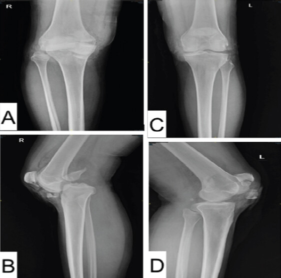
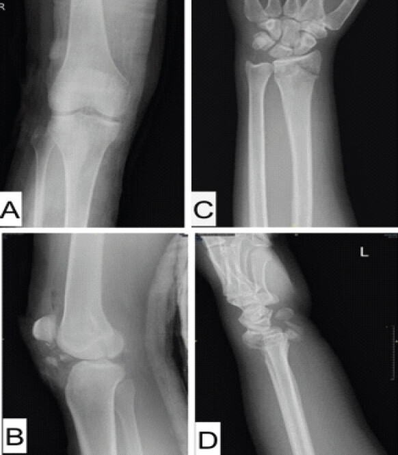
Radiographs of the right knee demonstrated posterior dislocation with a comminuted medial femoral condyle Hoffa fracture, intra-articular extension, and an inferior-pole fracture of the patella. The left knee showed a medial Hoffa fracture with a comminuted patellar fracture (Fig. 2).
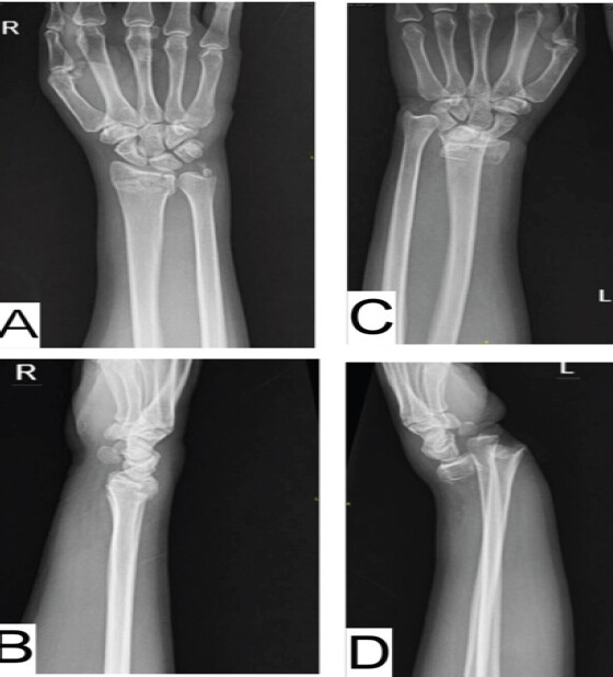
The right wrist showed a displaced, comminuted intra-articular distal radius fracture with an associated ulnar styloid fracture (AO/OTA 2R3B2). The left wrist showed a dorsally displaced, comminuted intra-articular distal radius fracture with disruption of the distal radioulnar joint (AO/OTA 2R3B2.3) (Fig. 3).
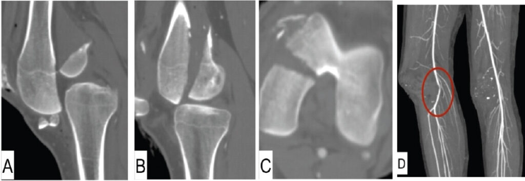
Computed tomography of both knees demonstrated the coronal fracture configuration, comminution, and articular extension of the medial femoral condylar fractures; the angiographic images also showed focal compression and severe stenosis of the right mid-popliteal artery at the level of the posteriorly dislocated knee, without arterial transection or contrast extravasation (Fig. 4A–D).
Management and Outcomes
Emergency closed reduction of the right knee was performed to relieve vascular compression and restore distal perfusion. Closed reduction of the left distal radius fracture was also performed to restore wrist alignment (Fig. 5). After reduction, arterial Doppler examination of the right lower limb demonstrated triphasic distal flow, confirming restoration of limb perfusion. Thereafter, serial clinical vascular examinations of right lower limb were performed throughout the period of hospitalization with regular assessment of distal pulses, limb perfusion, and capillary refill. The distal pulses remained palpable throughout the admission with no clinical evidence of recurrent or delayed vascular compromise.
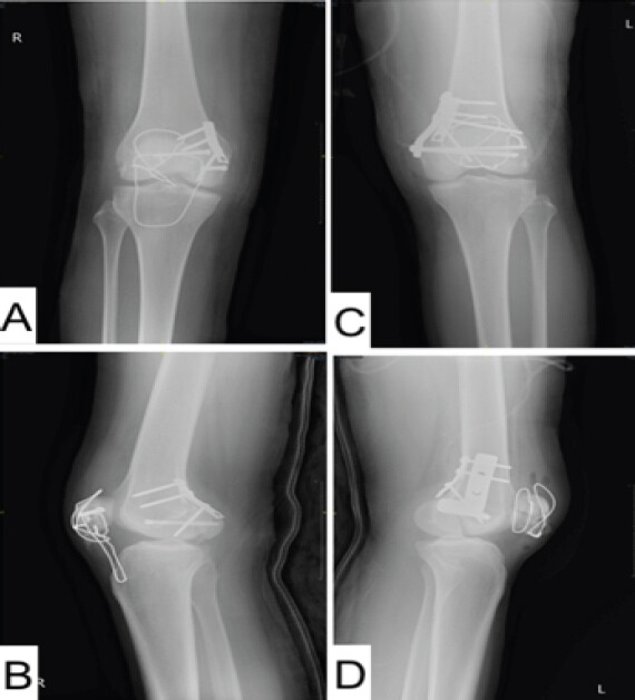
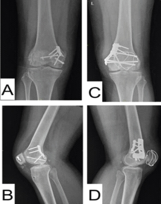
Following resuscitation, haemodynamic stability was achieved. Repeat arterial blood gas analysis showed a pH of 7.425, pCO2 of 42.6 mmHg, and pO2 of 95.6 mmHg. After restoration of right lower-limb perfusion, reassessment, and multidisciplinary evaluation, the patient was considered adequately stabilised to undergo single-stage definitive surgical management. Both contaminated open knee injuries and open left wrist injury underwent meticulous surgical exploration and debridement by the plastic surgery team, with the removal of contamination and devitalized tissue followed by thorough irrigation. Material was sent for gram stain and culture. Following satisfactory debridement and definitive fracture fixation, primary wound closure was achieved. No additional flap or skin graft coverage was required. Intravenous piperacillin-tazobactam was continued postoperatively as per institutional open fracture antibiotic protocol for seven days. The right medial Hoffa fracture was anatomically reduced and stabilised with an anteroposterior interfragmentary compression screw and a one-third semitubular buttress plate to resist shear across the coronal fracture plane. The inferior-pole patellar fracture was stabilised with tension-band wiring, while the associated patellar-tendon disruption was reconstructed using suture anchors and augmented with McLaughlin wiring. On the left side, the medial Hoffa fracture was stabilised with a one-third tubular plate and an additional L-shaped buttress plate to provide supplementary stability to the large medial femoral condylar fragment. The patellar fracture was stabilised with tension-band wiring (Fig. 6). For both knees, the existing traumatic open wounds were extended as required to obtain adequate exposure for debridement, fracture reduction, and fixation. The procedures were performed under tourniquet control.
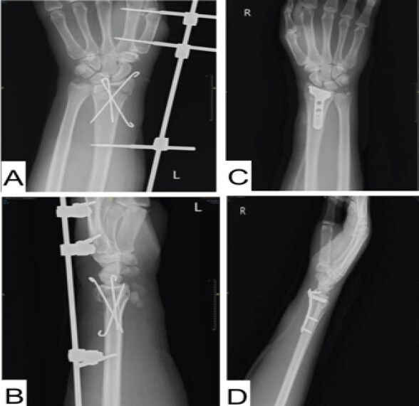
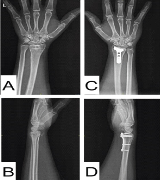
The closed right distal radius fracture was approached through a modified Henry approach and underwent open reduction and volar plate fixation. The open left distal radius fracture was debrided and stabilised with Kirschner wires and a wrist-spanning external fixator to limit additional soft-tissue injury while maintaining alignment and stability (Fig. 7). The entire single-stage procedure was completed in approximately 4 hours. Estimated intraoperative blood loss was less than 100 mL, and no additional intraoperative blood transfusion was required.
Postoperatively, rehabilitation was individualised because of the simultaneous bilateral knee and wrist injuries. In-bed range-of-motion and static muscle exercises were initiated during the immediate postoperative period. Deep-vein thrombosis prophylaxis was administered according to protocol. Tissue samples obtained during debridement showed no bacterial growth even in delayed culture. The primarily closed wounds were monitored closely for wound breakdown, skin necrosis, and infection and subsequently healed without the need for additional soft-tissue procedures. During the initial 2 weeks, supervised knee mobilisation was restricted to 0° to 30° of flexion. Thereafter, full active knee range-of-motion exercises were initiated and gradually progressed as tolerated. Weight-bearing was initially deferred bilaterally until satisfactory soft-tissue healing and radiographic assessment. Full weight-bearing was allowed on the left lower limb at 3 months; partial weight-bearing was begun on the right because of the associated extensor-mechanism reconstruction, progressing to full weight-bearing in both lower limbs at 4 months. The right McLaughlin wire and patellar tension-band wire were removed at 8 months to avoid implant-related complications. Following volar plate fixation of the right wrist, active wrist range-of-motion exercises were initiated immediately. The left wrist-spanning external fixator was removed at 2 months, after which supervised wrist mobilisation was initiated.
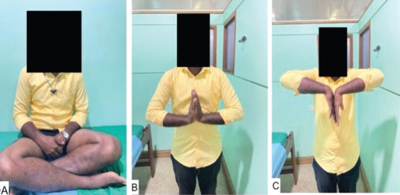
At 1-year follow-up, all wounds and surgical scars were well healed, with no evidence of infection. Distal neurovascular status was intact in all four limbs. Both wrists demonstrated 80° of palmar flexion, 70° of dorsiflexion, 80° of pronation, and 80° of supination, with preserved sensation and satisfactory grip; the Modified Mayo Wrist Score was 85 on each side (Fig. 8). Both knees had a range of motion from 0° to 120° and no extensor lag on either side, with Lysholm scores of 91 on the right and 90 on the left. The patient was independently ambulant and had returned to his pre-injury occupation on unrestricted duties without limitation related to the knees or wrists. Follow-up radiographs demonstrated union of all fractures with satisfactory alignment and stable implants (Fig. 9 and 10).
Discussion
The present case highlights the decision-making involved in same-day definitive fixation of complex multi-extremity trauma following successful resuscitation and restoration of limb perfusion. The principal management priorities were correction of haemodynamic instability, rapid restoration of limb perfusion, debridement of contaminated open injuries, stable reconstruction of multiple articular surfaces and the extensor mechanism, and selection of a surgical strategy that would permit coordinated rehabilitation.
The strategy for fracture fixation in polytrauma has evolved from Early Total Care (ETC) to Damage-Control Orthopaedics (DCO) and subsequently to Early Appropriate Care (EAC). ETC advocates early definitive fixation of major fractures to facilitate mobilisation and reduce complications associated with prolonged immobilisation [9,10]. However, prolonged definitive surgery in a physiologically unstable patient may contribute to a harmful ‘second-hit’ response. DCO was therefore introduced for unstable or borderline patients and involves temporary fracture stabilisation, commonly with external fixation, followed by definitive fixation after physiological recovery [11,12]. EAC provides a more individualised approach in which definitive fixation is undertaken once an adequate response to resuscitation has been achieved, using haemodynamic and metabolic parameters to assess readiness for surgery [13].
In the present case, the patient initially presented with hypovolemic shock but responded satisfactorily to ATLS-based resuscitation, including transfusion of 2 units of packed red blood cells. Post-resuscitation arterial blood gas analysis showed a pH of 7.425, pCO2 of 42.6 mmHg, and pO2 of 95.6 mmHg, and haemodynamic stability was achieved before surgery. Urgent reduction of the right knee restored distal perfusion, with triphasic arterial Doppler flow and maintained distal pulses on subsequent clinical surveillance. In addition, the patient had multiple unstable intra-articular injuries involving both knees and wrists, together with disruption of the right extensor mechanism. Adequate debridement and primary closure of the contaminated open wounds could be achieved, and multidisciplinary orthopaedic, plastic-surgery, vascular, anaesthesia, and critical-care support was available at a Level I trauma centre. Taken together, these factors supported proceeding with same-day definitive fixation in accordance with EAC principles rather than adopting a DCO strategy.
In this patient, single-stage definitive fixation was selected to avoid staged definitive procedures and repeated anaesthetic exposure and to permit a coordinated rehabilitation strategy. Fixation was individualised according to fracture morphology and soft-tissue status [14,15].
Buttress plating was selected for the medial Hoffa fragments to resist shear displacement. Associated patellar fractures increase the complexity of Hoffa fractures because restoration of the extensor mechanism and articular congruity must be achieved simultaneously. The fixation construct should be selected according to the degree of comminution, fragment size, tendon disruption, and soft-tissue condition [16–19]. In the present patient, tension-band fixation was used for both patellar fractures, while suture-anchor reconstruction and McLaughlin wire augmentation were added on the right because of the associated patellar-tendon injury. These methods provided sufficient stability for a staged rehabilitation programme. The absence of arterial transection, prompt restoration of triphasic distal flow after reduction, and maintenance of palpable distal pulses on serial clinical examinations supported continued vascular surveillance without arterial reconstruction.
Volar plate fixation of the closed right distal radius fracture provided rigid fixation and preserved the potential for early functional use of one upper limb [20]. By contrast, the contaminated open left wrist injury was treated with Kirschner wires and a spanning external fixator to reduce additional soft-tissue insult while maintaining fracture reduction.
Rehabilitation was particularly challenging because bilateral wrist fractures initially prevented effective use of walking aids. Hence, mobilisation was initially limited predominantly to in-bed exercises. Mobilisation was therefore individualised according to wound healing, stability of the fixation, extensor-mechanism recovery, and upper-limb function. Despite this limitation, the patient achieved fracture union, good functional range of movement in all four injured joints, and return to work at 1 year.
As this is a single case report, no firm conclusions can be drawn regarding the overall superiority or complication-reducing effect of single-stage fixation. The favourable outcome in this patient should be interpreted in the context of careful patient selection, successful resuscitation, restoration of limb perfusion, meticulous soft-tissue management, and multidisciplinary care..
Conclusion
Complex multiextremity trauma requires rapid identification of limb-threatening vascular compromise, meticulous debridement of open injuries, and fixation tailored to fracture morphology and soft-tissue condition. In an adequately resuscitated and physiologically stabilized patient, single-stage definitive fixation may reduce repeated surgical exposure and support coordinated rehabilitation. Careful multidisciplinary decision-making and structured follow-up were central to the favorable outcome in this case.
Clinical Message
In a polytrauma patient with bilateral periarticular knee injuries and bilateral wrist fractures, urgent reduction of a dislocated knee can restore perfusion and preserve the limb. Once physiological stability is achieved, fracture-specific single-stage fixation, meticulous soft-tissue management, and an individualized rehabilitation plan can produce satisfactory union and functional recovery.
Conflict of Interest:
Nil
Source of Support:
Nil
Consent
The authors confirm that informed consent was obtained from the patient for publication of this article
How to Cite this Article
Magadum SD, Jasti RP, Mohandas P, Ravichandran SB. Single-Stage Fixation of Bilateral Open Medial Hoffa Fractures Associated with Bilateral Patella Fracture and Bilateral Distal Radius Fractures: A Case Report. Journal of Orthopaedic Case Reports 2026 October;16(10): 154-160.
References
- Patel PB, Tejwani NC. The Hoffa fracture: Coronal fracture of the femoral condyle a review of literature. J Orthop 2018;15:726-31. [Google Scholar] | [PubMed]
- Zhou Y, Pan Y, Wang Q, Hou Z, Chen W. Hoffa fracture of the femoral condyle: Injury mechanism, classification, diagnosis, and treatment. Medicine (Baltimore) 2019;98:e14633. [Google Scholar] | [PubMed]
- Pathak S, Salunke A, Karn S, Ratna HV, Thivari PS, Sharma S. Hoffa's fracture with associated injuries around the knee joint: An approach to a rare injury. Cureus 2020;12:e7865. [Google Scholar] | [PubMed]
- Huang G, Zhang M, Zhang Y, Wang X, Zhang M, Liu G. Hoffa fracture combined with rotational dislocation of the knee joint: A novel case report. Medicine (Baltimore) 2021;100:e25253. [Google Scholar] | [PubMed]
- Tomar L, Govil G, Dhawan P. Open complex distal femur intraarticular fracture with Hoffa extension, extensor mechanism tear and post-recovery secondary traumatic peri implant fracture: Report on an uncommon chronology. Int J Res Orthop 2020;6:1335-40. [Google Scholar] | [PubMed]
- Wu CM, Liao HE, Lan SJ. Simultaneous bilateral floating knee: A case report. World J Clin Cases 2022;10:10172-9. [Google Scholar] | [PubMed]
- Akel A, Sarhan MY, Abu-Jeyyab M, Daradkeh ST, Moseley S, Dawoud MS. Medial hoffa fracture: A case report and literature review of approach and management. Am J Case Rep 2024;25:e943136. [Google Scholar] | [PubMed]
- Simpson P, Keating JF. The multiply injured patient. Found Years 2008;4:314-8. [Google Scholar] | [PubMed]
- Nicola R. Early total care versus damage control: Current concepts in the orthopedic care of polytrauma patients. ISRN Orthop 2013;2013:329452. [Google Scholar] | [PubMed]
- Vatkar A, Kale S, Jayaram R, Verma A, Pandey S, Kale S. 1-Year follow-up result of early definitive management of the trauma patient in a tertiary health care centre. J Orthop Case Rep 2025;15:183-8. [Google Scholar] | [PubMed]
- Cimbanassi S, O'Toole R, Maegele M, Henry S, Scalea TM, Bove F. Orthopedic injuries in patients with multiple injuries: Results of the 11th trauma update international consensus conference Milan, December 11, 2017. J Trauma Acute Care Surg 2020;88:e53-76. [Google Scholar] | [PubMed]
- Avraham D, Herman A, Oulianski M. A case of damage control after polytrauma and bilateral femur fracture. Trauma Case Rep 2024;52:101037. [Google Scholar] | [PubMed]
- Weinberg DS, Narayanan AS, Moore TA, Vallier HA. Assessment of resuscitation as measured by markers of metabolic acidosis and features of injury. Bone Joint J 2017;99-B:122-7. [Google Scholar] | [PubMed]
- Patil A, Vinayak U, Attarde D, Shyam A, Sancheti P, Rapole S. Single-stage definitive fixation for floating hip injuries: Evaluating functional outcomes and complications. Eur J Orthop Surg Traumatol 2025;35:309. [Google Scholar] | [PubMed]
- Bertrand ML, Andrés-Cano P, Pascual-López FJ. Periarticular Fractures of the Knee in Polytrauma Patients. Open Orthop J 2015;9:332-46. [Google Scholar] | [PubMed]
- Gadagoli BS, Raghavendra MS, Kubsad S, Suresha B, Nitish K, Pai HS. Observational study of clinical profile of fractures patella. Int J Pharm Clin Res 2023;15:203-9. [Google Scholar] | [PubMed]
- Matthews B, Hazratwala K, Barroso-Rosa S. Comminuted patella fracture in elderly patients: A systematic review and case report. Geriatr Orthop Surg Rehabil 2017;8:135-44. [Google Scholar] | [PubMed]
- Babar TK, Bele AW, Singh NC, Qureshi MI, Kovela RK, Kulkarni CA. A case report of comminuted patella fracture with open reduction. J Pharm Res Int 2021;33:1-5. [Google Scholar] | [PubMed]
- Luo TD, Marino DV, Pilson H. Patella fractures. In: StatPearls. Treasure Island, FL: StatPearls Publishing; 2025 [Google Scholar] | [PubMed]
- Drobetz H, Koval L, Weninger P, Luscombe R, Jeffries P, Ehrendorfer S. Volar locking distal radius plates show better short-term results than other treatment options: A prospective randomised controlled trial. World J Orthop 2016;7:687-94. [Google Scholar] | [PubMed]
© 2026 Journal of Orthopaedic Case Reports - Published by Indian Orthopaedic Research Group
About the Authors
-

Sunil D Magadum
-

Ram Prasad Jasti
-

Prithvi Mohandas
-

Surya Balaji Ravichandran
Related Articles in Journal of Orthopaedic Case Reports
 October 1, 2026 Evaluation of Functional Outcome of Extended Flexor Carpi Radialis Approach for Fracture Fixation of Distal End Radius Fracture
 October 1, 2026 Bilateral Cedell Fractures of the Talus Associated with Ankle Fractures in a Polytrauma Patient: A Rare Injury Managed with Damage Control Orthopedics and Staged Fixation
 October 1, 2026 Degloving in Disguise: Managing a Delayed Spinal Morel-Lavallée Lesion after Thoracic Fracture-Dislocation
 October 1, 2026 Bridge over Troubled Tendon? A Novel Case of Heterotopic Ossification of Lister’s Tubercle Causing Extensor Pollicis Longus Tenosynovitis