


Introduction
Osteochondromas are benign bone tumorscharacterisedby sessile or pedunculated bony projections arising from the external surface of bones and are most commonly diagnosed in individuals younger than 20 years of age[1]. They originate from bones that undergo endochondral ossification, with the region around the knee – particularly the distal femur and proximal tibia – representing the most frequent sites of occurrence. Approximately 85% of osteochondromas present as solitary lesions, whereas the remaining 15% occur as multiple lesions, typically in association with hereditary multiple exostoses (HME), an autosomal dominant disorder in which nearly 90% of cases are linked to pathogenic mutations in the EXT1 or EXT2 genes[1]. Any sudden increase in size or alteration in radiological appearance after skeletal maturity warrants careful evaluation, as such changes may suggest malignant transformation[1,2]. The risk of malignant degeneration is significantly higher in HME, with around 10% of affected individuals developing secondary chondrosarcoma, compared to approximately 1% in cases of solitary osteochondroma [1,3,4]. Cartilage cap thickness >2 cm in adults or >3 cm in children, as well as new onset of pain or growth, or rapid growth of the lesion, especially after the closure of the growth plate, might reflect cancerous transformation[5]. Osteochondroma has a characteristic histologic appearance similar to a normal growth plate. The cartilaginous cap is formed of primary trabeculae of hyaline cartilage and linear clusters of active chondrocytes with a surrounding well-defined perichondrium. However, malignant transformation into a chondrosarcoma presents a different microscopic picture. Microscopic examination of chondrosarcoma may reveal necrosis and mitotic activity. Nuclear enlargement, hyperchromasia, size variation, and binucleation are characteristic features observed in chondrocytes [6]. Osteochondromas involving the foot and ankle are uncommon, and those arising from the calcaneus are even rarer. Unlike lesions in other anatomical regions, which may remain asymptomatic until adulthood, calcaneal osteochondromas tend to become symptomatic early due to their interference with weight-bearing, leading to pain and difficulty during walking. Although rapid enlargement of an osteochondroma in adults often raises suspicion for malignant transformation, the present case describes a 38-year-old male with a progressively enlarging calcaneal osteochondroma that demonstrated no evidence of malignancy on histopathological examination.
Case Report
A 38-year-old male with no significant past medical, surgical, or family history presented to our outpatient department with long-standing complaints involving his left foot. He reported experiencing dull, aching pain for the past 8 years, which progressively worsened over time. Approximately 1 year after the onset of pain, he noticed the gradual appearance of a swelling over the plantar aspect of the foot, which continued to enlarge steadily over the next 5 years before stabilizing in size over the last 2 years. Initially, the discomfort was mild and occurred only while wearing slippers or after prolonged ambulation. However, during the last 4 years, the patient experienced a substantial escalation in symptoms, describing severe, excruciating pain during routine walking and prolonged standing, even with soft or cushioned footwear. This began to significantly impair his daily activities and mobility. He underwent treatment at several local health care centers and took over-the-counter medications, but his symptoms persisted, and finally he was referred to our center for further management.
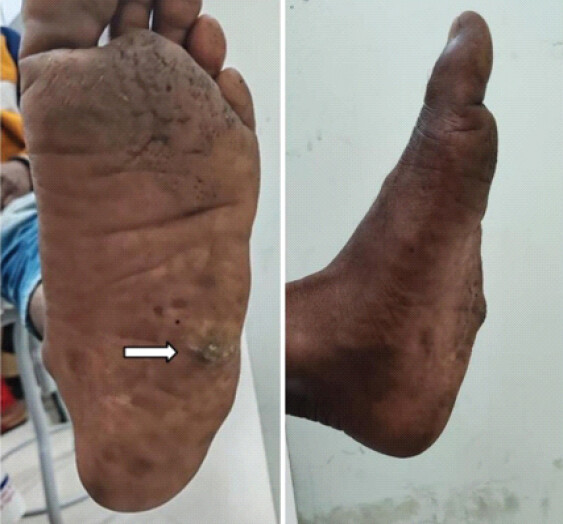
Clinical inspection revealed a large, prominent swelling arising from the plantolateral aspect of the left heel. The overlying skin exhibited marked hyperkeratosis with superficial excoriation at the apex – likely secondary to chronic friction and weight-bearing pressure (Fig. 1). On palpation, the mass was hard and tender, but there was no associated warmth, erythema, or fluctuance. Hindfoot movements, including inversion, eversion, plantar flexion, and dorsiflexion, remained unrestricted and painless.
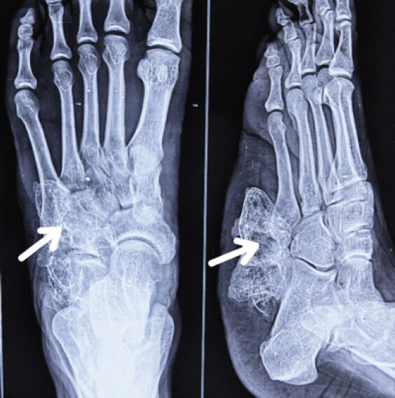
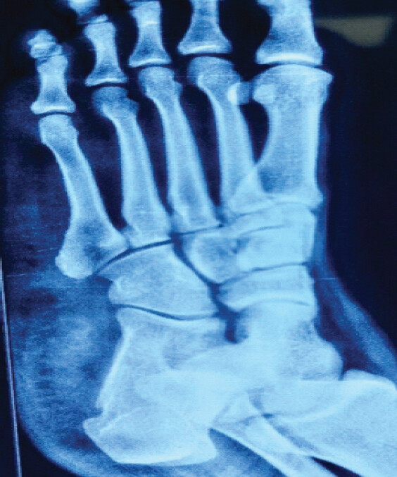
Standing radiographs of the left foot demonstrated a large sessile bony outgrowth projecting from the plantolateral surface of the calcaneus, consistent with an osteochondromatous lesion (Fig. 2). The other differential diagnoses that were considered were parosteal osteosarcoma, large calcaneal osteophytes. The patient presented with red flag signs of suspected secondary malignant transformation of the lesion, such as rapid growth and worsening pain. Magnetic resonance imaging (MRI) was performed, which showed characteristic cortical and medullary continuity with the underlying calcaneum, with a largest cartilage cap thickness of 8mm with no adjacent soft-tissue involvement. This further helped us to exclude the other differential diagnoses. Additional skeletal survey imaging was performed to exclude HME; no other lesions were identified.
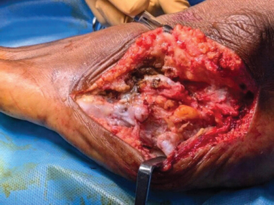
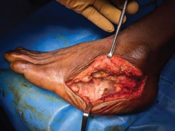
Given the progressive symptoms and mechanical impairment, the patient was planned for surgical excision. He was positioned in the floppy lateral position on the operating table. A curvilinear skin incision approximately 12 cm in length was made along the mucocutaneous junction of the lateral aspect of the foot (Fig. 3). Full-thickness flaps were elevated carefully to expose the tumor while protecting the peroneal tendons and adjacent neurovascular structures. The mass was excised en bloc, leaving behind a well-defined cavity in the calcaneal region (Fig. 4).
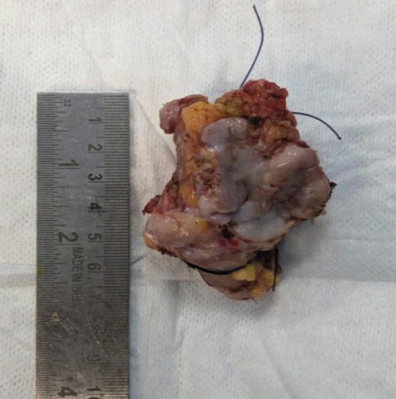
The excised tumor measured 7.5 × 5 × 3 cm (Fig. 5), measuring one of the largest in volume (112.5 cm3) among calcaneal osteochondromas that have been reported. A search in the literature yielded a maximum volume of approximately70.5 cm3 reported by Blitz and Lopez [3]. It had a maximum cartilage cap thickness of 9 mm. The wound was closed in layers using modified Allgöwer-Donati sutures.
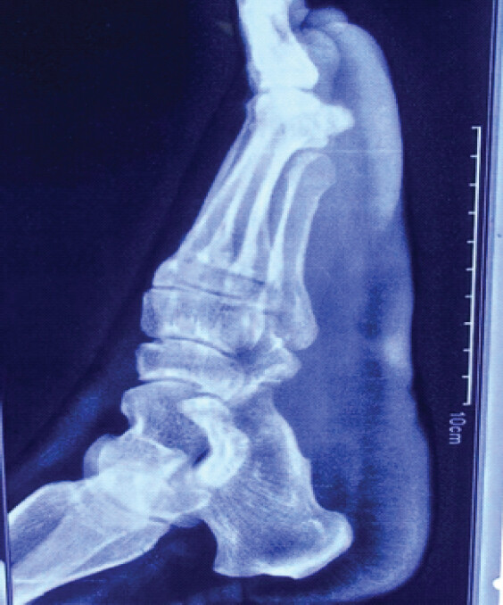
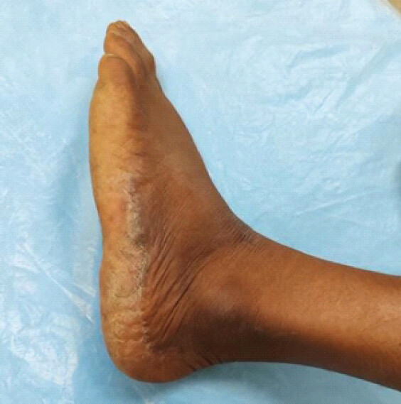
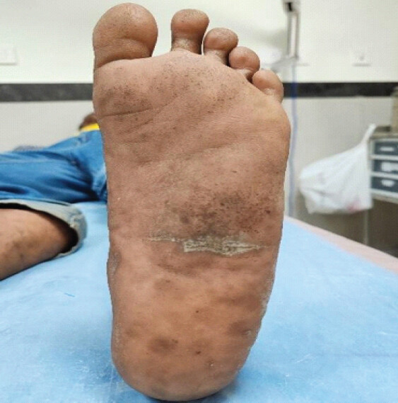
Immediate post-operative radiographs confirmed complete excision of the lesion with no residual bony projection (Fig. 6). The incision healed uneventfully, and sutures were removed at 21 days, revealing healthy wound recovery. At the 6-month follow-up, the patient reported complete resolution of pain and had returned to normal daily activities. Clinical examination at this stage showed a well-healed foot contour on both lateral and plantar aspects (Figs. 7 and 8). At 24 months postoperatively, radiographs demonstrated no recurrence of the lesion (Fig. 9).
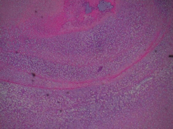
Histopathological examination confirmed the diagnosis of a benign osteochondroma. Microscopy revealed multiple fragments lined by a hyaline cartilage cap covered by a fibrous perichondrium. A distinct zone of enchondral ossification was evident at the interface between cartilage and bone. The osseous component displayed mature, laminated trabeculae interspersed with adipocytic marrow. No features of malignancy – such as necrosis, nuclear atypia, or abnormal mitotic activity – were identified (Fig. 10).
Discussion
Osteochondromas account for approximately 2–3% of the general population and represent the most common benign bone tumors[7,8]. Of all osteochondromas, only about 10% arise in the bones of the hands and feet, making these locations relatively uncommon sites of involvement[4,9]. Within this subset, calcaneal tumors constitute an estimated 3% of all foot and ankle neoplasms[1]. Most osteochondromas are asymptomatic and discovered incidentally; however, those arising from the calcaneus tend to become symptomatic earlier because of the mechanical stresses and weight-bearing forces acting on the hindfoot.
Malignant transformation is rare in solitary osteochondromas, occurring in <1% of cases, but the risk rises significantly to 5–25% in patients with HME [3,10]. Clinical warning signs suggestive of malignant degeneration include rapid or renewed growth of the lesion after skeletal maturity and the new onset of persistent or worsening pain. When malignant change occurs, it typically results in secondary low-grade chondrosarcoma, with the thickness of the cartilage cap serving as one of the most reliable predictors of transformation[9,11,12]. Cartilage cap thickness >2 cm in adults or >3 cm in children is considered suspicious of malignant transformation [5].
Diagnostic evaluation generally begins with standard plain radiographs, which are usually sufficient to identify the typical corticomedullary continuity characteristic of osteochondroma. Advanced imaging modalities such as computed tomography or MRI may be utilised for detailed pre-operative planning, assessment of neurovascular involvement, and evaluation of cartilage cap thickness to exclude sarcomatous degeneration. Bone scans may also be employed in select cases. Despite the relative prevalence of osteochondromas in long bones, very few cases of calcaneal osteochondroma have been reported in the literature[13, 14, 15, 16]. Moreover, the anatomical origins of these lesions vary widely across studies, including locations such as the medial tubercle, posteroinferior calcaneal surface, and areas near the medial malleolus. In our patient, the lesion arose from the plantolateral aspect of the calcaneum, similar to the cases described by Nogier et al.[4] and Avramidis et al.[17].
Definitive management involves complete surgical excision with a tumor-free margin. Achieving total removal is associated with a recurrence rate of <2%, whereas incomplete excision – particularly when remnants of the cartilage cap remain – can predispose to recurrence[3]. Based on the benign radiological characteristics of the lesion and the absence of features suggestive of HME, we elected to perform an en bloc excision. It is noteworthy that small osteophytes in the foot and ankle region may clinically mimic an osteochondroma and must be differentiated appropriately.
Nogier et al.[4] described a similar case involving a 36-year-old male with a large plantolateral calcaneal osteochondroma that continued to grow after skeletal maturity without malignant transformation, closely paralleling our findings. Koplay et al.[9] also reported a case of a 25-year-old female with a posteromedial calcaneal osteochondroma that enlarged during adulthood. Although the lesion recurred shortly after initial excision and prompted concern for malignant change, histopathology on re-excision again confirmed benign features.
In another comparable report, Avramidis et al.[17] presented a solitary, large calcaneal osteochondroma that demonstrated extensive post-maturity growth. Surgical excision was performed, and histopathology confirmed benign osteochondroma without malignancy. The patient remained recurrence-free at 3-year follow-up.
Our case aligns closely with the observations of Avramidis et al.[17] and Nogier et al.[4], featuring a large plantolateral calcaneal osteochondroma that continued to enlarge even after epiphyseal closure. The lesion was excised en bloc, and histopathological evaluation confirmed benign characteristics without evidence of malignant transformation. Postoperatively, the patient regained normal functional mobility within three weeks, with suture removal at 21 days and no wound-related complications. At 24 months of follow-up, there was no radiological or clinical evidence of recurrence.
Conclusion
Large solitary calcaneal osteochondromas may grow after skeletal maturity but can still remain benign. Any rapid increase in size or worsening symptoms should prompt evaluation to exclude malignant transformation. En bloc excision with clear margins remains the treatment of choice, with excellent long-term outcomes.
Clinical Message
Calcaneal osteochondromas are rare and may become symptomatic because of their weight-bearing location. Although growth after skeletal maturity and increasing pain are red flags for possible malignant transformation, they do not invariably indicate malignancy. Careful clinical and radiological assessment is important. When symptomatic, complete en bloc excision with adequate margins provides excellent functional outcomes and minimizes recurrence.
Conflict of Interest:
Nil
Source of Support:
Nil
Consent:
The authors confirm that informed consent was obtained from the patient for publication of this article
How to Cite this Article
Sahoo B, Kumar J, Bhaumik D, Kumar A, Roy M, Hasanuzzaman M. A Solitary Large Calcaneal Osteochondroma in a Skeletally Mature Patient: Case Report. Journal of Orthopaedic Case Reports 2026 October;16(10): 106-112.
References
- Unni KK, Inwards CY. Osteochondroma (Osteocartilaginous Exostosis). In: Dahlin's Bone Tumors: General Aspects and Data on 10,165 Cases. 6th ed Philadelphia, PA: Wolters Kluwer Health/Lippincott Williams and Wilkins; 2010; 9-21. [Google Scholar] | [PubMed]
- Garrison RC, Unni KK, McLeod RA, Pritchard DJ, Dahlin DC. Chondrosarcoma arising in osteochondroma. Cancer 1982;49:1890-7. [Google Scholar] | [PubMed]
- Blitz NM, Lopez KT. Giant solitary osteochondroma of the inferior medial calcaneal tubercle: A case report and review of the literature. J Foot Ankle Surg 2008;47:206-12. [Google Scholar] | [PubMed]
- Nogier A, De Pinieux G, Hottya G, Anract P. Case reports: Enlargement of a calcaneal osteochondroma after skeletal maturity. Clin Orthop Relat Res 2006;447:260-6. [Google Scholar] | [PubMed]
- Tepelenis K, Papathanakos G, Kitsouli A, Troupis T, Barbouti A, Vlachos K. Osteochondromas: An updated review of epidemiology, pathogenesis, clinical presentation, radiological features and treatment options. In Vivo 2021;35:681-91. [Google Scholar] | [PubMed]
- Erkılınç G, Tosyalı HK, Mavili HS, Atmış Ö, Örgüç İŞ, Temiz P. The role of histopathology in the diagnosis of osteochondroma: Our experience with eighty-eight cases. Süleyman Demirel Üniv Sağlık Bilimleri Derg 2025;16:17-28. [Google Scholar] | [PubMed]
- Karasick D, Schweitzer ME, Eschelman DJ. Symptomatic osteochondromas: Imaging features. AJR Am J Roentgenol 1997;168:1507-12. [Google Scholar] | [PubMed]
- Griffiths HJ, Thompson RCJr Galloway HR, Everson LI, Suh JS. Bursitis in association with solitary osteochondromas presenting as mass lesions. Skeletal Radiol 1991;20:513-6. [Google Scholar] | [PubMed]
- Koplay M, Toker S, Sahin L, Kilincoglu V. A calcaneal osteochondroma with recurrence in a skeletally mature patient: A case report. Cases J 2009;2:7013. [Google Scholar] | [PubMed]
- Mehta M, White LM, Knapp T, Kandel RA, Wunder JS, Bell RS. MR imaging of symptomatic osteochondromas with pathological correlation. Skeletal Radiol 1998;27:427-33. [Google Scholar] | [PubMed]
- Van Lerberghe E, Van Damme B, Van Holsbeeck M, Burssens A, Hoogmartens M. Case report 626: Osteosarcoma arising in a solitary osteochondroma of the femur. Skeletal Radiol 1990;19:594-7. [Google Scholar] | [PubMed]
- Nojima T, Yamashiro K, Fujita M, Isu K, Ubayama Y, Yamawaki S. A case of osteosarcoma arising in a solitary osteochondroma. Acta Orthop Scand 1991;62:290-2. [Google Scholar] | [PubMed]
- Kumar R, Anjana Kundan M. Retrocalcaneal bursitis due to rare calcaneal osteochondroma in adult male : Excision and outcome. J Orthop Case Rep 2016;6:16-9. [Google Scholar] | [PubMed]
- Malik R, Kapoor N, Malik R. Transformation of solitary osteochondroma calcaneum to chondrosarcoma: A case report. Indian J Pathol Microbiol 2004;47:42-3. [Google Scholar] | [PubMed]
- Akmaz I, Arpacioğlu MO, Pehlivan O, Solakoglu C, Kiral A, Kaplan H. Calcaneal osteochondroma. J Am Podiatr Med Assoc 2004;94:409-11. [Google Scholar] | [PubMed]
- Karakurum G, Oznur A, Atilla B. Bilateral peroneal tubercle osteochondroma of the calcaneus: Case report. Foot Ankle Int 1998;19:259-61. [Google Scholar] | [PubMed]
- Avramidis K, Katounis C, Krikis P, Skoufogiannis P. A solitary, large calcaneal osteochondroma growing extensively after skeletal maturity: A case report and review of the literature. Cureus 2023;15:e42570. [Google Scholar] | [PubMed]
© 2026 Journal of Orthopaedic Case Reports - Published by Indian Orthopaedic Research Group
About the Authors
-

Bismaya Sahoo
-

Jitendra Kumar
-

Debraj Bhaumik
-

Amit Kumar
-

Mainak Roy
-

Md Hasanuzzaman
Related Articles in Journal of Orthopaedic Case Reports
 April 1, 2026 Osteochondroma of Dorsal Spine: A Rare Clinical Entity
 February 1, 2026 Isolated Osteochondromas of the Inner and Outer Tables of the Ilium: A Report of two Rare Cases
 February 1, 2026 A Rare Case of Calcaneal Osteochondroma: Case Report
 June 10, 2024 Pedunculated Osteochondroma Presenting as Verruca Vulgaris: A Diagnosis Requiring a High Index of Suspicion: Case Report