


Introduction
Congenital limb reduction defects occur when a part or the entire upper limb or lower limb cannot form normally during pregnancy [1]. The prevalence of fetal limb anomalies is approximately 6 in 10,000 live births and in general, upper extremity defects are more common than lower extremity ones [2]. Congenital limb reduction defects are subdivided into “isolated” when only limb anomalies are present and “associated” when one or more additional major non-limb malformations are recognized as part of a syndrome [3]. The aim of this case report is to describe a rare case of asymmetrical multiple congenital limb deficiency in a 1-year-old male child, classify the limb deficiencies using the International Society for Prosthetics and Orthotics (ISPO) classification system, and highlight the importance of comprehensive antenatal screening, early diagnosis, and appropriate parental counseling.
Case Report
A 1-year-old male child was brought by his mother to the outpatient department with deformity in both upper and lower limbs since birth. He was the third child in the family, born after two healthy female siblings. He was delivered at full term by normal vaginal delivery with the assistance of a local village birth attendant (dai). There was no history of consanguinity. No family history of congenital birth defects was present. The mother had no history of infection or exposure to known teratogenic drugs during pregnancy and was a non-smoker and non-alcoholic. Antenatal care was inadequate. The mother attended only two antenatal visits and underwent only one antenatal ultrasonographic scan during pregnancy.
On examination, the baby had a right-sided deficiency of the upper limb distal to the mid-arm and a left upper limb with a shortened forearm and cleft hand; both lower limbs were shortened with equino-valgus foot deformity and with an absent 5th toe in both feet, with syndactyly of the 1st and 2nd toes in the right foot (Fig. 1). The child was otherwise healthy. The anteroposterior radiograph of both upper limbs (Fig. 2) shows the absence of the right upper limb distal to the mid humerus; the left upper limb shows hypoplasia of the radius and ulna with absence of the central 2nd, 3rd, and 4th rays and scoliosis of the lower dorsal spine with convexity to the left side. The anteroposterior radiograph of the pelvis with both lower limbs (Fig. 3) shows a normal pelvis, absent femur, absent fibula with an absent 5th ray in the right lower limb; hypoplasia of femur, hypoplasia of fibula, absent 5th ray in the left lower limb.
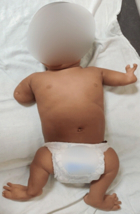
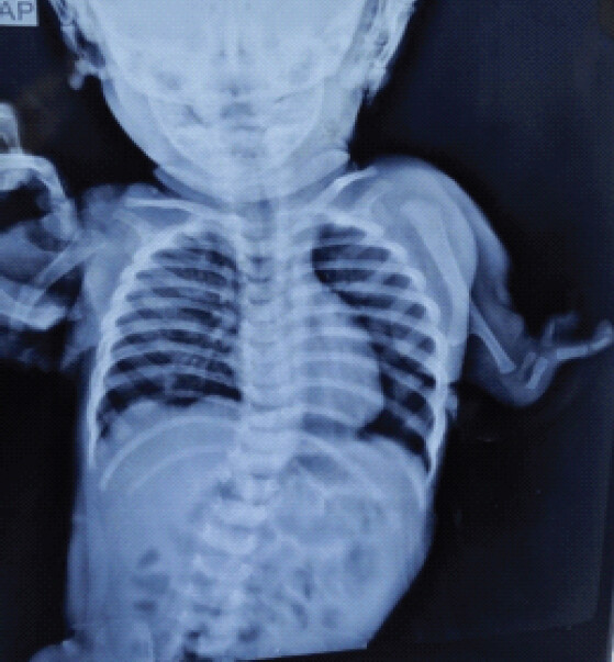
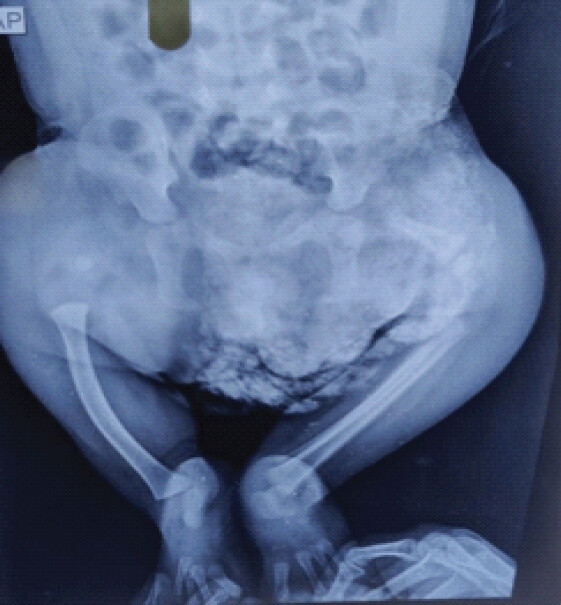
The following diagnosis was made: Congenital asymmetrical multiple limb deficiency with right upper limb-transverse upper arm middle third; left upper limb-longitudinal radius partial, ulna partial, 2nd, 3rd, and 4th ray total, 1st and 5th ray partial; right lower limb-longitudinal femur total, fibula total, 5th ray total; left lower limb-longitudinal femur partial, fibula partial, 5th ray total as per the ISPO Classification system [4] with syndactyly of 1st and 2nd toe in right foot and scoliotic deformity lower dorsal spine. Longitudinal deficiency may be classified as either total or partial. Total longitudinal deficiency denotes the complete absence of the affected bone, whereas partial longitudinal deficiency indicates that the affected bone is partially absent or hypoplastic (reduced in size). On clinical examination, there were no obvious abnormalities involving the cardiovascular, respiratory, gastrointestinal, or central nervous systems apart from the described limb deficiencies. In view of the multiple limb deficiencies, investigations were performed to assess for associated systemic abnormalities. Complete blood count, C-reactive protein, liver function tests, and kidney function tests were within normal limits. Two-dimensional echocardiography, ultrasonography of the kidneys, ureters, and bladder, and magnetic resonance imaging of the spine were performed and reported as normal. The child was referred to the genetics department for further evaluation of a possible underlying genetic or syndromic etiology. Despite the fact that the most common cause of congenital limb deficiencies is idiopathic, the child was evaluated for secondary causes such as chromosomal aberrations, amniotic band syndrome, and Fanconi anemia in consultation with the Department of Paediatrics and Genetics. The parents were counseled regarding the congenital nature and extent of the limb deficiencies and the associated spinal deformity. They were informed that the precise underlying etiology could not be established. The importance of regular follow-up was explained, particularly for monitoring spinal alignment, limb function, growth, and overall development. The parents were also counseled regarding the potential need for multidisciplinary management, including pediatric orthopedics, rehabilitation, physiotherapy, and prosthetic assessment according to the child’s functional requirements and growth. They were advised regarding the importance of appropriate antenatal care and detailed anomaly ultrasonography in subsequent pregnancies.
Discussion
Limb development begins at the end of the 4th week of gestation, when the limb buds appear as small ectodermal outgrowths overlying a mesodermal core. Vascularization of this core, followed by invasion of somatic mesoderm and peripheral nerves, drives subsequent growth and differentiation. Upper limb development typically precedes that of the lower limbs, and the external limb architecture is fully established by the end of the embryonic period, at the 8th week of gestation [5]. Any insult that disrupts the normal sequence of limb bud formation, growth, or differentiation during this critical window can result in a spectrum of congenital limb deficiencies. Recognized causes include genetic disorders, amniotic band syndrome, maternal metabolic disease such as diabetes, and exposure to teratogens including alcohol and drugs such as thalidomide [6].
The current and preferred classification system for congenital limb defects is that of the ISPO [4], which divides these anomalies into transverse and longitudinal deficiencies. Transverse deficiencies denote absence of all skeletal elements distal to a given level, although there may be digital buds. Longitudinal deficiencies involve reduction or absence of an element or elements within the long axis of the limb, and there may be normal skeletal elements distal to the affected bone or bones [4].
Literature showing association between congenital limb reduction defects and scoliosis remains scarce. Lester et al. reported scoliosis in a cohort of patients with congenital upper limb deficiency [7], whereas Powers et al., in a retrospective study, found a higher incidence of idiopathic scoliosis among patients with amelia [8]. These findings suggest a possible link between severe limb reduction defects and spinal deformity.
This case underscores the value of antenatal ultrasound in the early diagnosis of congenital limb malformations. Routine anomaly scanning is best performed during the second trimester, between 18 and 22 weeks of gestation [9]. Andrikopoulou et al. reported that even a slight increase in the time spent performing the ultrasound examination can substantially improve the detection of fetal limb abnormalities [10].
The precise etiology of the limb deficiency in our patient could not be established. Overall, this case emphasizes the importance of antenatal screening, ISPO-based classification of limb deficiencies, and screening for scoliosis in patients with major limb reduction defects.
Conclusion
This case describes a rare presentation of congenital asymmetrical multiple limb deficiency involving all four limbs, with mixed transverse and longitudinal deficiencies. Despite evaluation, no definite syndromic or secondary cause could be confirmed. This case highlights the importance of antenatal check-ups and anomaly scans in the early diagnosis and counseling of congenital anomalies.
Clinical Message
Asymmetrical limb deficiency involving all four limbs is a rare congenital anomaly that may occur as an isolated condition or in association with other syndromes. This case highlights the importance of antenatal ultrasonographic screening, preferably during the late first or early second trimester, for the early detection of congenital anomalies and timely decision-making regarding management. The ISPO classification system provides a simple and practical framework for describing congenital limb deficiencies.
Conflict of Interest:
Nil
Source of Support:
Nil
Consent
The authors confirm that informed consent was obtained from the patient for publication of this article
How to Cite this Article
Shukla R, Dubey A, Sahu R, Jain V. Congenital Asymmetrical Multiple Limb Deficiency in 1-Year-Old Male Child: A Case Report. Journal of Orthopaedic Case Reports 2026 October;16(10): 120-123.
References
- Vasluian E, Van Der Sluis CK, Van Essen AJ, Bergman JE, Dijkstra PU, Reinders-Messelink HA. Birth prevalence for congenital limb defects in the northern Netherlands: A 30-year population-based study. BMC Musculoskelet Disord 2013;14:323. [Google Scholar] | [PubMed]
- Ceausu I, Iliescu D, Poalelungi C, Posea C, Bacalbasa N, Dobritoiu D. The antenatal detection of fetal limb anomalies. In: Congenital Anomalies from the Embryo to the Neonate. United Kingdom: Intech Open; 2018. [Google Scholar] | [PubMed]
- Jaruratanasirikul S, Tangtrakulwanich B, Rachatawiriyakul P, Sriplung H, Limpitikul W, Dissaneevate P. Prevalence of congenital limb defects: Data from birth defects registries in three provinces in Southern Thailand. Congenit Anom (Kyoto) 2016;56:203-8. [Google Scholar] | [PubMed]
- Day HJ. The ISO/ISPO classification of congenital limb deficiency. Prosthet Orthot Int 1991;15:67-9. [Google Scholar] | [PubMed]
- Hill MA. Musculoskeletal System-Limb Development-Embryology. Available from: https://embryology.med.unsw.edu.au/embryology/index.php/musculoskeletal_system_-_limb_development. [Last accessed on 2018 Dec 11]. [Google Scholar] | [PubMed]
- Froster UG, Baird PA. Maternal factors, medications, and drug exposure in congenital limb reduction defects. Environ Health Perspect 1993;101 Suppl 3:269-74. [Google Scholar] | [PubMed]
- Lester DK, Painter GL, Berman AT, Skinner SR. “Idiopathic” scoliosis associated with congenital upper-limb deficiency. Clin Orthop Relat Res 1986;202:205-10. [Google Scholar] | [PubMed]
- Powers TA, Haher TR, Devlin VJ, Spencer D, Millar EA. Abnormalities of the spine in relation to congenital upper limb deficiencies. J Pediatr Orthop 1983;3:471-4. [Google Scholar] | [PubMed]
- Leung KY, Poon CF, Teotico AR, Hata T, Won H, Chen M. Recommendations on routine mid-trimester anomaly scan. J Obstet Gynaecol Res 2015;41:653-61. [Google Scholar] | [PubMed]
- Andrikopoulou M, Vahanian SA, Chavez MR, Murphy J, Hanna N, Vintzileos AM. Improving the ultrasound detection of isolated fetal limb abnormalities. J Mater Fetal Neonatal Med 2017;30:46-9. [Google Scholar] | [PubMed]
© 2026 Journal of Orthopaedic Case Reports - Published by Indian Orthopaedic Research Group
About the Authors
-

Rohit Shukla
-

Ankita Dubey
-

Rupendra Sahu
-

Vibhor Jain
Related Articles in Journal of Orthopaedic Case Reports
 October 1, 2026 Tibial Plateau Fracture in a Patient with Fibular Hemimelia: A Case Report
 May 1, 2026 Complications of Limb Salvage in Concordant Bilateral Fibular Hemimelia and Tetramelic Ectrodactyly: A Case Report
 March 10, 2024 Isolated Bilateral Upper Limb Amelia – A Rare Case Report
 April 11, 2012 Article Template Osteochondritis Dessicans- Primary Fixation using Bioabsorbable Implants.