


Introduction
Femoral neck fractures in young patients present a challenging clinical problem, with preservation of the native hip traditionally favored through internal fixation. However, displaced fracture patterns, poor bone quality, and patient-specific risk factors may predispose to fixation failure, nonunion, and avascular necrosis. In select cases, arthroplasty may offer a more predictable outcome despite concerns regarding implant longevity.
Performing total hip arthroplasty (THA) in patients with ipsilateral above-the-knee amputations introduces additional technical complexity. The absence of a distal lever arm complicates limb manipulation, femoral preparation, and assessment of stability and version [1]. Several techniques have been described to address these challenges, though many require specialized equipment or extensive soft-tissue dissection, given common hip flexor and abductor contractures [2].
We present a case of THA for a displaced femoral neck fracture in a young above-the-knee amputee, highlighting a simple intraoperative technique using a Steinmann pin to facilitate controlled limb manipulation. The patient’s written informed consent to publish this case report was obtained.
Case Report
A 27-year-old male presented with acute left hip pain following a first-time seizure. His medical history was significant for a traumatic left above-the-knee amputation following a motorcycle collision in 2019 after failed limb salvage surgeries. Before the current injury, he ambulated independently with a prosthesis and reported occasional phantom limb pain without residual stump site complications or contractures.
Radiographs and computed tomography (CT) demonstrated a displaced left transcervical femoral neck fracture with approximately 24° of posterior tilt on CT imaging (Fig. 1).
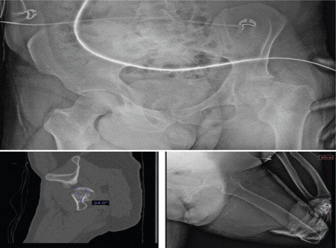
The patient reported daily tobacco and occasional alcohol use with no further medical history. On physical examination, he was neurovascularly intact with no other associated injuries. He was subsequently cleared for surgery from a multidisciplinary standpoint after a normal appearing electroencephalogram and further diagnostic testing. A shared patient decision-making discussion was held regarding operative management, including risks and benefits of open reduction and internal fixation versus THA. Given the degree of displacement, posterior tilt, smoking history, and desire for definitive surgical management, THA was selected.
Surgical technique
The patient received intravenous antibiotics preoperatively for infection prophylaxis and was positioned in the right lateral decubitus position under general anesthesia. A modified anterolateral approach to the hip was performed through a longitudinal incision centered over the anterior tip of the greater trochanter. Approximately 30% of the anterior abductor complex was subperiosteally elevated to allow capsular exposure.
Due to the absence of a distal limb for manipulation, a small stab incision was made along the distal anterolateral aspect of the femoral stump. This location was proximal enough to engage the diaphyseal bone while also avoiding contact with his prosthesis. Blunt dissection was carried down to the femoral cortex, and a fully threaded Steinmann pin was drilled into the anterolateral cortex of the femoral shaft. This provided a temporary lever arm, allowing controlled external rotation, flexion, and positioning of the femur throughout the procedure (Fig. 2).
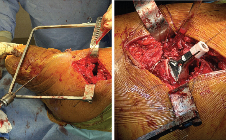
A T-type capsulotomy was performed, and the hip was dislocated anteriorly, revealing the femoral neck fracture and associated hematoma. A standard femoral neck osteotomy was performed, and the femoral head was removed. The acetabulum was sequentially reamed to 54 mm, revealing osteoporotic bone. A 54-mm acetabular shell was implanted with 40–45° of abduction and approximately 20° of anteversion, supplemented with a single screw for fixation. A 40-mm liner was inserted.
Using the Steinmann pin for controlled manipulation, the femoral canal was prepared and broached to size 10. Trial reduction demonstrated excellent stability and restoration of leg length. A small posterior-medial calcar cortical delamination was noted and secured with a cerclage cable. A size 10 high-offset femoral stem was implanted with 10–15° of anteversion, and a 40-mm ceramic head with neutral neck length was impacted.
The hip was reduced and demonstrated excellent stability through a full range of motion, utilizing the fully threaded Steinmann pin. The wound was irrigated, the capsule and abductors were repaired, and layered closure was performed. A negative-pressure wound dressing was applied. Distally, the former Steinmann pin site was irrigated and closed with 3-0 nylon suture.
Postoperative course
The patient tolerated the procedure well and was transferred to the post-anesthesia care unit in stable condition. Post-operative radiographs demonstrated appropriate component positioning without evidence of complication. Plans were made for protected weight-bearing, early mobilization, and inpatient seizure evaluation (Fig. 3).
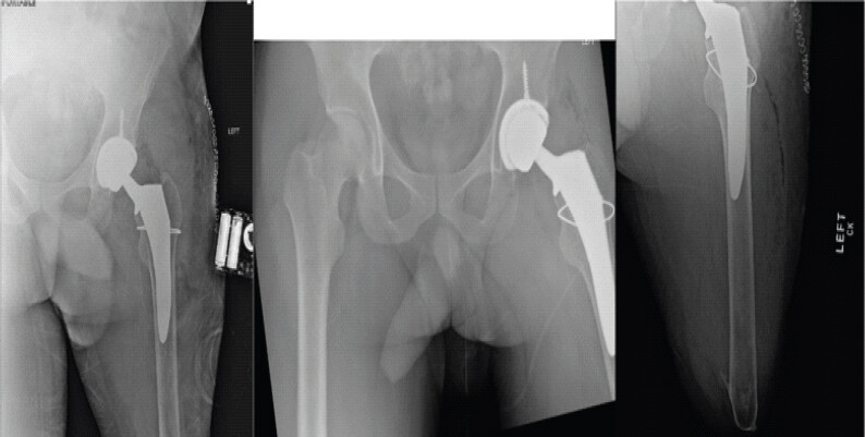
At 6-month follow-up, the patient’s wounds had completely healed without sequela. The patient was back to using his prosthesis for ambulation without assisted devices. Both stationary pain and pain with ambulation had been alleviated in the previously fractured left hip. The patient reported no additional concerns, pain in thigh, or additional adverse effects.
Discussion
Management of displaced femoral neck fractures in young patients traditionally favors reduction and internal fixation to preserve the native hip despite substantial fixation failure rates remaining substantial with fracture displacement, poor bone quality, and comorbidities [3,4,5,6]. These challenges are amplified in patients with ipsilateral lower extremity amputation, who experience profound musculoskeletal and biomechanical adaptations following limb loss [7,8,9]. Lateral shifting of the center of gravity after amputation increases shear forces across the femoral neck, creating a biomechanically unfavorable environment for fracture healing, even when reduction appears satisfactory [1,10]. Consequently, the likelihood of revision surgery may be high in select cases.
To the best of our knowledge, reports of THA in amputee patients are limited, and those that are reported are almost exclusively limited to below-the-knee amputees. In patients with more proximal levels of amputation, the absence of a distal lever arm complicates intraoperative procedures such as hip dislocation, femoral preparation, and accurate component positioning. In this case, a fully threaded Steinmann pin in the anterolateral femoral stump provided a temporary lever arm, allowing controlled manipulation of the residual limb during acetabular exposure, femoral preparation, and trialing.
The decision to proceed with primary THA was influenced by factors associated with fixation failure, including complete fracture displacement, significant posterior tilt on axial CT imaging, active tobacco use, and compromised bone quality [10,11,12,13,14]. While arthroplasty in young patients raises concerns regarding implant longevity and future revision, these risks were weighed against the high probability of non-union or avascular necrosis following internal fixation in a biomechanically unfavorable environment [7,15]. THA-specific risks, including instability and infection, were mitigated through careful component positioning, use of a large femoral head, meticulous soft-tissue repair, and application of negative-pressure wound therapy.
This case highlights the complexity of managing femoral neck fractures in amputee patients and the paucity of literature-guided surgical decision-making. While limited by its single-case design, it demonstrates that primary THA, combined with a simple limb-control technique, may be a reasonable and reproducible option in select above-the-knee amputees with high-risk fracture patterns.
Conclusion
Primary THA is a reasonable option for select above-the-knee amputees who sustain displaced femoral neck fractures with multiple risk factors for fixation failure. A fully threaded Steinmann pin placed in the residual femoral stump provided simple, reproducible intraoperative limb control without extensive soft-tissue dissection or specialized instrumentation. In this case, the patient had an uncomplicated recovery with return to independent prosthetic ambulation and resolution of hip pain by 6-month follow-up.
Clinical Message
Displaced femoral neck fractures in young above-the-knee amputees present unique biomechanical and technical challenges. This case demonstrates that primary total hip arthroplasty can be considered when multiple risk factors for fixation failure are present, including fracture displacement, significant posterior tilt, tobacco use, and poor bone quality. The use of a fully threaded Steinmann pin placed within the residual femoral stump provides a simple, safe, and effective method for intraoperative limb control, facilitating femoral preparation, component positioning, and stability assessment without extensive soft-tissue dissection or specialized instrumentation. This technique may serve as a valuable adjunct for surgeons managing complex hip arthroplasty cases in patients with proximal lower-extremity amputations.
Conflict of Interest:
Nil
Source of Support:
Nil
Consent:
The authors confirm that informed consent was obtained from the patient for publication of this article
How to Cite this Article
Starcher NJ, Patel J, Golden AR, Kirk CA. Primary Total Hip Arthroplasty for a Displaced Femoral Neck Fracture in an Above-the-Knee Amputee: A Case Report. Journal of Orthopaedic Case Reports 2026 October;16(10): 142-146.
References
- Masmoudi K, Rbai H, Fradj AB, Saâdena J, Boughattas A. Primary total hip replacement for a femoral neck fracture in a below-knee amputee. J Orthop Case Rep 2016;6:63-6. [Google Scholar] | [PubMed]
- Sathappan SS, Wee J, Ginat D, Teicher M, Meere P, Di Cesare PE. Total hip arthroplasty in above-knee amputees: A case report. Am J Orthop (Belle Mead NJ) 2011;40:E17-9. [Google Scholar] | [PubMed]
- Girard J, Glorion C, Bonnomet F, Fron D, Migaud H. Risk factors for revision of hip arthroplasties in patients younger than 30 years. Clin Orthop Relat Res 2011;469:1141-7. [Google Scholar] | [PubMed]
- Slobogean GP, Sprague SA, Scott T, Bhandari M. Complications following young femoral neck fractures. Injury 2015;46:484-91. [Google Scholar] | [PubMed]
- Varghese VD, Boopalan PR, Titus VT, Oommen AT, Jepegnanam TS. Indices affecting outcome of neglected femoral neck fractures after valgus intertrochanteric osteotomy. J Orthop Trauma 2014;28:410-6. [Google Scholar] | [PubMed]
- Duckworth AD, Bennet SJ, Aderinto J, Keating JF. Fixation of intracapsular fractures of the femoral neck in young patients: Risk factors for failure. J Bone Joint Surg Br 2011;93:811-6. [Google Scholar] | [PubMed]
- Amanatullah DF, Trousdale RT, Sierra RJ. Total hip arthroplasty after lower extremity amputation. Orthopedics 2015;38:e394-400. [Google Scholar] | [PubMed]
- Burke MJ, Roman V, Wright V. Bone and joint changes in lower limb amputees. Ann Rheum Dis 1978;37:252-4. [Google Scholar] | [PubMed]
- Sadeghi H, Allard P, Duhaime PM. Muscle power compensatory mechanisms in below-knee amputee gait. Am J Phys Med Rehabil 2001;80:25-32. [Google Scholar] | [PubMed]
- Haleem S, Yousaf S, Hamid T, Nagappa S, Parker MJ. Characteristics and outcomes of hip fractures in lower limb amputees. Injury 2021;52:914-7. [Google Scholar] | [PubMed]
- Yeranosian M, Horneff JG, Baldwin K, Hosalkar HS. Factors affecting the outcome of fractures of the femoral neck in children and adolescents: A systematic review. Bone Joint J 2013;95-B:135-42. [Google Scholar] | [PubMed]
- Honkanen JS, Ekman EM, Huovinen VK, Mäkelä KT, Koivisto M, Karvonen MP. Preoperative posterior tilt increases the risk of later conversion to arthroplasty after osteosynthesis for femoral neck fracture. J Arthroplasty 2021;36:3187-93. [Google Scholar] | [PubMed]
- Sjöholm P, Otten V, Wolf O, Gordon M, Karsten G, Sköldenberg O. Posterior and anterior tilt increases the risk of failure after internal fixation of Garden I and II femoral neck fracture. Acta Orthop 2019;90:537-41. [Google Scholar] | [PubMed]
- Barnes R, Brown JT, Garden RS, Nicoll EA. Subcapital fractures of the femur. A prospective review. J Bone Joint Surg Br 1976;58:2-24. [Google Scholar] | [PubMed]
- Salai M, Amit Y, Chechik A, Blankstein A, Dudkiewicz I. Total hip arthroplasty in patients with below-knee amputations. J Arthroplasty 2000;15:999-1002. [Google Scholar] | [PubMed]
© 2026 Journal of Orthopaedic Case Reports - Published by Indian Orthopaedic Research Group
About the Authors
-

Nathaniel J Starcher
-

Jignesh Patel
-

Andrew R Golden
-

Cameron A. Kirk
Related Articles in Journal of Orthopaedic Case Reports
 August 1, 2026 Posterior Acetabular Wall Fracture Mimicking a Subcapital Femoral Neck Fracture on Plain Radiographs: A Diagnostic Pitfall and the Role of Intraoperative Fluoroscopy: A Case Report
 June 1, 2026 Managing Periprosthetic Fracture of the Proximal Femur Using Total Hip Arthroplasty in a Patient with Sequelae of Poliomyelitis: Case Report and Literature Review
 April 1, 2026 Outcomes of Total Hip Arthroplasty for Traumatic versus Non-Traumatic Hip Pathology: A Prospective Cohort Study
 March 1, 2026 Managing Hip Arthritis in an Above-Knee Amputee: A Case Report of Total Hip Arthroplasty with Surgical Technique and Rehabilitation Considerations