


Introduction
Arteriovenous fistula accounts for only a small proportion of traumatic vascular injuries and is among the most easily missed, because the classic hard signs of vascular injury – absent distal pulses, an expanding hematoma, a pulsatile mass, a bruit or a thrill – are frequently absent in the acute phase [1,2]. Diagnosis is therefore commonly delayed; with some series describing the majority recognized only after weeks to months, while large historical series report substantially lower rates of major delay [2,3]. Because arterial inflow continues to perfuse the limb through the distal vessels, the foot stays warm and the pulses remain palpable, lending the patient a deceptive “mask of comfort” [1].
When a penetrating injury causes a long-bone fracture, attention is often directed toward the skeletal injury, potentially delaying recognition of an associated vascular lesion. We report a superficial femoral arteriovenous fistula with pseudoaneurysm following a gunshot wound and comminuted subtrochanteric femoral fracture. The only objective clues were a transfusion-refractory fall in hemoglobin with persistent tachycardia and tachypnea, despite an initially normal color Doppler study. This case highlights a pragmatic bedside diagnostic approach for orthopedic surgeons and contributes to the limited Indian literature on occult vascular injury. It is reported according to the SCARE 2023 guidelines.
Case Report
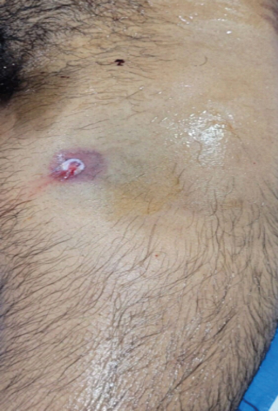
A 28-year-old man presented to the casualty department following a single gunshot wound to the left upper thigh. He was conscious and oriented, with a Glasgow Coma Scale of 15, and was hemodynamically stable. Examination showed a solitary entry wound in the proximal thigh near the groin (Fig. 1), with local pain, swelling, and tenderness, but he was otherwise comfortable. The left femoral, popliteal, dorsalis pedis, and posterior tibial pulses were all palpable, and there was no expanding hematoma, bruit or thrill. Distal sensation and toe movements were intact, and the compartments were soft.
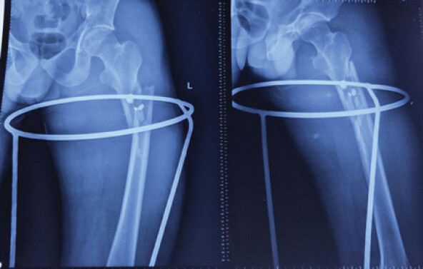
Plain radiographs demonstrated a comminuted subtrochanteric fracture of the left femur with an adjacent retained bullet fragment (Fig. 2). An extended focused assessment with sonography for trauma was normal. The limb was stabilized in a Thomas splint, and the patient was admitted under orthopedics for resuscitation and fracture fixation.
The admission hemoglobin was 9.5 g/dL, and one unit of packed red cells was transfused. Rather than rising, the post-transfusion hemoglobin fell to 7.3 g/dL, a result confirmed on a repeat sample. Re-examination of the limb remained unremarkable. By the 3rd day, the hemoglobin had fallen further to 6.4 g/dL, and the patient had developed persistent tachycardia and tachypnea while continuing to appear comfortable. A color Doppler study of the injured limb was reported as normal.
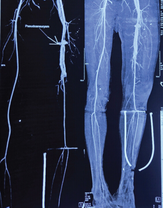
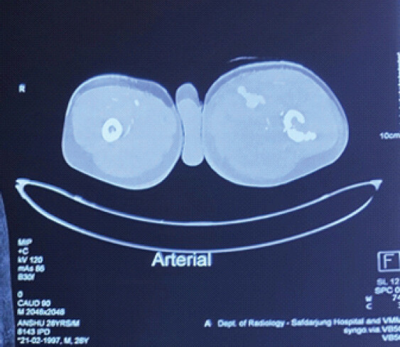
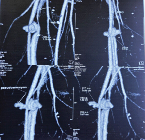
Faced with a transfusion-refractory anemia and unexplained systemic signs in proximity to a major vascular territory, computed tomography (CT) angiography was performed. This demonstrated an arteriovenous fistula with an associated pseudoaneurysm arising from the proximal superficial femoral vessels at the level of the wound, with preserved and normal distal run-off (Fig. 3, 4, 5).
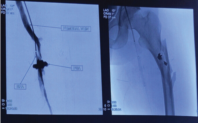
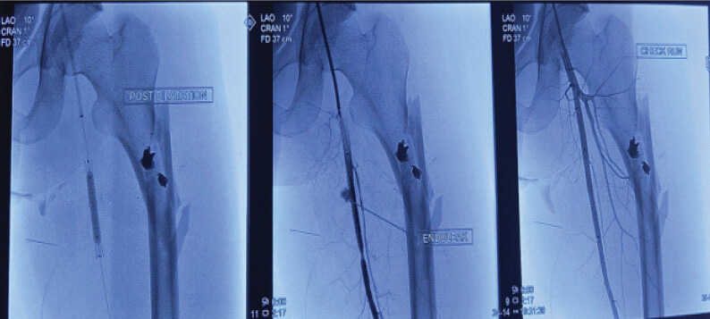
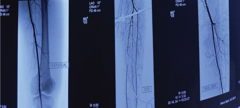
Vascular surgery and interventional radiology opinions were obtained, and an endovascular approach was selected. Under fluoroscopic guidance, a covered stent graft was deployed across the fistula in the superficial femoral artery, with angiographic resolution of the fistula and pseudoaneurysm and restoration of normal distal flow (Fig. 6, 7, 8). The patient was started on dual antiplatelet therapy with aspirin and clopidogrel. A follow-up color Doppler confirmed stent patency with a normal venous waveform.
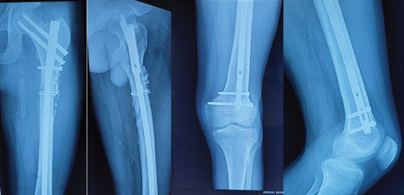
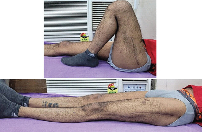
Definitive fixation of the femoral fracture was deferred, both because manipulation carried a risk to the freshly treated vessel and because the patient was receiving dual antiplatelet therapy. The limb was maintained in a custom Thomas splint and regular review was advised. At the patient’s request, further care was transferred to a center nearer his home; the subsequent operative details and follow-up imaging were obtained from those records and from direct review at follow-up. Approximately 2 months after the injury, he underwent open reduction, removal of the metallic fragments, and internal fixation with a reconstruction nail supplemented by cerclage wiring and bone grafting. Recovery was uneventful; knee and patellar mobilization began early, and protected weight-bearing with a walker started about a week later. By roughly 2 months after fixation, he mobilized without support, with near-normal hip and knee movement, and radiographs showed progressing union with distal pulses equal in character to the opposite limb. At 6 months after injury (approximately 4 months after fixation), he was symptom-free and had returned to work, with a united fracture and a full range of motion (Fig. 9 and 10).
Discussion
The deceptive presentation of this injury is rooted in its hemodynamics. An abnormal communication between a high-pressure artery and a low-pressure vein creates a low-resistance shunt. In long-standing fistulae, this lowers systemic vascular resistance and drives a compensatory rise in heart rate and cardiac output, ultimately risking high-output cardiac failure and pulmonary hypertension [4]. In the acute setting of our patient, however, the systemic signs are most parsimoniously explained by blood loss rather than by an established high-output state. The progressive, transfusion-refractory fall in hemoglobin is best attributed to continued bleeding into the pseudoaneurysm sac and the surrounding thigh – a compartment that can sequester a large volume before becoming clinically obvious – compounded by hemodilution from resuscitation, while the tachycardia and tachypnea are most plausibly responses to hypovolemia and reduced oxygen-carrying capacity.
Crucially, a comminuted subtrochanteric fracture is itself a major source of occult blood loss and can, on its own, produce a falling hemoglobin and tachycardia; a transfusion-refractory anemia is therefore not, in isolation, specific for vascular injury. What raised suspicion in our patient was the combination of features: A fall in hemoglobin disproportionate to, and persisting beyond, what the fracture and its hematoma could reasonably explain, together with persistent systemic signs and the proximity of the wound to a major vessel. We regard this combination, rather than the hemoglobin trend alone, as the appropriate trigger for definitive imaging.
Color Doppler, although the usual first-line vascular investigation, was non-diagnostic in our patient. Its sensitivity falls when only soft signs of injury are present and is further reduced by operator dependence, an overlying hematoma, and the depth of the proximal femoral vessels [5]; we cannot exclude that the initial study was technically limited. CT angiography, by contrast, is the imaging modality of choice for extremity vascular trauma, with reported sensitivities of approximately 90–95% and specificities approaching 100% for the detection of arterial injury [6], and it reliably identifies clinically unsuspected post-traumatic fistulae [7]. It was diagnostic here. We therefore regard a normal Doppler as insufficient to exclude this injury when clinical suspicion is high. In retrospect, several technical and operator-related factors may have contributed to the initial false-negative study: The proximal superficial femoral vessels lie deep near the groin; overlying hematoma and soft-tissue swelling degrade the acoustic window; an examination confined to the standard arterial stations can miss a short fistulous tract unless the wound trajectory itself is deliberately interrogated; and the characteristic findings – an arterialized, pulsatile venous waveform, low-resistance high-diastolic arterial inflow, and perivascular tissue vibration – are apparent only when spectral sampling is performed at and around the site of injury [5]. We could not retrospectively determine which of these factors operated in our patient, and we have therefore treated the false-negative result as unexplained rather than attributing it to a single cause.
The orthopedic relevance is considerable, because most peripheral arterial injuries are accompanied by a fracture and so present first to the orthopedic surgeon. In a large Indian series, Joshi reported that the majority of peripheral arterial injuries followed road-traffic trauma and that bony injury was associated in roughly two-thirds of cases [8]. Norman and colleagues, studying gunshot wounds that fractured a long bone, found occult vascular injury on arteriography in a notable minority of patients with a normal physical examination, a small proportion of which were major lesions such as a pseudoaneurysm or fistula [9]. Robbs and colleagues, in 202 patients with traumatic fistulae, showed that intervention within a week of injury carried lower morbidity, because later repair was complicated by fibrosis and venous dilatation [10]. Together these data justify a low threshold for angiography whenever a penetrating wound or fracture lies in proximity to a major vessel.
We do not claim this to be the first report of a traumatic femoral fistula after a gunshot wound; the entity is well described. What we wish to highlight is the combination of an occult presentation with a proximal superficial femoral lesion – rather than the more commonly reported profunda femoris pseudoaneurysm associated with proximal femoral fractures [11] – and the value of a simple bedside triad (transfusion-refractory anemia, persistent tachycardia, and persistent tachypnea) in prompting definitive imaging. We emphasize that this triad is proposed from a single case as a hypothesis-generating observation; its sensitivity, specificity, and predictive value are unknown, and it requires prospective validation in larger series before it can be recommended as a screening rule.
Treatment has shifted from open resection and reconstruction toward endovascular options such as covered stents and coil embolization, which were well suited to our patient and avoided dissection in a possibly contaminated, freshly injured field [1]. Two cautions arise from our own course. First, the diagnosis took 3 days; in retrospect, the failure of the hemoglobin to rise after transfusion was the signal that should have prompted earlier angiography. Second, the agents used after stenting are antiplatelets, not anticoagulants, and their use forces a deliberate decision about the timing of fracture fixation: Manipulating a comminuted subtrochanteric fracture during dual antiplatelet therapy carries a real bleeding and re-injury risk. Covered stent grafts typically require 1–3 months of dual antiplatelet therapy; rather than default to prolonged splinting and inter-hospital transfer, as occurred here, a planned fixation interval agreed with the vascular team is preferable. The long-term consequence of an untreated fistula is illustrated by Chaudry et al, who described one presenting 52 years after injury with iliofemoral aneurysmal change and pulmonary hypertension [12].
The strategy adopted here – endovascular repair followed by deferred fixation – was a pragmatic choice rather than one supported by comparative evidence, and at least two alternatives deserve explicit mention. Open repair of the fistula would have permitted immediate or early fracture fixation, at the price of a dissection through a freshly injured and potentially contaminated field [1,10]. Alternatively, fixation could have been undertaken earlier despite antiplatelet therapy, accepting a higher bleeding risk, with aspirin continued through surgery and the clopidogrel briefly interrupted in agreement with the vascular team, as is commonly practiced for urgent surgery in other stented vascular territories. No trial evidence addresses this specific scenario, and the choice must be individualized by a multidisciplinary team according to wound contamination, fracture pattern, patient physiology, and local endovascular expertise. In our patient, fixation followed approximately 2 months after stenting, at a second center, and the perioperative antiplatelet handling there could not be reconstructed in detail; a fixation date agreed prospectively with the vascular team at the time of stenting, together with a written antiplatelet plan, would have been preferable and is what we now recommend.
Limitations
This report describes a single patient, and its findings cannot be generalized. There was no control or comparison group, so no conclusion can be drawn about the relative merits of endovascular repair with deferred fixation as against open vascular repair or immediate fracture fixation; the alternative pathways discussed above remain untested in this setting. The mechanism of vascular injury is inferred from imaging alone, because endovascular treatment meant that the exact arterial and venous wall lesions were never confirmed operatively.
Follow-up was limited to 6 months, which may be too short to identify late complications of covered stent grafting such as in-stent restenosis, thrombosis, stent fracture, infection or recurrence of the fistula, and these were not systematically evaluated. Beyond the early post-procedural color Doppler confirming stent patency, structured long-term surveillance with serial duplex ultrasonography or CT angiography is not yet available, although continued periodic duplex surveillance is recommended. Functional outcome was assessed clinically and radiologically only; validated instruments, such as the Harris hip score or the lower extremity functional scale and patient-reported outcome or quality-of-life measures such as the SF-36, were not administered. Blood loss was not quantified volumetrically, and because definitive fixation took place at a second center the total transfusion requirement could not be fully reconstructed, so serial hemoglobin values are the only quantitative index of blood loss presented. Similarly, the initial false-negative color Doppler study could not be re-analyzed to isolate the specific technical or operator-related factors responsible, and a detailed patient-specific protocol for perioperative antiplatelet management was not prospectively defined.
Finally, the index injury was penetrating; these observations may not extend to blunt trauma or closed femoral fractures. The proposed diagnostic triad of transfusion-refractory anemia, persistent tachycardia and persistent tachypnea is derived from a single case and must not be treated as a validated screening rule. Larger multicenter studies or prospective registries are needed to establish the true incidence of occult traumatic arteriovenous fistula after limb trauma, to validate the diagnostic algorithm and imaging triggers proposed here, and to define evidence-based timing of fracture fixation in patients requiring antiplatelet therapy after endovascular repair.
Conclusion
Traumatic arteriovenous fistula may present without classic vascular signs. In penetrating limb trauma or fractures near major vessels, transfusion-refractory hemoglobin decline with unexplained tachycardia and tachypnea, disproportionate to expected fracture-related blood loss, should raise suspicion for vascular injury and prompt CT angiography despite normal distal pulses or color Doppler. Early recognition permits minimally invasive endovascular treatment and may prevent limb- and life-threatening complications. After vascular control, fracture management should follow standard principles, with fixation timing individualized according to antiplatelet therapy and coordinated with the vascular team. These recommendations are provisional and require validation in larger studies.
Clinical Message
In any patient with a penetrating injury or a fracture near a major vessel, a transfusion-refractory fall in hemoglobin with unexplained tachycardia and tachypnea – even with palpable distal pulses and a normal color Doppler – should prompt CT angiography to exclude an occult arteriovenous fistula or pseudoaneurysm. A normal color Doppler does not exclude the diagnosis.
Conflict of Interest:
Nil
Source of Support:
Nil
Consent
The authors confirm that informed consent was obtained from the patient for publication of this article
How to Cite this Article
Khalil ZA, Mangal KK, Mahapatra S, Jaiman A. Occult Femoral Arteriovenous Fistula and Pseudoaneurysm after a Gunshot-Associated Subtrochanteric Fracture: Transfusion-Refractory Anemia as the Sentinel Sign – A Case Report. Journal of Orthopaedic Case Reports 2026 October;16(10): 147-153.
References
- Nagpal K, Ahmed K, Cuschieri R. Diagnosis and management of acute traumatic arteriovenous fistula. Int J Angiol 2008;17:214-6. [Google Scholar] | [PubMed]
- Young CJ, Dardik A, Sumpio B, Indes J, Muhs B, Ochoa Chaar CI. Venous ulcer: Late complication of a traumatic arteriovenous fistula. Ann Vasc Surg 2015;29:836.e1-3. [Google Scholar] | [PubMed]
- Rich NM, Hobson RW 2nd, Collins GJJr. Traumatic arteriovenous fistulas and false aneurysms: A review of 558 lesions. Surgery 1975;78:817-28. [Google Scholar] | [PubMed]
- Narechania S, Tonelli AR. Hemodynamic consequences of a surgical arteriovenous fistula. Ann Am Thorac Soc 2016;13:288-91. [Google Scholar] | [PubMed]
- Wani ML, Sheikh MT, Nayeem-Ul-HassanIrshad I, Ahangar AG, Ganie FA. Evaluating peripheral vascular injuries: Is color doppler enough for diagnosis?. Int Cardiovasc Res J 2014;8:15-7. [Google Scholar] | [PubMed]
- Miller-Thomas MM, West OC, Cohen AM. Diagnosing traumatic arterial injury in the extremities with CT angiography: Pearls and pitfalls. RadioGraphics 2005;25 Suppl 1:S133-42. [Google Scholar] | [PubMed]
- hen JK, Johnson PT, Fishman EK. Diagnosis of clinically unsuspected posttraumatic arteriovenous fistulas of the pelvis using CT angiography. AJR Am J Roentgenol 2007;188:W269-73. [Google Scholar] | [PubMed]
- Joshi SS. Peripheral arterial injuries: An Indian experience. Indian J Surg 2016;78:187-91. [Google Scholar] | [PubMed]
- Norman J, Gahtan V, Franz M, Bramson R. Occult vascular injuries following gunshot wounds resulting in long bone fractures of the extremities. Am Surg 1995;61:146-50. [Google Scholar] | [PubMed]
- Robbs JV, Carrim AA, Kadwa AM, Mars M. Traumatic arteriovenous fistula: Experience with 202 patients. Br J Surg 1994;81:1296-9. [Google Scholar] | [PubMed]
- Lo SP, Hsieh CC, Yang CY, Lin CJ, Chang CW. Delayed superficial femoral artery pseudoaneurysm following distal femoral shaft fracture: A case report. Kaohsiung J Med Sci 2011;27:242-6. [Google Scholar] | [PubMed]
- Chaudry M, Flinn WR, Kim K, Neschis DG. Traumatic arteriovenous fistula 52 years after injury. J Vasc Surg 2010;51:1265-7. [Google Scholar] | [PubMed]
© 2026 Journal of Orthopaedic Case Reports - Published by Indian Orthopaedic Research Group
About the Authors
-

Zeeshan Ahmad Khalil
-

Krishna Kumar Mangal
-

Sandip Mahapatra
-

Aryaman Jaiman
Related Articles in Journal of Orthopaedic Case Reports
 October 1, 2026 Superficial Femoral Artery Pseudoaneurysm after Total Knee Arthroplasty: An Uncommon Vascular Complication
 July 1, 2026 Tuberculosis of the Elbow in a Chronic Kidney Disease Patient with Arteriovenous Fistula: A Case Report
 June 1, 2026 Pseudoaneurysm of the Medial Femoral Circumflex Artery following Surgical Fixation of Trochanteric Femur Fracture: A Case Report
 June 1, 2026 Delayed Pseudoaneurysm of the Subclavian Artery Following Clavicle Fracture Fixation: A Case Report