


Introduction
Humeral shaft fractures account for roughly 3–5% of all fractures [1] and, in the majority of patients, heal reliably with conservative or standard surgical management. Non-union is relatively uncommon – reported at approximately 1–10% of cases [2] – but when it arises, particularly in the setting of osteoporosis or other metabolic bone disease, it presents a substantial clinical problem. Osteoporosis impairs screw purchase and overall fixation integrity [2,3], and any construct applied to osteopenic bone must be capable of providing stability until biologic healing occurs, even in the absence of robust cortical support.
The general principle in managing humeral non-union is to achieve rigid mechanical stability while simultaneously addressing the biologic environment at the fracture site. This typically involves open reduction, debridement of the non-union, refreshing of the fracture ends to viable bone, and fixation with a plate and screws augmented by bone graft [2, 4]. Where the bone stock is severely deficient, however, plate fixation alone may not confer sufficient stability. In these circumstances, structural strut allografts – either applied as cortical onlay grafts or delivered intramedullary – have been employed to reinforce screw purchase, bridge defects, and supplement load sharing across the fracture site [3].
Published series have demonstrated encouraging results with this approach. Van Houwelingen and McKee reported six osteopenic humeral non-union cases managed with a 4.5-mm compression plate and a humeral cortical onlay allograft strut supplemented by autograft, with all six-achieving union at a mean of 3.4 months [3]. Vidyadhara et al. described six osteoporotic humeral non-union patients treated with an intramedullary fibular strut graft combined with dynamic compression plate (DCP) fixation, again with universal union [4]. More recently, Fink Barnes et al. reported 10 of 13 humeral shaft non-unions (77%) achieving union following revision open reduction and internal fixation (ORIF) with a fibular allograft strut [5]. Collectively, these series support the concept that combining rigid plate fixation with a structural strut graft effectively addresses both the mechanical and biologic requirements of non-union in osteoporotic bone.
The application of strut allograft augmentation is not restricted, however, to established non-union. Osteogenesis imperfecta (OI) represents a distinct form of metabolic bone disease in which cortical fragility and compromised bone architecture create challenges directly analogous to those seen in osteoporotic non-union. In OI, the primary structural deficiency is present from the outset, such that even an acute fracture may behave biologically as though in poor-quality bone. The same mechanical principles – load sharing, screw purchase augmentation, and bridging of structural defects – apply equally in this context.
We report a case series of three patients who underwent humeral ORIF augmented by structural strut allograft, representing two distinct forms of metabolically compromised bone: osteoporosis with established non-union (Cases 1 and 2) and OI with an acute distal humeral fracture (Case 3). The series illustrates both the shared principles underlying the technique and the adaptations required to accommodate differing fracture locations, bone pathologies, and fixation implants.
Case Report
Surgical technique
Cases 1 and 2 (non-union)
Under general anesthesia with the patient in lateral decubitus and the arm supported, a standard anterolateral, extended deltopectoral, or posterior approach was used depending on the fracture location. The non-union site was exposed, and any fibrous tissue and sclerotic bone were excised until healthy, bleeding bone ends were encountered. Medullary canals were opened with reamers. An onlay cortical strut allograft was used to span the fracture site and bridge any segmental defect; residual cavities were filled with morselized cancellous bone chips. A 4.5-mm locking compression plate was contoured and applied. Where the intraoperative stability of a single plate construct was deemed insufficient, supplementary double condylar 3.5-mm locked anatomical plates with cerclage wires were added around the strut. At least three bicortical screws were placed in each major fragment, with some screws engaging both host bone and the allograft strut to maximise purchase. All wounds were closed in layers. Postoperatively the arm was rested in a sling, and gentle shoulder and elbow mobilization under physiotherapy supervision was encouraged after 2–3 weeks, with lifting restrictions maintained until radiographic bridging callus was confirmed.
Case 3 (acute fracture in OI)
The operative approach for this case differed from the standard non-union technique, reflecting both the acute nature of the injury and the unique biomechanical properties of OI bone. Standard rigid cortical fixation carries a risk of iatrogenic fracture through stress concentration in OI bone, making flexible intramedullary fixation the preferred primary modality. Under general anesthesia, a distal-to-proximal elastic (titanium) intramedullary nail (titanium elastic nail [TEN]) was inserted with multiple intramedullary wires. An onlay allograft strut was applied alongside the nail to supplement rotational stability and provide cortical buttressing in the deficient OI bone. The radial nerve was explored intraoperatively given the pre-operative sensory palsy; no structural nerve disruption was identified. The wound was closed in layers, and early active mobilization of fingers and wrist was commenced on Day 1, with formal physiotherapy and occupational therapy input from the outset.
Case 1
A 72-year-old female presented with persistent pain and restricted arm function 1 year after sustaining a shaft humeral fracture that had failed to unite following conservative management. Radiographs demonstrated a hypertrophic non-union with marked osteopenia and a small segmental defect (Fig. 1). After thorough evaluation, the patient underwent ORIF with a long proximal humerus locking plate augmented by a cortical onlay strut allograft. The non-union site was debrided to healthy bleeding bone ends, and the defect was packed with cancellous bone chips. The structural allograft was secured to the host bone and plate using bicortical screws, ensuring that some screws engaged both graft and native cortex for enhanced stability. The procedure was uneventful, and standard post-operative rehabilitation with early passive motion was initiated after 2 weeks.
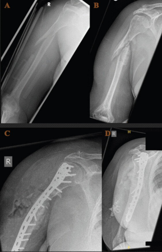
Wound healing was initially delayed, and the patient required a short course of oral antibiotics for superficial wound weeping, with no evidence of deep infection. Radiographs at 6 months demonstrated progressive integration of the strut graft and solid union (Fig. 1). Functionally, her Oxford Shoulder Score improved from 20 preoperatively to 39 postoperatively, and her Disabilities of the Arm, Shoulder and Hand (DASH) score improved from 40 to 15, reflecting substantial gains in both pain relief and limb function. At 1 year, the graft had fully incorporated, and the patient reported satisfaction with arm use for routine activities of daily living.
Case 2
A 68-year-old female sustained a left humeral shaft fracture after tripping over untied shoelaces in her kitchen and falling directly onto her arm. She remained on the floor for approximately 90 min before assistance arrived. Her medical history included hypothyroidism and a previous right proximal humeral fracture that had healed uneventfully. Initially managed conservatively at another institution, she presented 6 months post-injury with pain, deformity, and poor arm function. Radiographs revealed an atrophic non-union with osteoporotic bone and cortical loss (Fig. 2). Surgical planning incorporated open reduction, debridement to viable bone ends, and structural allograft-augmented ORIF.
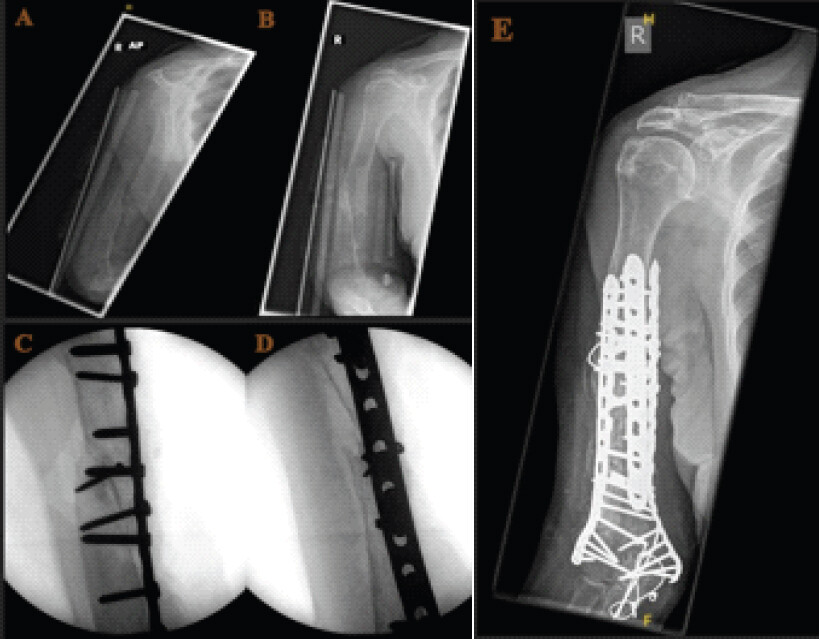
Through a posterior approach, the non-union was exposed and debrided. The medullary canal was reopened, and an onlay strut allograft was applied spanning the defect. A pre-contoured 4.5-mm locking plate was applied with bicortical screws engaging both graft and host bone proximally and distally. Residual defect was filled with morselized cancellous graft. Intraoperative assessment of construct stability indicated that the single plate was insufficient, and supplementary double condylar 3.5-mm locked anatomical plates were added with cerclage wires around the strut. The wound was closed in layers. The patient followed a structured rehabilitation program with gradual shoulder and elbow mobilization. At 6-month follow-up, radiographs confirmed bridging callus formation (Fig. 2) and allograft incorporation, and the patient had regained independent function with pain-free range of motion and progressive improvement in daily activities.
Case 3
A 78-year-old female with a background of OI and a previous left supracondylar humeral fracture – the latter having resulted in a pre-existing ulnar nerve deficit – presented following a low-energy ground-level fall, sustaining an acute left distal third humerus fracture. An initial clinical concern regarding possible skin penetration by a bony fragment prompted urgent emergency department review, though this was not confirmed on assessment. History was obtained directly from the patient.
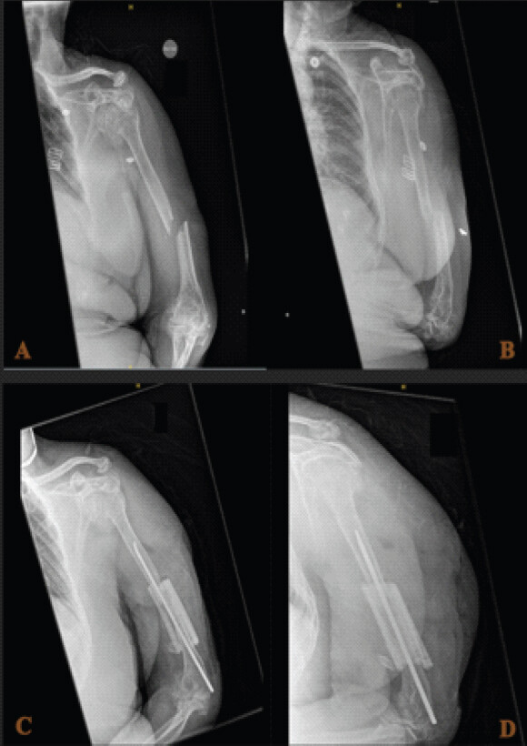
Pre-operative examination demonstrated tingling over the dorsum of the hand consistent with sensory involvement of the radial nerve; no overt motor weakness in the radial nerve distribution was identified. The pre-existing ulnar nerve deficit was carefully documented as a baseline finding. Distal vascularity was intact. Imaging confirmed a displaced distal third humerus fracture (Fig. 3). In addition, a concurrent undisplaced proximal humerus fracture was suspected clinically, a recognised phenomenon in OI given the tendency for multi-level osseous injury from a single traumatic event.
Pre-operative discussion with the patient covered several fixation options, including flexible intramedullary (TEN) nail fixation – selected as the primary modality given the OI diagnosis and the associated cortical fragility – supplementary small plate fixation if intraoperative stability was insufficient, and planned exploration of the radial nerve. The patient was consented, marked, and prepared according to the standard the World Health Organization surgical safety checklist protocol. All regular medications were continued perioperatively.
Intraoperatively, the diagnosis of a distal third humerus fracture with a sensory radial nerve palsy was confirmed. Exploration of the radial nerve revealed no structural disruption, consistent with a neurapraxia at the fracture site. An elastic intramedullary nail was inserted from distal to proximal using multiple intramedullary wires, and an onlay allograft strut was secured alongside the nail to supplement rotational control and cortical load sharing – the same biological principles applied in the osteoporotic non-union cases, adapted here to an acute fracture in OI bone.
On post-operative Day 1, the patient was alert, comfortable, and free from new complaints. Active finger mobilization was intact, and neurovascular assessment confirmed no intraoperative further nerve damage. The wound was clean and dry. Blood investigations and a post-operative radiograph were both satisfactory (Fig. 3). Physiotherapy and occupational therapy input was commenced immediately, with early active finger and wrist exercises prioritized to prevent stiffness in the context of a pre-existing arthritic elbow. Follow-up in the fracture clinic was arranged at 2 weeks.
Discussion
The three cases presented here share a common thread: humeral fractures occurring in bone of structurally compromised quality, where standard fixation alone would be unlikely to provide adequate stability. In Cases 1 and 2, the deficit arises from osteoporosis compounded by established non-union, with the remodeling, sclerosis, and medullary obliteration that accompany a prolonged failure of healing. In Case 3, the problem is intrinsic to the underlying diagnosis of OI, where cortical architecture is constitutionally deficient regardless of the acuity of the injury. The unifying surgical response in all three patients was the application of a structural strut allograft to address these shortfalls – augmenting screw purchase, distributing load across a wider bony surface, and providing a scaffold for biologic incorporation.
The evidence base for strut allograft in osteoporotic humeral non-union, while drawn largely from small case series, is consistent in its conclusions. Van Houwelingen and McKee demonstrated universal union in six osteopenic non-unions treated with a 4.5-mm compression plate and humeral cortical onlay strut allograft [3], and Vidyadhara et al. similarly reported 100% union rates using an intramedullary fibular strut graft with DCP fixation in osteoporotic cases [4]. Fink Barnes et al., in the largest series to date, achieved union in 77% of 13 humeral shaft non-unions using revision ORIF with fibular allograft [5]. These series reflect a range of graft types (fibular vs. humeral cortical) and fixation constructs but are united by the principle that structural strut augmentation effectively compensates for deficient native bone in achieving mechanically stable fixation. Cases 1 and 2 in our series are consistent with these published outcomes: Case 1 achieved solid union with graft incorporation by 6 months, and Case 2 similarly demonstrated bridging callus at the same interval, albeit requiring supplementary double plating with cerclage in response to intraoperative assessment of construct insufficiency.
The decision to supplement the primary locking plate in Case 2 with double condylar locked plates and cerclage wires merits comment. The initial single-plate construct, while radiologically positioned well, was judged intraoperatively to provide insufficient rotational stability against the background of severe cortical thinning and the atrophic quality of the non-union. This reflects a practical reality of operating in severely osteoporotic bone: the intraoperative feel of stability is unreliable, and surgeons must be prepared to escalate the construct. The addition of a second plate orthogonal to the first, secured around the strut graft with cerclage, increased torsional and bending resistance substantially and was the approach ultimately responsible for the satisfactory outcome in this case.
Case 3 introduces a different dimension to the series. OI, arising from mutations in collagen type I genes, produces bone that is not simply osteopenic but fundamentally abnormal in its matrix composition, with cortices that are thin, brittle, and prone to propagating fractures along lines of stress [6]. This creates a surgical environment in which the standard approach to internal fixation – rigid plate fixation with bicortical screw purchase – carries risks that do not apply to osteoporotic bone. Stress concentration at screw entry points and plate ends can precipitate iatrogenic fractures in OI cortex, making the more compliant mechanics of flexible intramedullary fixation an attractive alternative for acute humeral fractures in this population. The TEN nail, by virtue of its intramedullary position and load-sharing mechanics, avoids the cortical stress risers inherent to plating while still providing adequate alignment control. The allograft strut in this context served to supplement rotational stability and provide a biological scaffold for healing rather than to replace the structural contribution of a plate, as in the non-union cases.
The pre-operative sensory radial nerve palsy in Case 3 required careful management both clinically and from a documentation perspective. Holstein-Lewis fractures – fractures at the junction of the middle and distal thirds of the humerus – are recognised to carry a risk of radial nerve injury, with reported rates of associated nerve palsy ranging from 2% to 22% depending on the series [7, 8]. Radial nerve injury in humeral fractures is a complication which, although predictable and partly preventable, is not easily avoidable [9]. In the majority of cases, the deficit is a neurapraxia secondary to traction or contusion at the fracture site, and spontaneous recovery can be expected in the absence of structural disruption [10]. The importance of meticulous pre-operative documentation cannot be overstated: Any deficit that is not recorded clearly prior to surgery risks being misattributed to a post-operative complication, with significant implications for both the patient and the operating team. In this case, the pre-existing ulnar nerve deficit from the prior supracondylar fracture added further complexity to the neurological baseline, and both deficits were formally documented before the patient reached theatre. Intraoperative exploration confirmed an intact radial nerve, and post-operative assessment at Day 1 demonstrated recovery of sensory function distally, consistent with resolution of neurapraxia following fracture reduction.
There are limitations to this series that should be acknowledged. The patient numbers are small, and the cases represent a convenience series rather than a prospectively collected cohort, so no formal comparison can be drawn between cases. The follow-up available for Case 3 is limited to the immediate post-operative period; longer-term data on fracture healing, nerve recovery, and functional outcomes will be required before conclusions can be drawn about the success of the operative approach in this patient. Formal functional outcome measures (DASH, Oxford Shoulder Score) were not recorded for Case 3 in the period covered here, in contrast to Case 1 where validated scores demonstrated clear functional benefit. Future reporting of Case 3 at fracture clinic follow-up will allow a more complete assessment. Nonetheless, the radiological satisfactoriness of the immediate post-operative construct and the neurological recovery observed on Day 1 are encouraging early indicators.
Taken together, the three cases illustrate that structural strut allograft augmentation is not a technique confined to a single indication or fixation implant. It can be delivered as an onlay graft alongside a locking plate for established non-union or adapted to accompany a flexible intramedullary nail for an acute fracture in OI. The unifying principle is that it compensates for the structural deficit of the host bone, whether that deficit is acquired through years of failed healing or is constitutionally present from the outset. Surgical teams managing humeral fractures in elderly patients with metabolic bone disease should be familiar with the technique and prepared to employ it across this range of presentations.
Conclusion
Structural strut allograft augmentation of humeral ORIF represents an effective and adaptable technique for managing fractures in bone of compromised quality. In osteoporotic non-union, it provides the mechanical and biologic environment necessary to achieve union when standard fixation has failed or is at risk of failing. In OI, it can be combined with flexible intramedullary nail fixation to supplement cortical stability in an acute fracture setting while respecting the inherent fragility of the underlying bone. The cases presented here support the broader application of this technique across the spectrum of humeral fractures in metabolically deficient bone and highlight the importance of individualizing both the fixation implant and the allograft strategy to the specific bone biology and fracture pattern encountered.
Clinical Message
Humeral fractures and non-unions in metabolically compromised bone remain difficult to treat because conventional fixation may fail in deficient cortical bone. Structural strut allograft augmentation provides both mechanical reinforcement and biological support, allowing stable fixation and progression to healing across a spectrum of humeral fracture patterns, underlying bone pathologies, and implant constructs. These cases highlight the importance of tailoring fixation strategies to bone quality and demonstrate that strut allograft augmentation can be a valuable option when managing fractures in osteoporotic bone and OI.
Conflict of Interest:
Nil
Source of Support:
Nil
Consent:
The authors confirm that informed consent was obtained from the patient for publication of this article
How to Cite this Article
Nagy M, Aldanyowi SN, Erdman P, Ashwood N. Humeral Fractures in Metabolically Compromised Bone: Outcomes of Structural Strut Onlay Allograft with Open Reduction and Internal Fixation – A Case Series of Three Patients. Journal of Orthopaedic Case Reports 2026 October;16(10): 186-192.
References
- Ekholm R, Adami J, Tidermark J, Hansson K, Törnkvist H, Ponzer S. Fractures of the shaft of the humerus. An epidemiological study of 401 fractures. J Bone Joint Surg Br 2006;88:1469-73. [Google Scholar] | [PubMed]
- Dar SA, Butt MF, Dar TA, Dar RA, Ali Z. Management of humeral fracture nonunion in severe osteoporosis by a combination of locking plating and intramedullary fibular grafting. Chin J Traumatol 2016;19:298-301. [Google Scholar] | [PubMed]
- Van Houwelingen AP, McKee MD. Treatment of osteopenic humeral shaft nonunion with compression plating, humeral cortical allograft struts, and bone grafting. J Orthop Trauma 2005;19:36-42. [Google Scholar] | [PubMed]
- Vidyadhara S, Vamsi K, Rao SK, Gnanadoss JJ, Pandian S. Use of intramedullary fibular strut graft: A novel adjunct to plating in the treatment of osteoporotic humeral shaft nonunion. Int Orthop 2009;33:1009-14. [Google Scholar] | [PubMed]
- Fink Barnes LA, Ruig DF, Freibott CE, Rajfer R, Rosenwasser MP. Treatment of nonunions of the humeral shaft with nonvascularized fibular strut allograft: Postoperative outcomes and review of a surgical technique. JSES Int 2020;4:739-44. [Google Scholar] | [PubMed]
- Rauch F, Glorieux FH. Osteogenesis imperfecta. Lancet 2004;363:1377-85. [Google Scholar] | [PubMed]
- Shao YC, Harwood P, Grotz MR, Limb D, Giannoudis PV. Radial nerve palsy associated with fractures of the shaft of the humerus: A systematic review. J Bone Joint Surg Br 2005;87:1647-52. [Google Scholar] | [PubMed]
- Ekholm R, Ponzer S, Törnkvist H, Adami J, Tidermark J. The Holstein-Lewis humeral shaft fracture: Aspects of radial nerve injury, primary treatment, and outcome. J Orthop Trauma 2008;22:693-7. [Google Scholar] | [PubMed]
- Basile G, Fozzato S, Prevot LB, Giorgetti A, Gallina M, Basile M. Radial nerve injuries in humeral fractures: Case series and medico-legal implications. Injury 2024;55:111497. [Google Scholar] | [PubMed]
- Feng D, Zhang J, Zhu Y, Wu S, Shan J, Ye A. Plate fixation with autogenous bone grafting for longstanding humeral shaft nonunion: A retrospective study of 6 cases. Medicine (Baltimore) 2018;97:e11974. [Google Scholar] | [PubMed]
© 2026 Journal of Orthopaedic Case Reports - Published by Indian Orthopaedic Research Group
About the Authors
-

Mohamed Nagy
-

Saud N Aldanyowi
-

Paul Erdman
-

Neil Ashwood
Related Articles in Journal of Orthopaedic Case Reports
 September 1, 2024 Functional Outcomes of Gap Non-Union Humerus Fractures Treated with Fibular Strut Graft, Iliac Crest Graft, and Locking Compression Plate
 February 10, 2024 Outcome of Osteosynthesis of Late-Presenting Proximal Humerus Non-union: A Case Series
 September 1, 2026 Surgical Management of Exceptionally Severe Cubitus Varus Deformity in Osteogenesis Imperfecta: A Case Report
 September 1, 2026 Multifactorial Non-union After First Metatarsophalangeal Arthrodesis Revised With Structural Iliac Crest Autograft: A Case Report and Comprehensive Literature Review