


Introduction
Type II odontoid fractures are among the most common injuries of the upper cervical spine, and anterior odontoid screw fixation is a widely used technique because it preserves atlantoaxial rotation while achieving direct fracture stabilization [1,2,3,4,5]. The procedure is generally regarded as safe, although dysphagia, screw back-out and non-union are recognized complications [5,6,7,8,9]. Vascular or brainstem injury following this operation is exceptional and, to our knowledge, has not previously been reported.
Medial medullary syndrome results from infarction of the paramedian medulla, an area supplied by perforating branches of the vertebral and anterior spinal arteries, and classically produces contralateral limb weakness, contralateral loss of proprioception, and an ipsilateral hypoglossal palsy [10–13]. Bilateral involvement is considerably rarer and has mainly been reported in association with vertebral artery dissection, atherosclerosis, or arteritis [12,14]. We describe a patient who developed bilateral medial medullary syndrome after anterior fixation of a type II odontoid fracture and discuss a possible mechanistic link to perioperative manipulation of the cervical spine.
Case Report
Presentation and initial imaging
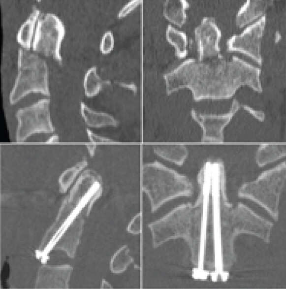
A 28-year-old man presented with neck pain after a road traffic accident. Neurological examination at admission was entirely normal. Computed tomography (CT) of the cervical spine demonstrated a posteriorly displaced type II odontoid fracture together with a fracture of the C2 spinous process, and surgery was planned for later the same day.
His vascular risk factors and co-morbidity profile are summarized in Table 1.
Patient co-morbidity and vascular risk factor profile
| Risk factor/co- morbidity | Details |
|---|---|
| Hypertension | Diagnosed 2 years prior; on amlodipine 5 mg OD. |
| Dyslipidemia | Total cholesterol 248 mg/dL, LDL 168 mg/dL; on atorvastatin 20 mg nocte |
| Smoking | Smoker for 10 years, ~10 cigarettes/day |
| Alcohol | Occasional alcohol use |
| Diabetes mellitus | No. |
| Known vascular disease | No prior history. |
| Family history | Father had myocardial infarction at age 52. |
| Other | No known autoimmune or coagulation disorders. |
| Medications at admission | Amlodipine, atorvastatin. |
| Allergies | None known. |
Severe atherosclerosis (grade IV plaques in the bilateral vertebral and basilar arteries) was identified on CT angiography performed as part of the etiological work-up.
Perioperative course
During induction of anesthesia, the patient developed anisocoria with left-sided mydriasis. Anesthesia was stopped and urgent CT angiography was performed, which showed no vascular abnormality – in particular, the vertebral artery was normal – and no change in fracture position. Once he had woken, the neurological examination was again normal and the pupillary asymmetry had resolved. Surgery was therefore postponed, and halo-ring traction (5 kg) was applied to allow gradual, controlled reduction of the fracture over the following 3 days.
Definitive fixation was carried out on day 3, at the completion of the halo-ring traction period. The operative technique is detailed below.
The patient was positioned supine on a radiolucent table with the neck in neutral alignment. General anesthesia was induced with orotracheal intubation under fiber-optic guidance, and manual in-line stabilization was maintained throughout. Halo-ring traction (5 kg) was applied, and gentle longitudinal traction with slight neck extension achieved anatomical reduction, confirmed on lateral and open-mouth fluoroscopy. A right-sided Smith–Robinson anterior cervical approach was used at the C1–C2 level; the prevertebral fascia was opened and the longus colli retracted laterally to expose the C2 body. Two 3.5 mm partially threaded cannulated cancellous screws (DePuy Synthes) were inserted anterior-to-posteriorly along the axis of the odontoid under biplanar fluoroscopic guidance, with entry points in the inferior half of the C2 body as a paramedian pair on either side of the midline, directed superiorly and posteriorly to the tip of the odontoid. Screw positions were confirmed on anteroposterior and lateral fluoroscopy, with no canal breach. Intraoperative neuromonitoring (somatosensory evoked potential and motor evoked potential) remained stable throughout. The operative time was 110 min, with an estimated blood loss of <50 mL and no intraoperative complications. Closure was performed in standard layered fashion with a suction drain.
Postoperative course and neurological deterioration
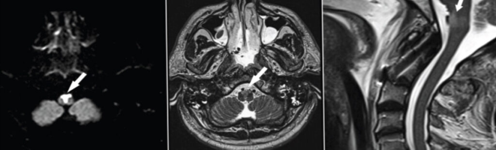
Weaning from ventilation was prolonged and complicated. CT of the head and cervical spine confirmed satisfactory fracture reduction and correct screw positions, with unremarkable brain parenchyma. Because the clinical course remained unexplained, cerebral and cervical magnetic resonance imaging (MRI) was obtained and revealed acute bilateral ischemia of the medial medulla. Tracheostomy and percutaneous endoscopic gastrostomy were subsequently performed, after which weaning was achieved successfully.
Neurological findings
Examination showed a bilateral incomplete tetraparesis, predominantly left-sided, with pyramidal signs. Cranial nerve examination was otherwise normal apart from deviation of the tongue to the right, in keeping with a right-sided hypoglossal nerve palsy. Formal sensory testing (light touch and pinprick) and anorectal examination were performed once the patient was extubated; voluntary anal contraction and deep anal pressure were both preserved. Detailed International Standards for Neurological Classification of Spinal Cord Injury (ISNCSCI) motor and sensory scores, neurological level, and ASIA grade at postoperative day 2 (the time of worst neurological deficit) and at 6-month follow-up are summarized in Table 2.
ISNCSCI motor and sensory examination, neurological level, and ASIA grade at postoperative day 2 (time of worst neurological deficit) and at 6-month follow-up
| Domain | Postoperative (day 2) – at time of worst deficit | 6-month follow-up | ||
|---|---|---|---|---|
| R | L | R | L | |
| Motor examination (key muscles) | ||||
| C5 (Elbow flexors) | 3 | 1 | 5 | 3 |
| C6 (Wrist extensors) | 3 | 1 | 5 | 3 |
| C7 (Elbow extensors) | 3 | 1 | 5 | 3 |
| C8 (Finger flexors) | 2 | 0 | 4 | 2 |
| T1 (Finger abductors) | 2 | 0 | 4 | 2 |
| L2 (Hip flexors) | 2 | 1 | 4 | 2 |
| L3 (Knee extensors) | 2 | 1 | 4 | 2 |
| L4 (Ankle dorsiflexors) | 1 | 0 | 3 | 1 |
| L5 (Long toe extensors) | 1 | 0 | 3 | 1 |
| S1 (Ankle plantar flexors) | 2 | 1 | 4 | 2 |
| Light Touch (Max: 112) | 56/112 | 32/112 | 96/112 | 68/112 |
| Pin Prick (Max: 112) | 54/112 | 30/112 | 94/112 | 66/112 |
| Neurological level | C8 | C8 | C7 | C7 |
| ASIA grade | C | C | D | D |
| Zone of partial preservation | S2-S4 | S2-S4 | S2-S4 | S2-S4 |
| VAC (Anal Contraction) | Present | Present | Present | Present |
| DAP (Deep Anal Pressure) | Present | Present | Present | Present |
R: Right, L: Left, ISNCSCI: International standards for neurological classification of spinal cord injury
Clinical timeline
The overall sequence of injury, surgery, deterioration, and recovery is summarized in Table 3.
Clinical timeline from injury to rehabilitation discharge
| Time point | Event |
|---|---|
| Day 0 | Road traffic accident; neck pain; neurologically intact. CT: Posteriorly displaced type II odontoid fracture with C2 spinous process fracture. Surgery planned same day. |
| Day 0 | Anisocoria with left mydriasis during anesthetic induction. Anesthesia stopped. CT angiography normal (no vertebral artery pathology, no fracture displacement). Findings resolved after waking; surgery postponed. |
| Day 3 | Anterior odontoid screw fixation performed uneventfully (two 3.5 mm cannulated lag screws) at the end of a 3-day period of preoperative halo-ring traction (5 kg) and fluoroscopic reduction. |
| Postop (early) | Prolonged and complicated weaning from ventilation. CT head/cervical spine unremarkable apart from confirming good reduction and screw positions. |
| Postop (early) | Cerebral and cervical MRI obtained because of unexplained course: Acute bilateral medial medullary ischemia identified. |
| Postop (early) | Tracheostomy and percutaneous endoscopic gastrostomy performed; successful weaning achieved. Enoxaparin 1 mg/kg SC twice daily started (postoperative day 2); aspirin 150 mg once daily added (postoperative day 3); no unfractionated heparin used. Neurorehabilitation initiated. |
| 3 weeks | Discharged from acute care to inpatient rehabilitation. ASIA C; wheelchair-bound; tube feeding; intermittent catheterization. |
| 3 months | Tracheostomy successfully decannulated. |
| 6 months | Discharge from rehabilitation (final follow-up). ASIA D maintained; ambulating short distances with a single cane; normal diet; volitional voiding; independent bowel function; mild residual bilateral distal (hand/toe) weakness. |
ASIA: American Spinal Injury Association, CT: Computed tomography, MRI: Magnetic resonance imaging
Imaging findings
Preoperative CT confirmed the displaced type II odontoid fracture, and postoperative CT confirmed satisfactory reduction with correctly positioned screws (Fig. 1a and b). MRI obtained after the failed weaning attempts showed restricted diffusion with a high diffusion-weighted imaging (DWI) signal and corresponding high T2 signal in the anterior medulla, consistent with acute bilateral medial medullary ischemia (Fig. 2). The infarcted area lay close to both the surgical field and the vertebral artery.
Treatment and etiological work-up
Enoxaparin (Clexane) 1 mg/kg subcutaneously twice daily was started on postoperative day 2, and aspirin 150 mg once daily was added on postoperative day 3 once a thrombotic vascular mechanism was considered likely; no unfractionated heparin was used at any stage. The decision to institute this antithrombotic regimen was made after weighing the risk of extending the medullary infarct against the risk of epidural or wound hemorrhage so soon after cervical instrumentation; a prophylactic-dose regimen was chosen for this reason, and no hemorrhagic transformation was seen on subsequent imaging. Neurorehabilitation began promptly, and the tracheostomy was successfully decannulated three months after the injury. Screening for an underlying cause was otherwise unremarkable except for severe atherosclerotic disease, with grade IV atheromatous plaques identified in the bilateral vertebral and basilar arteries on CT angiography (Table 1). Given the patient’s young age, dedicated screening for causes of premature vasculopathy – including familial hypercholesterolemia and other inherited dyslipidemias – was undertaken as part of this etiological work-up; no such disorder was identified, and the atherosclerotic risk-factor profile summarized in Table 1 remained the only identifiable contributor.
Outcome
He was discharged from acute care to inpatient rehabilitation 3 weeks after injury, remaining ASIA C at that point, wheelchair-bound, tube-fed, and requiring intermittent catheterization, with recurrent aspiration pneumonia during this early period. His ISNCSCI grade had improved to ASIA D by 3 months, at which stage he mobilized with a walker and tolerated a soft diet following tracheostomy decannulation. By 6-month follow-up, he was ambulating short distances with a single cane, tolerating a normal diet, voiding volitionally, and had regained independent bowel function, although mild bilateral distal weakness (hand and toe) persisted (Table 2). Detailed functional, mobility, and continence outcomes at each time point are summarized in Table 4.
Functional outcome – objective measures at discharge, 3 months, and 6 months
| Parameter | At Discharge (3 weeks post-op) | 3 Months | 6 Months |
|---|---|---|---|
| ASIA Grade | C | D | D |
| UEMS (Max 50) | 22 | 36 | 44 |
| LEMS (Max 50) | 18 | 32 | 40 |
| 10-Meter Walk Test | — | 0.38 m/s (with aid) | 0.72 m/s (with cane) |
| Walking Aid | Wheelchair | Walker | Single cane (short distances) |
| Functional independence measure | 64/126 | 86/126 | 98/126 |
| Barthel index | 45/100 | 65/100 | 85/100 |
| Swallowing | Tube feeding | Soft diet | Normal diet |
| Tracheostomy | Present | Decannulated | Decannulated |
| Bladder function | Intermittent catheterization | Intermittent catheterization | Volitional voiding |
| Bowel function | Assisted | Assisted | Independent |
ASIA: American Spinal Injury Association
Discussion
The central lesson of this case is that a normal postoperative CT does not exclude significant brainstem ischemia. Here, the diagnosis only became apparent once MRI with DWI was obtained in response to unexplained weaning failure. We would recommend a low threshold for MRI, including DWI sequences, whenever a patient fails to wean or develops new neurological signs after anterior fixation of an odontoid fracture, and CT does not account for the deterioration [15,16].
Differential diagnosis
The transient anisocoria and mydriasis during induction initially raised concern for an occult vertebral or basilar artery injury; however, CT angiography at the time excluded dissection or occlusion of the basilar artery, and the pupillary asymmetry had resolved by the time the patient woke, making it unlikely to be mechanistically linked to the medullary infarction that followed. Isolated basilar artery occlusion was also considered, but this typically produces a broader pattern of cranial nerve, cerebellar, and consciousness disturbance that was not seen here. A demyelinating or inflammatory process was thought unlikely given the acute, vascular-territory-restricted appearance on DWI and the absence of any preceding systemic illness. The bilateral, paramedian distribution of the lesion, together with its close temporal relationship to surgery, was most consistent with a perioperative ischemic event.
Proposed mechanism
The etiology in this patient remained uncertain, and the only vascular abnormality identified on systematic work-up was advanced, grade IV atherosclerosis of the bilateral vertebral and basilar arteries, identified on CT angiography performed as part of the etiological work-up (Table 1). Given the anatomical proximity of the medullary infarct to both the surgical field and the vertebral artery (Fig. 3), we consider it plausible that perioperative mechanical manipulation of the upper cervical spine – during intubation, positioning, fracture reduction, or sustained traction – contributed to atherothrombotic occlusion of small perforating branches of the vertebral or anterior spinal artery in a vessel already compromised by atherosclerotic disease. Experimental work on cervical manipulation in atherosclerotic animal models has shown that rotational and manipulative forces can destabilize plaque and reduce arterial tensile properties [17,18], and manipulation of the neck has separately been linked to stroke in patients with underlying vascular disease [19]. Perioperative ischemic stroke is itself a recognized, if uncommon, complication of surgery unrelated to the cervical spine [10,11], which lends further plausibility to a perioperative vascular mechanism in this case.
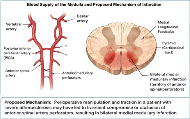
The specific biomechanical steps used in this case merit closer consideration. Sustained halo-ring traction combined with slight neck extension during fracture reduction and positioning is mechanistically distinct from the head rotation implicated in most manipulation-related vertebral artery injury: Extension preferentially loads the V3 segment of the vertebral artery as it curves around the lateral mass of the atlas, and a systematic review of craniocervical positioning found that combined maximal extension and rotation, more than either movement alone, was associated with a measurable reduction in vertebral artery hemodynamics [20]. Isolated case reports have similarly documented reversible positional occlusion of the vertebral artery in extension [21], and intraoperative traction, even within conventional limits, has been implicated in vertebral artery dissection during cervical spine surgery [22]. This combination of sustained axial traction and extension differs from posterior cervical procedures, in which the neck is generally maintained in a neutral or flexed position and reported vertebral artery injuries are more commonly attributed to direct instrumentation – lateral mass or pedicle screw malposition – than to traction- or extension-related hemodynamic compromise [23]. In a vessel already narrowed by grade IV atherosclerotic plaque, even a modest, sub-critical reduction in flow of the kind described with extension may have been sufficient to precipitate thrombosis of small perforating branches, whereas the same maneuver in a normal artery would be unlikely to have hemodynamic consequences. This distinction has practical relevance for surgical teams managing trauma in vascularly compromised patients: where feasible, minimizing the duration and degree of neck extension during traction and positioning, and maintaining a low threshold to reassess vertebral artery patency if traction is prolonged, may help reduce the risk of this rare but serious complication.The clinical picture – bilateral, left-predominant limb weakness with pyramidal signs and a unilateral (right-sided) hypoglossal palsy – is consistent with bilateral medial medullary infarction, in which the corticospinal tracts on both sides are affected, producing quadriparesis that was more marked on the left, while hypoglossal fascicular involvement can remain asymmetric depending on the precise distribution of the paramedian lesions [13]. Bilateral disease, as seen here, has previously been reported almost exclusively with dissection, atherosclerosis, or vasculitis as the underlying cause [12,14], rather than as a perioperative event, which is what makes this presentation unusual.
Literature comparison
Table 5 compares the clinical, radiological, and treatment features of this case with previously reported cases of bilateral medial medullary infarction.
Literature comparison – bilateral medial medullary infarction
| Author (Year) | Age/Sex | Etiology | Vascular Abnormality | Key Neurological Features | Imaging | Treatment | Outcome |
|---|---|---|---|---|---|---|---|
| Casey et al. (1997) [24] | 67/F | Hypertensive vasculopathy | Small vessel disease | Quadriparesis, pseudobulbar palsy | MRI: bilateral medial medullary lesions | Supportive | Partial recovery (ASIA D) |
| Shibata et al. (2003) [25] | 58/M | Vertebral artery occlusion | Left vertebral artery occlusion | Quadriparesis, dysphagia, loss of vibration | MRI: bilateral medial medullary infarction | Antiplatelet | Good recovery |
| Kwon et al. (2006) [26] | 72/M | Atherothrombotic | Basilar artery stenosis | Tetraparesis, respiratory failure | MRI: bilateral medial medullary infarction | Antiplatelet | Partial recovery |
| Gallo et al. (2012) [27] | 61/M | Small vessel disease | No large vessel occlusion | Quadriparesis, dysarthria | MRI-DWI positive | Supportive | Good recovery |
| Rishi et al. (2017) [28] | 55/M | Thromboembolic | Cardioembolic source (Atrial fibrillation) | Quadriparesis, dysphonia | MRI-DWI positive | Anticoagulation | Partial recovery |
| Sato et al. (2020) [29] | 69/F | Hypoperfusion | Severe atherosclerosis | Quadriparesis, ataxia | MRI-DWI positive | Supportive | Partial recovery |
| Suga et al. (2022) [30] | 63/M | Vertebrobasilar insufficiency | High-grade basilar stenosis | Tetraparesis, dysphagia | MRI: bilateral medial medullary infarction | Antiplatelet | Partial recovery |
| Current Case (2026) | 28/M | Post-op after odontoid screw fixation | Severe atherosclerosis (grade IV plaques) | Quadriparesis, respiratory failure, bulbar dysfunction | MRI-DWI: bilateral medial medullary infarction | Aspirin + Enoxaparin 1 mg/kg BD | Improved to ASIA D at 6 months |
DWI: Diffusion-weighted imaging, MRI: Magnetic resonance imaging, ASIA: American Spinal Injury Association
Limitations
This report has several limitations. It describes a single patient, and the proposed link between cervical manipulation and medullary infarction is inferential rather than proven; no dedicated imaging of the vertebral artery or its perforating branches was obtained at the presumed time of infarction, so a focal dissection or occlusion at that level cannot be entirely excluded. The relative contribution of each perioperative maneuver – intubation, positioning, fracture reduction, or sustained traction – cannot be separated out from this single case, and longer-term functional and swallowing outcomes beyond 6 months are not available.
Conclusion
Bilateral medial medullary syndrome is a rare but serious complication that should be considered after anterior screw fixation of a type II odontoid fracture, particularly in patients with vascular risk factors who fail to wean from ventilation or develop new neurological signs postoperatively. Because CT can be falsely reassuring, MRI with DWI is essential for timely diagnosis. Awareness of this possible association may help surgical and anesthetic teams limit unnecessary cervical manipulation in at-risk patients and pursue early MRI when the postoperative course is unexplained.
Clinical Message
In a patient with a normal neurological examination before surgery, a severe brainstem syndrome can still emerge after anterior odontoid fixation. When postoperative weaning fails or new deficits appear and CT is unrevealing, MRI with DWI should be obtained promptly, and coexisting atherosclerosis should be sought as a potential contributing factor.
Conflict of Interest:
Nil
Source of Support:
Nil
Consent:
The authors confirm that informed consent was obtained from the patient for publication of this article
How to Cite this Article
Raj S, Kumar D, Kumar V, Sen AK, Kumar SR, Goni VG. Bilateral Medial Medullary Syndrome Following Anterior Odontoid Screw Fixation for a Type II Fracture: A Case Report. Journal of Orthopaedic Case Reports 2026 October;16(10): 252-259.
References
- Golob JFJr, Claridge JA, Yowler CJ, Como JJ, Peerless JR. Isolated cervical spine fractures in the elderly: A deadly injury. J Trauma 2008;64:311-5. [Google Scholar] | [PubMed]
- Pommier B, Ollier E, Pelletier JB, Castel X, Vassal F, Tetard MC. Conservative versus surgical treatment for odontoid fracture: Is the surgical treatment harmful? Systematic review and meta-analysis. World Neurosurg 2020;141:490-9.e2. [Google Scholar] | [PubMed]
- Collins I, Min WK. Anterior screw fixation of type II odontoid fractures in the elderly. J Trauma 2008;65:1083-7. [Google Scholar] | [PubMed]
- Yuan S, Wei B, Tian Y, Yan J, Xu W, Wang L . The comparison of clinical outcome of fresh type II odontoid fracture treatment between anterior cannulated screws fixation and posterior instrumentation of C1-2 without fusion: A retrospective cohort study. J Orthop Surg Res 2018;13:3. [Google Scholar] | [PubMed]
- Pal D, Sell P, Grevitt M. Type II odontoid fractures in the elderly: An evidence-based narrative review of management. Eur Spine J 2011;20:195-204. [Google Scholar] | [PubMed]
- Marciano RD, Seaman B, Sharma S, Wood T, Karas C, Narayan K. Incidence of dysphagia after odontoid screw fixation of type II odontoid fracture in the elderly. Surg Neurol Int 2018;9:84. [Google Scholar] | [PubMed]
- Joaquim AF, Patel AA. Surgical treatment of type II odontoid fractures: Anterior odontoid screw fixation or posterior cervical instrumented fusion. Neurosurg Focus 2015;38:E11. [Google Scholar] | [PubMed]
- Harrop JS, Sharan AD, Przybylski GJ. Epidemiology of spinal cord injury after acute odontoid fractures. Neurosurg Focus 2000;15:e4. [Google Scholar] | [PubMed]
- Kepler CK, Vaccaro AR, Dibra F, Anderson DG, Rihn JA, Hilibrand AS . Neurologic injury because of trauma after type II odontoid nonunion. Spine J 2014;14:903-8. [Google Scholar] | [PubMed]
- Bateman BT, Schumacher HC, Wang S, Shaefi S, Berman MF. Perioperative acute ischemic stroke in noncardiac and nonvascular surgery: Incidence, risk factors, and outcomes. Anesthesiology 2009;110:231-8. [Google Scholar] | [PubMed]
- Wang H, Li SL, Bai J, Wang DX. Perioperative acute ischemic stroke increases mortality after noncardiac, nonvascular, and non-neurologic surgery: A retrospective case series. J Cardiothorac Vasc Anesth 2019;33:2231-6. [Google Scholar] | [PubMed]
- Farooqui A, Kolikonda M, Bokka SK, Liu W. Bilateral medial medullary syndrome with atypical clinical presentation and brain MRI findings. Neurology 2019;92. P5.3-065. [Google Scholar] | [PubMed]
- Bassetti C, Bogousslavsky J, Mattle H, Bernasconi A. Medial medullary stroke: Report of seven patients and review of the literature. Neurology 1997;48:882-90. [Google Scholar] | [PubMed]
- Deshpande A, Chandran V, Pai A, Rao S, Shetty R. Bilateral medial medullary syndrome secondary to Takayasu arteritis. BMJ Case Rep 2013;2013:bcr0120125600. [Google Scholar] | [PubMed]
- Srinivasan A, Goyal M, Al Azri F, Lum C. State-of-the-art imaging of acute stroke. Radiographics 2006;26 Suppl 1S75-95. [Google Scholar] | [PubMed]
- Weidauer S, Nichtweiß M, Hattingen E, Berkefeld J. Spinal cord ischemia: Aetiology, clinical syndromes and imaging features. Neuroradiology 2015;57:241-57. [Google Scholar] | [PubMed]
- Zhang S, Qi J, Zhang L, Chen C, Mondal S, Ping K . Cervical rotatory manipulation decreases uniaxial tensile properties of rabbit atherosclerotic internal carotid artery. Evid Based Complement Alternat Med 2017;2017:5189356. [Google Scholar] | [PubMed]
- Qi J, Ping R, Zhang S, Xu Y, Wu K, Li Y. Effects of cervical rotatory manipulation (CRM) on carotid atherosclerosis plaque in vulnerability: A histological and immunohistochemical study using animal model. Biomed Res Int 2019;2019:3793840. [Google Scholar] | [PubMed]
- Lee KP, Carlini WG, McCormick GF, Albers GW. Neurologic complications following chiropractic manipulation: A survey of California neurologists. Neurology 1995;45:1213-5. [Google Scholar] | [PubMed]
- Kranenburg HA, Tyer R, Schmitt M, Luijckx GJ, Van Der Schans C, Hutting N . Effects of head and neck positions on blood flow in the vertebral, internal carotid, and intracranial arteries: A systematic review. J Orthop Sports Phys Ther 2019;49:688-97. [Google Scholar] | [PubMed]
- Garg A, Gaikwad SB, Kanodia A, Suri A, Gupta V, Mishralf NK. Positional occlusion/stasis of vertebral arteries in a case of cervical rheumatoid arthritis presenting with multiple posterior circulation infarcts: A case report with angiographic demonstration. Spine (Phila Pa 1976) 2004;29:E321-5. [Google Scholar] | [PubMed]
- Dickerman RD, Zigler JE. Atraumatic vertebral artery dissection after cervical corpectomy: A traction injury?. Spine (Phila Pa 1976) 2005;30:E658-61. [Google Scholar] | [PubMed]
- Sakellariou E, Benetos IS, Evangelopoulos DS, Galanis A, Alevrogianni F, Vavourakis M . Incidence of vertebral artery injury in patients undergoing cervical spine trauma surgery in correlation with surgical approach: A review. Medicine (Baltimore) 2023;102:e34653. [Google Scholar] | [PubMed]
- Casey SO, Feldman EL, Brown RDJr, Compére V, Cros D. Bilateral medial medullary infarction: Report of a case and review of the literature. Neurology 1997;48:1395-7. [Google Scholar] | [PubMed]
- Shibata M, Kawano H, Sato N, Kimura K. Bilateral medial medullary infarction due to vertebral artery occlusion. J Neurol Sci 2003;208:73-7. [Google Scholar] | [PubMed]
- Kwon SU, Kim JS, Kim YH, Lee MC. Bilateral medial medullary infarction associated with basilar artery stenosis. J Clin Neurol 2006;2:74-8. [Google Scholar] | [PubMed]
- Gallo P, Costa F, Resta F, Bertaccini B. Bilateral medial medullary syndrome: Case report and review of the literature. Neurol Sci 2012;33:701-4. [Google Scholar] | [PubMed]
- Rishi P, Singh MB, Goyal V, Bhatia R. Bilateral medial medullary infarction secondary to cardioembolism. BMJ Case Rep 2017;2017:bcr2017219252. [Google Scholar] | [PubMed]
- Sato K, Uchiyama K, Yoshimoto T, Toi S. Hypoperfusion-related bilateral medial medullary infarction. Intern Med 2020;59:523-7. [Google Scholar] | [PubMed]
- Suga T, Fujii T, Nakamura T, Sakai K. Vertebrobasilar insufficiency causing bilateral medial medullary infarction: A case report. Neurol Med Chir (Tokyo) 2022;62:265-89. [Google Scholar] | [PubMed]
© 2026 Journal of Orthopaedic Case Reports - Published by Indian Orthopaedic Research Group
About the Authors
-

Sharath Raj
-

Deepak Kumar
-

Vishal Kumar
-

Aashish Kumar Sen
-

Saloni Rani Kumar
-

Vijay G Goni
Related Articles in Journal of Orthopaedic Case Reports
 September 1, 2026 Blunt Traumatic Posterior Cord Syndrome Following a Hangman Fracture: A Case Report
 August 1, 2026 Radiologically Devastating Yet Neurologically Intact: A Rare Case of Ambulatory C6–C7 Translational Injury with Bilateral Facet Dislocation and Vertebral Artery Thrombosis
 April 1, 2026 A Rare Case of C5–C6 Bilateral Facetal Dislocation with Central Cord Syndrome Managed by Staged Stabilization
 July 1, 2025 Case Report – Surgical Management and Neurological Recovery in an 18-Year-Old Male with C4-C5 Cervical Spine Fracture