


Introduction
Talar fractures are uncommon, accounting for <1% of all fractures. Posterior process fractures are a rare subset, divided into posterolateral (Shepherd) and posteromedial (Cedell) types [1]. Posteromedial talar process fractures result from complex rotational ankle forces, typically forced dorsiflexion combined with pronation, producing avulsion at the posterior talotibial ligament attachment [2]. They are frequently misdiagnosed due to subtle radiographic findings and clinical overlap with soft-tissue injuries, particularly in polytrauma patients. Missed diagnosis risks non-union, chronic pain, flexor hallucis longus (FHL) dysfunction, and subtalar arthritis. Bilateral involvement is exceedingly rare and sparsely documented [3]. We report a case of bilateral Cedell fracture in a polytrauma patient managed with a staged protocol, with the aim of raising awareness of this injury and demonstrating the efficacy of damage control orthopedic (DCO) principles in complex hindfoot trauma.
Case Report
A 20-year-old man presented to the emergency department following a fall from height. He reported severe back pain and bilateral ankle pain with swelling. There was no loss of consciousness or head injury. On arrival, he was hemodynamically stable (Glasgow Coma Scale 15, Injury Severity Score 16). Neurological examination was intact with preserved anal tone (American Spinal Injury Association grade E). Lumbar spine palpation revealed tenderness at L3. Bilateral ankle examination demonstrated tenderness, swelling, and abnormal mobility. Distal neurovascular status of both lower limbs was intact.
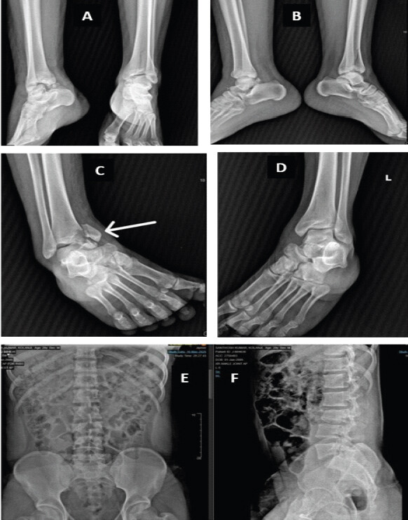
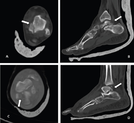
Radiographs demonstrated a right ankle fracture-dislocation with medial malleolus fracture, left distal tibial plafond fracture, and an L3 bony Chance fracture. Computed tomography (CT) was performed for all injured regions and was essential in delineating bilateral posteromedial talar process fractures, confirming articular involvement of the subtalar joint, and guiding operative planning.
The Left side showed an anterior pilon injury with central plafond impaction.
On the right side, there was a Herscovici Type B medial malleolus fracture with plafond marginal impaction near the anteromedial aspect of the plafond as shown in the axial CT scan (Figs. 1 and 2).
Management
A staged DCO approach was adopted.
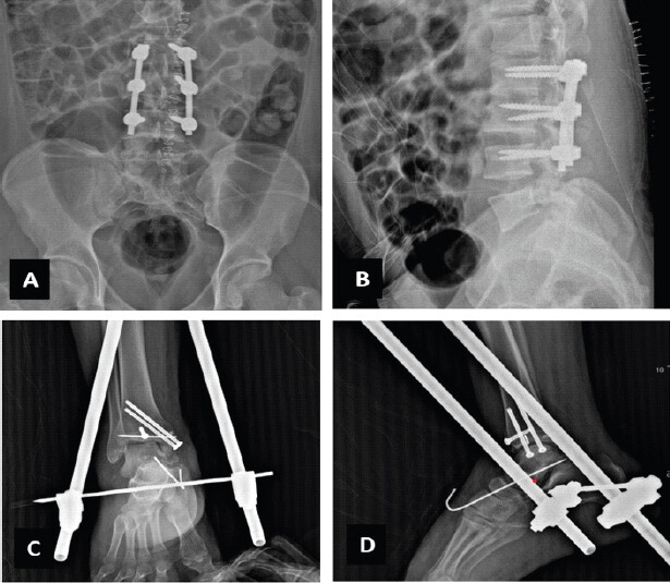
Stage 1 addressed the most urgent injuries. Posterior spinal instrumentation from L2 to L4 was performed for the L3 bony Chance fracture, achieving restoration of spinal alignment, as the injury was a purely osseous flexion-distraction lesion without neurological compromise or significant anterior comminution (Fig. 3a and b). Surgical fixation of the right medial malleolus fracture through anteromedial approach interval was created through the fracture and marginal impaction was corrected and cancellous bone from distal tibia was used as local bone graft . Provisional fixation was achieved with a Kirschner wire, placed subchondral. The anterior plafond rim also was fixed using an anterior-to-posterior screw using cancellous screws, supplemented by temporary ankle-spanning external fixation for the associated fracture-dislocation of the ankle (Fig. 3c and d).
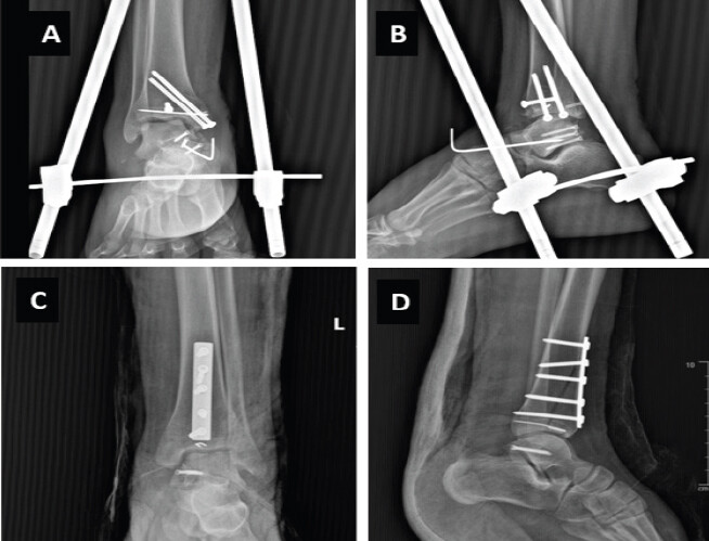
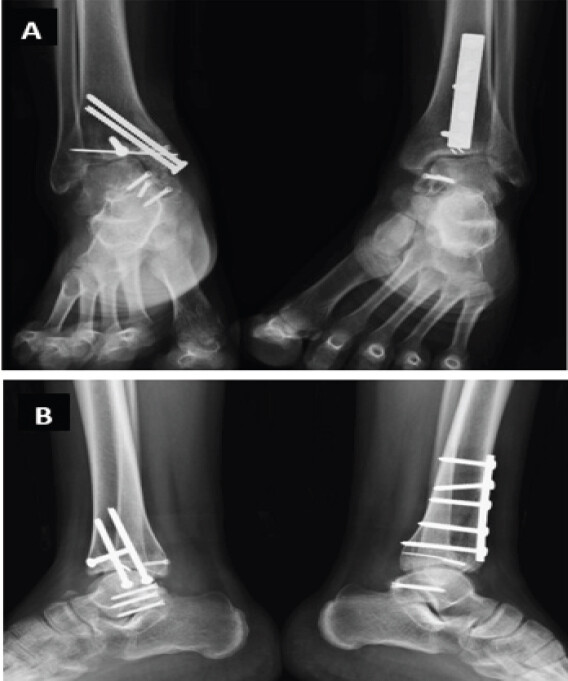
Stage 2 was performed 1 week after the initial surgery, following physiological optimization and recovery of the soft tissues around the ankle after 1 week. The patient was positioned prone with an ipsilateral thigh tourniquet. Both the right and left talar fractures were addressed through a posteromedial approach using an incision just medial to the Achilles tendon. The FHL tendon was identified and retracted medially to expose the posteromedial talus, as described by Swords et al. [4]. Careful handling of the FHL was ensured throughout the procedure to minimize the risk of post-operative Checkrein deformity [5]. The right talar fracture was fixed with three 4-mm Herbert screws through a posterior-to-anterior trajectory, with care taken to avoid subtalar joint penetration (Fig. 4a and b). The left talar fracture was fixed with a single 4-mm headless compression screw through the same trajectory. The left pilon fracture was addressed through an anteromedial approach, providing direct access to the anterior plafond and central articular impaction. The anterior cortical fragment was opened, the impacted articular surface anatomically reduced, supported with local cancellous bone graft, temporarily stabilized using subchondral Kirschner wires, and fixed with an anterior buttress recon locking plate [6]. (Fig. 4c and d show the left ankle’s headless compression screw fixation of the talar fracture together with plate and screw fixation of the tibial plafond) (Fig. 4c and d) [6]. The approach was selected based on the fracture morphology demonstrated on preoperative CT imaging [7].
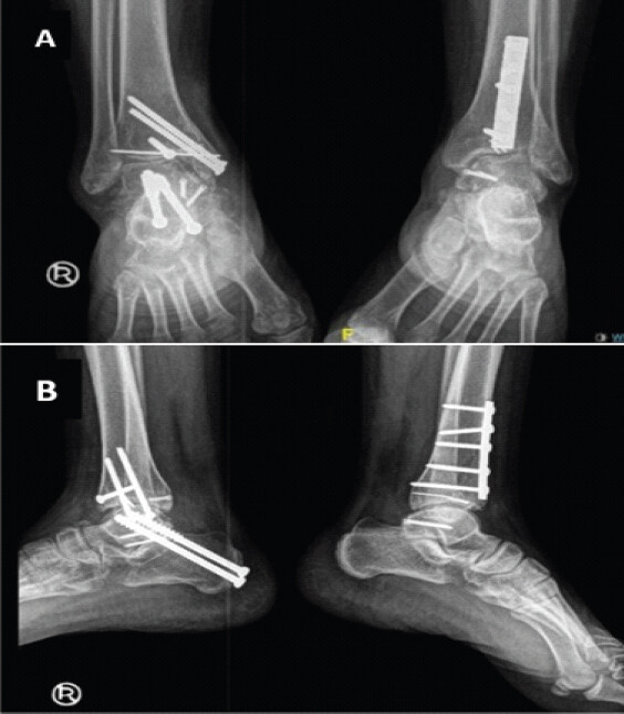
At follow-up, the left ankle united uneventfully with full range of motion. The patient developed persistent right hindfoot pain during weight-bearing. CT demonstrated post-traumatic subtalar arthritis with one Herbert screw breaching the subtalar joint. As the pain was attributed to both the initial articular injury and screw penetration, the screw was removed. The subtalar arthrodesis through the sinus tarsi approach with an incision from the lower end of the fibula to the fourth metatarsal base, and through the same approach, transosseous lateral ligament repair was performed. The subtalar joint was fixed with 2 partially threaded 6.5 cannulated cancellous screws. The patient went on to do well, with his subtalar pain relieved and ankle plantar and dorsiflexion was preserved.
Results
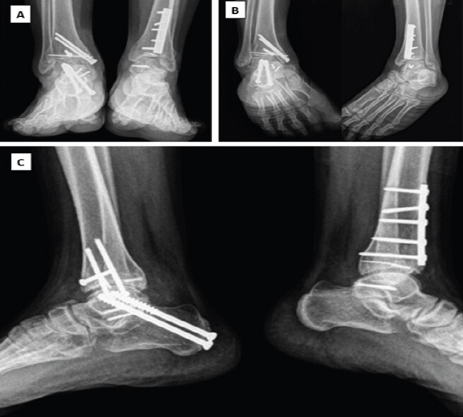
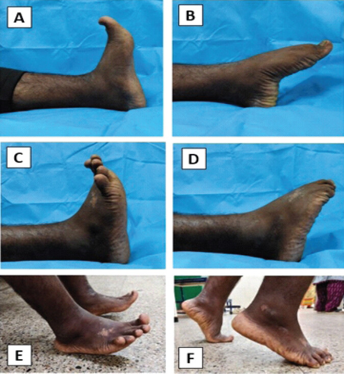
Post-operative course was uneventful, with satisfactory wound healing and no early wound-related complications. Postoperatively, both lower limbs were immobilized in a below-knee splint with strict non-weight-bearing for 6 weeks. Progressive ankle range-of-motion exercises were initiated after soft-tissue healing, followed by gradual weight-bearing based on clinical and radiographic evidence of fracture union. At 6 months, radiographs demonstrated complete union of the bilateral posteromedial talar process fractures and associated fractures (Fig. 5). During subsequent follow-up, the patient developed persistent right subtalar pain due to post-traumatic subtalar arthritis, managed as described above with revision fixation and subtalar arthrodesis (Fig. 6). At the 1-year follow-up, radiographs demonstrated complete union of the bilateral distal tibial and posteromedial talar process fractures with maintained reduction, stable implant position, restoration of the ankle mortise, preserved tibiotalar joint congruity, successful right subtalar arthrodesis, and no evidence of implant loosening or talar avascular necrosis (AVN)(Fig. 7). At the 1-year follow-up, the patient was pain-free, had preserved ankle dorsiflexion and plantarflexion bilaterally, intact FHL function, and was able to perform toe-standing and a single-leg heel rise. He returned to his pre-injury functional level with satisfactory hindfoot stability and an American Orthopaedic Foot and Ankle Society ankle-hindfoot score of 95 (Fig. 8).
Discussion
Cedell fractures remain a diagnostically challenging subset of talar injuries. Their mechanism – forced dorsiflexion and pronation generating avulsion at the posterior talotibial ligament – differs from the axial compression mechanisms underlying most talar body fractures [2]. In polytrauma, distracting injuries compound the risk of missed diagnosis; CT should be strongly considered when clinical suspicion persists despite inconclusive radiographs.
Displaced fractures with subtalar joint involvement are best managed operatively. A systematic review by Engelmann et al. reported superior functional outcomes following open reduction and internal fixation compared with non-operative treatment (33.3% vs. 64.7% impaired outcomes), supporting surgical fixation for displaced posterior process fractures [3]. Similarly, Zwiers et al. reported fragment excision as a reasonable alternative for small, comminuted, or chronic symptomatic fragments not amenable to stable fixation [8]. Long-term complications including subtalar arthritis and AVN require extended surveillance; although no AVN was detected at 6 months, late AVN may occur up to 2 years after injury, given the talus’s tenuous retrograde blood supply [9].
The posteromedial approach between the neurovascular bundle and FHL tendon provided excellent exposure while protecting critical structures [4, 10]. Headless compression screws through a posterior-to-anterior trajectory afforded stable fixation with minimal hardware prominence.
The staged DCO strategy was critical in this case. Temporizing the ankle with external fixation while addressing the spinal injury in Stage 1 allowed soft-tissue recovery and patient optimization before Stage 2 definitive hindfoot and pilon surgery, contributing to the favorable outcome.
Although the patient was hemodynamically stable, a staged DCO strategy was chosen due to the associated spinal injury, marked peri-ankle soft-tissue swelling, and the anticipated prolonged operative time for bilateral definitive fixation. Temporary stabilisation permitted soft-tissue recovery before definitive reconstruction 1 week later.
Previous reports have demonstrated satisfactory outcomes following operative treatment of isolated posteromedial talar process fractures; however, bilateral Cedell fractures remain exceedingly uncommon, making the present case a valuable addition to the existing literature [11, 12].
Limitations include short-term follow-up and the absence of additional patient-reported functional assessment tools such as the Manchester-Oxford Foot Questionnaire. Patient remains under continued follow-up surveillance for late complications including AVN and subtalar arthritis.
Conclusion
Bilateral Cedell fractures in polytrauma are exceptionally uncommon and can be easily overlooked. Careful clinical assessment, early CT imaging, and staged fixation based on DCO principles can provide excellent functional and radiological outcomes. Early anatomical reduction and stable fixation of displaced fragments minimise the risk of non-union, subtalar arthritis and FHL dysfunction. Even when a secondary procedure like subtalar arthrodesis becomes necessary, staged fixation followed by targeted salvage can still restore excellent hindfoot function.
Clinical Message
Posteromedial talar process (Cedell) fractures should be actively sought with computed tomography imaging in high-energy polytrauma, as they are easily missed on plain radiographs; even when subtalar arthritis develops post-fixation, staged damage control orthopedic management followed by targeted salvage procedures such as subtalar arthrodesis can still achieve a functionally excellent outcome.
Conflict of Interest:
Nil
Source of Support:
Nil
Consent:
The authors confirm that informed consent was obtained from the patient for publication of this article
How to Cite this Article
Sivakumar S, Pattabiraman K, Salam N, Chereykkatt N, Dwajan A, Patel S. Bilateral Cedell Fractures of the Talus Associated with Ankle Fractures in a Polytrauma Patient: A Rare Injury Managed with Damage Control Orthopedics and Staged Fixation. Journal of Orthopaedic Case Reports 2026 October;16(10): 264-269.
References
- Majeed H, McBride DJ. Talar process fractures: An overview and update of the literature. EFORT Open Rev 2018;3:85-92. [Google Scholar] | [PubMed]
- Yang H, Liao L, Xue F, Li Y, Hu G. Anatomical observation, classification, fracture and finite element analysis of the posterior process of the Asian adult talus. J Orthop Surg Res 2022;17:444. [Google Scholar] | [PubMed]
- Engelmann EW, Wijers O, Posthuma JJ, Schepers T. Systematic review: Diagnostics, management and outcome of fractures of the posterior process of the talus. Injury 2020;51:2414-20. [Google Scholar] | [PubMed]
- Swords M, Shank J, Benirschke S. Surgical treatment of posteromedial talus fractures: Technique description and results of 10 cases. Indian J Orthop 2018;52:269-75. [Google Scholar] | [PubMed]
- McLaughlin WM, Moran J, Brand J, Kahan JB, Yoo B. Posterior talar body fracture with flexor Hallucis longus incarceration: A case report & literature review. JBJS Case Connect 2021;11 4. [Google Scholar] | [PubMed]
- Mair O, Pflüger P, Hoffeld K, Braun KF, Kirchhoff C, Biberthaler P . Management of pilon fractures-current concepts. Front Surg 2021;8:764232. [Google Scholar] | [PubMed]
- Tornetta P 3rd, Gorup J. Axial computed tomography of pilon fractures. Clin Orthop Relat Res 1996;323:273-6. [Google Scholar] | [PubMed]
- Zwiers R, De Leeuw PA, Wiegerinck EM, Van Dijk CN. Surgical treatment for posteromedial talar process fractures. Foot Ankle Surg 2020;26:911-7. [Google Scholar] | [PubMed]
- Pearce DH, Mongiardi CN, Fornasier VL, Daniels TR. Avascular necrosis of the talus: A pictorial essay. Radiographics 2005;25:399-410. [Google Scholar] | [PubMed]
- Steffensmeier AM, Matar R, Chung D, Yue RA, Dixon TL, Sagi C . Fracture of the posteromedial tubercle of the talus: Case series of fifteen patients along with cadaveric dissection and anatomic considerations for surgical approach. Foot Ankle Orthop 2020;5 Suppl 42473011420S00458. [Google Scholar] | [PubMed]
- Sallent A, Pereira SN, Maled I, Duarri G, Busquets R. Fractures of the posteromedial process of the talus (Cedell's fracture): 3 cases and review of the literature. Foot Ankle Surg Tech Rep Cases 2022;2:100127. [Google Scholar] | [PubMed]
- Gutierres M, Cabral T, Miranda A, Almeida L. Fractures of the posteromedial process of the talus. A report of two cases. Int Orthop 1998;22:394-6. [Google Scholar] | [PubMed]
© 2026 Journal of Orthopaedic Case Reports - Published by Indian Orthopaedic Research Group
About the Authors
-

Sanjey Sivakumar
-

Kirubakaran Pattabiraman
-

Nabiel Salam
-

Nibras Chereykkatt
-

Anirudh Dwajan
-

Sandeep Patel
Related Articles in Journal of Orthopaedic Case Reports
 October 1, 2026 Degloving in Disguise: Managing a Delayed Spinal Morel-Lavallée Lesion after Thoracic Fracture-Dislocation
 October 1, 2026 Posterior Fibular Entrapment in a Bosworth Fracture-Dislocation: A Case Report and Review of Diagnostic and Operative Considerations
 October 1, 2026 Isolated Posterior Malleolus Fracture with Early Symptom Resolution: A Case Report
 October 1, 2026 Single-Stage Fixation of Bilateral Open Medial Hoffa Fractures Associated with Bilateral Patella Fracture and Bilateral Distal Radius Fractures: A Case Report