


Introduction
The distal femur comprises the femoral condyles and trochlea, forming a critical component of the knee joint and playing a vital role in stability and motion. Fractures of the distal femur are challenging to manage due to their anatomical complexity and functional significance [1].
These fractures extend from the supracondylar region to the articular surface and commonly result from low-energy trauma in osteoporotic elderly patients and high-energy injuries in younger individuals. They are broadly classified as intra-articular or extra-articular, with the latter involving the metaphyseal region without joint surface disruption. Extra-articular fractures may be transverse, oblique, spiral, or comminuted, and their classification is essential for guiding treatment strategies [1, 2].
Distal femur fractures account for approximately 1% of all fractures and 6–7% of femoral fractures, with an incidence of around 37/100,000 population. Due to their complexity, most cases require surgical intervention [3].
The primary goals of management include restoration of alignment, stable fixation, preservation of vascularity, and early mobilization to optimize functional outcomes. Surgical options include ORIF, Minimally Invasive Percutaneous Plate Osteosynthesis (MIPPO), and Retrograde Intramedullary Nailing (RIMN) [4, 5].
RIMN provides stable fixation along the mechanical axis with minimal soft-tissue disruption and is associated with shorter operative time, reduced infection rates, and early mobilization. However, it has limitations such as intra-articular entry complications, difficulty in distal fragment control, and potential malalignment [6, 7].
Conventional open plating techniques, although effective, are associated with extensive soft-tissue dissection and periosteal stripping, increasing the risk of infection and delayed union. To address these concerns, minimally invasive plating techniques such as MIPPO have been developed, focusing on biological fixation and preservation of blood supply [8].
MIPPO allows indirect fracture reduction and promotes callus formation through controlled micromotion, leading to improved healing and reduced complications. It is particularly advantageous in osteoporotic bone; however, it carries risks of malalignment, technical difficulty, and a steep learning curve [9, 10].
While both RIMN and MIPPO are widely used for extra-articular distal femur fractures, the optimal method of fixation remains controversial, with each technique offering distinct advantages and limitations [11, 12].
Therefore, this study was conducted to compare preoperative, intraoperative, and post-operative outcomes – including blood loss, duration of surgery, and functional outcome (NEER score) – between retrograde femoral nailing (RFN) and minimally invasive plate osteosynthesis in the management of extra-articular distal femur fractures.
Materials and Methods
Study design
This prospective comparative study was conducted at the Department of Orthopaedics, Guru Gobind Singh Medical College and Hospital, Faridkot, Punjab, from May 2023 to June 2025. A single-center study design was utilized, enrolling 60 consecutive eligible patients with AO type 33A2 and 33A3 fractures.
Group allocation
The study was conducted after obtaining approval from the Institutional Ethics Committee (BFUHS/2K24p-Th-2223). Patients were allocated using an alternate allocation method (quasi-randomization). Neither the patients, operating surgeons, nor outcome assessors were blinded to the surgical procedure. Introducing potential performance and observation bias:
-
Group A: RFN (n = 30)
-
Group B: MIPPO with distal femur locking plate (n = 30).
Inclusion criteria
-
Age >18 years
-
Extra-articular distal femur fractures (AO type A2–A3).
Exclusion criteria
-
Intra-articular distal femur fractures
-
Pathological fractures
-
Open fractures
-
Polytrauma requiring other priority surgeries
-
Patients unfit for surgery.
Surgical technique
RFN
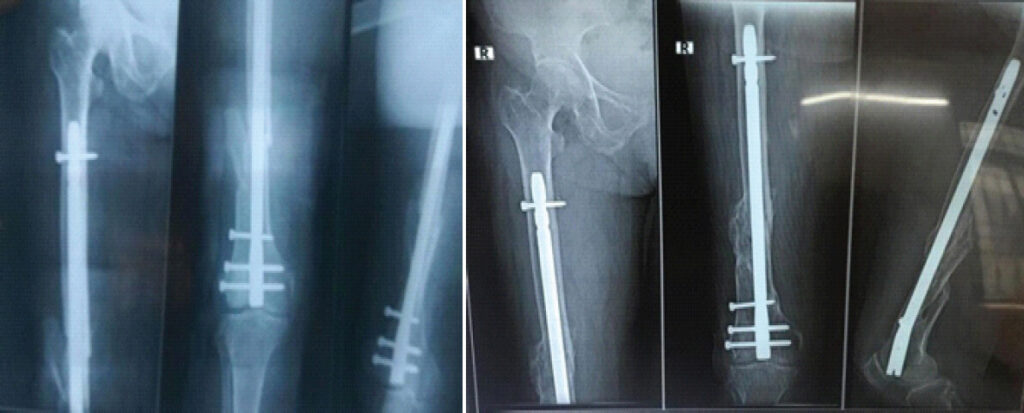
The procedure was performed with the patient in the supine position. Entry was made through the intercondylar notch. Closed reduction was attempted, and the nail was inserted retrograde into the femoral canal. Proximal and distal locking screws were inserted for stable fixation (Fig. 1).
MIPPO technique
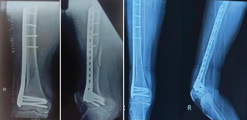
In the MIPPO group, a distal femoral locking compression plate was inserted through small incisions with minimal soft-tissue dissection. The plate was slid submuscularly and fixed with locking screws proximally and distally under fluoroscopic guidance (Fig. 2).
All surgeries were performed by experienced orthopedic trauma surgeons; however, surgeon experience was not strictly standardized or stratified between groups.
Post-operative protocol
-
Early quadriceps exercises
-
Knee range of motion exercises
-
Gradual progression to partial and full weight-bearing depending on fracture stability and radiological signs of union.
Outcome measures
Patients were assessed based on:
-
Radiological union time
-
Time to partial weight-bearing
-
Time to full weight-bearing
-
Functional outcome using NEER’s scoring system
-
Complications
-
Surgical time
-
Fluoroscopy exposure.
Follow-ups were conducted at:
-
6 weeks
-
3 months
-
6 months.
Statistical analysis
Data were entered and compiled using Microsoft Excel. Statistical analysis was performed using IBM Statistical Package for the Social Sciences Statistics version 22. Continuous variables were expressed as mean and standard deviation, while categorical variables were expressed as frequencies and percentages. Comparison between the two groups for continuous variables was carried out using the Student’s t-test. Categorical variables were analysed using the Pearson Chi-square test. A P < 0.05 was considered statistically significant.
Results
Demographic profile
The mean age was 49.67 ± 10.54 years, with 35 males and 25 females. Road traffic accidents accounted for 71.6% of injuries, followed by falls (25%) and assault (3.33%) (Table 1).
Demographic profile of participants
| Parameter | RFN group | MIPPO group | Total (n =60) |
|---|---|---|---|
| Mean age (years) | 48.67±10.32 | 50.67±10.76 | 49.67±10.54 |
| Gender (Male:Female) | 1.7:1 | 1.1:1 | 1.4:1 |
| Mechanism (RTA/fall/assault) | 22-07-2001 | 21-08-2001 | 43/15/2 |
RTA: Road-traffic accidents, RFN: Retrograde femoral nailing, MIPPO: Minimally invasive percutaneous plate osteosynthesis
Radiological union time (weeks)
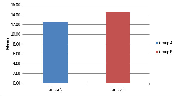
Patients in the RFN group had a mean fracture union time of 12.42 weeks, which was significantly less than that of patients in the MIPPO group (14.50 weeks) (Fig. 3).
Time to partial weight-bearing (weeks)
The current study discovered that the mean partial weight-bearing period was 5.97 weeks for the RFN group and 7.30 weeks for the MIPPO group. The P< 0.05 indicates a statistically significant difference between the groups’ mean times for partial weight-bearing (Table 2).
Time to partial weight-bearing (weeks)
| Time to partial weight-bearing (weeks) | Group A | Group B |
|---|---|---|
| Mean | 5.97 | 7.3 |
| Standard deviation | 1.47 | 1.73 |
| P-value | 0.001 (Significant) | |
Timing of full weight-bearing (in weeks) (Table 3)
Timing of full weight-bearing (in weeks)
| Time to full weight-bearing (weeks) | Group A | Group B |
|---|---|---|
| Mean | 13.03 | 15.73 |
| Standard deviation | 2.39 | 2.75 |
| P-value | 0.001 (Significant) | |
In the RFN group, the mean time of full weight-bearing was 13.03 weeks, with a standard deviation of 2.39, while in the MIPPO group; it was 15.73 weeks, with a standard deviation of 2.75. Results are statistically significant on comparison.
NEER score
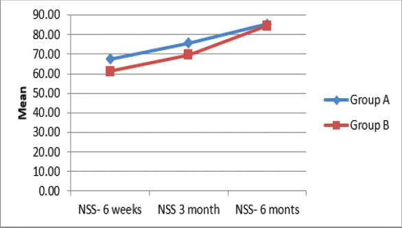
At 6 weeks, the mean NEER score for participants in the RFN group was 67.43, compared to 61.13 for patients who had MIPPO. At 3 months, participants in the RFN group’s mean NEER score was 75.57, compared to 69.57 for patients who had MIPPO. Mean NEER score was 85.40 in the RFN group and 84.50 in MIPPO group at 6 months.
At 6 weeks and 3rd months after surgery, the RFN group’s mean NEER score was found to be significantly higher, but at 6 months, both groups were statistically insignificant (Fig. 4).
Functional outcome
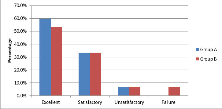
In the patients of RFN group, excellent, satisfactory, unsatisfactory, and failure results were seen in 60%, 33.3%, 6.7%, and 0% of the patients, respectively. Among patients in MIPPO group, excellent, satisfactory, unsatisfactory, and failure results were observed in 53.3%, 33.3%, 6.7%, and 6.7% of the patients, respectively. Non-significant results were found when evaluating and contrasting the functional outcomes between the two study groups (Fig. 5).
Complications
Among patients in the RFN group, 3.3%, 6.7%, 0%, 6.7%, and 10% of patients, respectively, experienced superficial infection, knee stiffness, non-union, knee pain, and malunion. Out of the patients in the MIPPO group, 6.7% experienced superficial infection, 3.3% experienced knee stiffness, 10% experienced non-union, 0% experienced knee discomfort, and 3.3% experienced malunion. Patients in the RFN group experienced greater rates of knee discomfort and malunion, whereas those in the MIPPO group saw higher rates of non-union.
Duration of surgery (minutes)
In the RFN group, the mean surgery time was 89.17 min, with a standard deviation of 25.70; in the group receiving MIPPO, the mean surgery time was 109.50 min, with a standard deviation of 12.06. Results are statistically significant on comparison.
Fluoroscopy exposure (cGy)
Patients in the MIPPO group had an average fluoroscopy exposure of 16.67 cGy, while those in the RFN group had an average of 21.17 cGy. In terms of statistical comparison, patients in the RFN group had a considerably higher mean fluoroscopy exposure than those in the MIPPO group.
Discussion
Management of extra-articular distal femur fractures remains a challenging aspect of orthopedic trauma due to the complex anatomy, biomechanical forces, and frequent association with osteoporotic bone. The present study compares functional and radiological outcomes between RFN and MIPPO, two widely accepted modalities that aim to balance mechanical stability with biological preservation.
Distal femur fractures account for approximately 6–7% of all femoral fractures and demonstrate a bimodal distribution, affecting elderly osteoporotic individuals following low-energy trauma and younger patients after high-energy injuries. This dual pattern necessitates individualized treatment strategies based on fracture configuration, bone quality, and patient factors.
The current study observed comparable functional outcomes between RFN and MIPPO, which aligns with several previous studies. Markmiller et al. reported no significant difference in knee range of motion or complication rates between less invasive stabilization system plating and retrograde nailing, suggesting both techniques are equally effective in managing distal femur fractures [13]. Similarly, Jankowski et al., in a systematic review, found no statistically significant differences in union rates, functional outcomes, or complication profiles between intramedullary nailing and locked plating [14]. These findings support the hypothesis that both implants can achieve satisfactory results when applied appropriately.
RFN offers the advantage of a load-sharing construct, which facilitates earlier weight-bearing and promotes faster union. In the present study, patients treated with RFN demonstrated relatively shorter operative times and earlier mobilization, which is consistent with findings by Jillala et al., who reported reduced surgical duration and earlier fracture union in the nailing group [15]. In addition, Singh et al. highlighted earlier weight-bearing and improved knee flexion with retrograde nailing, although it was associated with a higher incidence of anterior knee pain [16]. The intra-articular entry point of the nail remains a concern, as it may contribute to post-operative knee discomfort and stiffness.
On the other hand, MIPPO with locking compression plates emphasizes biological fixation by preserving periosteal blood supply and minimizing soft-tissue disruption. The current study demonstrated slightly better alignment and fewer malalignment-related complications in the plating group. This observation is supported by Haider et al., who reported improved fracture alignment and earlier union with MIPPO due to better preservation of fracture biology [17]. Similarly, Krettek et al. emphasized that MIPPO techniques reduce soft-tissue trauma and enhance fracture healing through indirect reduction and callus formation [7].
Despite these advantages, MIPPO is technically demanding and associated with a learning curve. Malalignment, particularly varus deformity, remains a recognized complication due to indirect reduction techniques. Henderson et al. reported non-union and hardware failure associated with inadequate mechanical stability in locked plating [18], underscoring the importance of surgical expertise and appropriate implant selection.
Union rates in both groups in the present study were comparable, consistent with previous literature. Studies by Gellman et al. and Handolin et al. demonstrated high union rates with retrograde nailing, often exceeding 90% [19, 20]. Similarly, studies evaluating MIPPO techniques have reported excellent union rates and functional outcomes, particularly in osteoporotic bone [21, 22]. Aggarwal et al., in a meta-analysis, concluded that both plating and nailing provide similar union rates, with minor differences in complication profiles [23].
One of the important findings of this study is that implant selection should be guided by fracture morphology and patient-specific factors rather than a one-size-fits-all approach. Retrograde nailing may be more suitable for simple extra-articular fractures with good canal anatomy, whereas MIPPO is preferable in comminuted fractures, osteoporotic bone, or where preservation of soft-tissue envelope is critical.
The strengths of this study include its comparative design and focus on functional outcomes using standardized scoring systems. However, certain limitations must be acknowledged. The sample size is relatively small, and the follow-up period may not be sufficient to assess long-term complications such as post-traumatic osteoarthritis. In addition, surgeon experience and technical variability may influence outcomes.
To establish clearer evidence-based guidelines for implant selection, a prospective, larger multicenter randomized controlled trial with extended follow-up is necessary.
Limitations
This study has several limitations that should be considered when interpreting the results:
-
Sample Size and Generalizability: The sample size of 60 patients (30/group) was determined based on eligible consecutive admissions during the study timeframe
-
Single-Center Design: Conducting the study at a single tertiary care facility reduces external validity and applicability across different healthcare settings
-
Follow-Up Duration: The 6-month follow-up period was chosen to evaluate short-term fracture union and early functional recovery; however, it is insufficient to evaluate late outcomes such as post-traumatic osteoarthritis, implant fatigue/failure, or functional decline
-
Cohort Selection: Inclusion was restricted to closed AO type 33A2 and 33A3 fractures, limiting applicability to intra-articular (types B/C), open, periprosthetic, or highly comminuted fractures
-
Allocation Method and Selection Bias: Patients were allocated using an alternate allocation technique (quasi-randomization) rather than strict computer randomization, introducing potential selection bias
-
Blinding: Neither patients nor outcome assessors were blinded to the intervention, increasing the risk of observer and performance bias.
-
Surgical and Bone Variations: Surgeon experience and technical expertise were not standardized or analyzed, which may influence surgical duration and reduction quality. In addition, bone mineral density (DEXA) was not routinely measured despite its impact on construct stability
-
Rehabilitation Tracking: Post-operative rehabilitation protocols were standardized on paper, but patient compliance was not objectively monitored
-
Outcome Assessments: Functional evaluation relied primarily on the NEER score and range-of-motion measurements without validated patient-reported outcome measures (PROMs) such as KOOS, Oxford Knee Score, SF-36, or EQ-5D, or objective assessments like gait analysis and muscle strength testing
-
Knee range of motion (flexion degrees) was measured and reported at 6 weeks, 3 months, and 6 months. However, objective gait analysis, formal dynamometric muscle strength testing, and detailed return-to-work metrics were not performed
-
Radiographic and Economic Scope: Radiographic analysis was focused on gross union and clinical alignment rather than standardized 3D coronal/sagittal/rotational metrics or limb length discrepancy. Furthermore, economic variables (hospital cost, implant pricing, cost-effectiveness) were not evaluated
-
While fluoroscopy exposure (cGy) was measured, sub-analyses controlling for specific fracture subtypes and cumulative dose parameters were not performed.
Conclusion
Both RFN and minimally invasive plate osteosynthesis are effective treatment modalities for extra-articular distal femur fractures, with comparable functional and radiological outcomes. RFN offers advantages of shorter operative time and earlier mobilization, whereas MIPPO provides superior biological fixation and better alignment in selected cases. Therefore, treatment should be individualized based on fracture characteristics, bone quality, and surgeon expertise. Future multicenter randomized controlled trials with extended follow-up are recommended to validate these findings and establish definitive clinical guidelines.
Clinical Message
Retrograde nailing and MIPPO yield similar outcomes in extra-articular distal femur fractures; treatment should be tailored to fracture morphology and patient factors.
Conflict of Interest:
Nil
Source of Support:
Nil
Consent:
The authors confirm that informed consent was obtained from the patient for publication of this article
How to Cite this Article
Singla N, Dahuja A, Gulbadhar L, Bansal K, Singh K, Singh M. Functional Outcome of Retrograde Femoral Nailing Versus Minimally Invasive Percutaneous Plate Osteosynthesis in Extra-Articular Distal Femur Fractures: A Prospective Quasi-Randomized Comparative Study. Journal of Orthopaedic Case Reports 2026 October;16(10): 392-399.
References
- Arastu MH, Kokke MC, Duffy PJ, Buckley RE. Fractures of the distal femur. Orthop Clin North Am 2013;44:55-71. [Google Scholar] | [PubMed]
- Martinet O, Cordey J, Harder Y, Maier A, Buhler M, Barraud GE. The epidemiology of fractures of the distal femur. Injury 2000;31 Suppl 3:C62-3. [Google Scholar] | [PubMed]
- Ehlinger M, Ducrot G, Adam P, Bonnomet F. Minimally-invasive internal fixation of extra-articular distal femur fractures using a locking plate: Tricks of the trade. Orthop Traumatol Surg Res 2012;98:929-35. [Google Scholar] | [PubMed]
- Krettek C, Schandelmaier P, Rudolf J, Tscherne H. Distal femoral nailing of femur fractures with the unreamed femoral nail (UFN). Injury 2001;32 Suppl 3:SC48-54. [Google Scholar] | [PubMed]
- Ricci WM, Streubel PN, Morshed S, Collinge C, Gardner MJ. Risk factors for failure of locked plate fixation of distal femur fractures: An analysis of 335 cases. J Orthop Trauma 2009;23:349-54. [Google Scholar] | [PubMed]
- Hierholzer C, Von Rüden C, Pötzel T, Woltmann A, Bühren V. Outcome analysis of retrograde nailing and less invasive stabilization system in distal femoral fractures: A retrospective analysis. Indian J Orthop 2011;45:243-50. [Google Scholar] | [PubMed]
- Krettek C, Müller M, Miclau T. Evolution of minimally invasive plate osteosynthesis (MIPO) in the femur. Injury 2001;32 Suppl 3:SC14-23. [Google Scholar] | [PubMed]
- Kumar KG, Sharma G, Farooque K, Sharma V, Ratan R, Yadav S. Locking compression plate in distal femoral intra-articular fractures: Our experience. Int Sch Res Notices 2014;2014:372916. [Google Scholar] | [PubMed]
- Ronga M, Longo UG, Maffulli N. Minimally invasive locked plating of distal tibia fractures is safe and effective. Clin Orthop Relat Res 2010;468:975-82. [Google Scholar] | [PubMed]
- Zlowodzki M, Williamson S, Zardiackas LD, Kregor PJ. Biomechanical evaluation of the less invasive stabilization system and the 95-degree angled blade plate for the internal fixation of distal femur fractures in human cadaveric bones with high bone mineral density. J Trauma 2006;60:836-40. [Google Scholar] | [PubMed]
- Kregor PJ, Stannard JA, Zlowodzki M, Cole PA. Treatment of distal femur fractures using the less invasive stabilization system: Surgical experience and early clinical results in 103 fractures. J Orthop Trauma 2001;15:485-95. [Google Scholar] | [PubMed]
- Meena RC, Meena UK, Gupta GL, Gahlot N, Gaba S. Intramedullary nailing versus proximal plating in the management of closed extra-articular proximal tibial fracture: A randomized controlled trial. J Orthop Traumatol 2015;16:203-8. [Google Scholar] | [PubMed]
- Markmiller M, Konrad G, Südkamp N. Femur-LISS and distal femoral nail for fixation of distal femoral fractures: Are there differences in outcome and complications? Clin Orthop Relat Res 2004;426:252-7. [Google Scholar] | [PubMed]
- Jankowski JM, Szukics PF, Shah JK, Keller DM, Pires RE, Liporace FA. Comparing intramedullary nailing versus locked plating in the treatment of native distal femur fractures: Is one superior to the other? Indian J Orthop 2021;55:646-54. [Google Scholar] | [PubMed]
- Jillala SR, Waseem Ahmed SM, Shruthi A, Gajul R, Katikitala A, Rakesh K. A comparative study of supracondylar nail versus locking compression plate in distal femur fractures. Ann Int Med Dent Res 2017;3:OR35-41. [Google Scholar] | [PubMed]
- Singh S, Baghel PK, Rastogi D, Shantanu K, Sharma V. Distal femoral locked plating versus retrograde nailing for extra articular distal femur fractures: A comparative study. Int J Orthop Sci 2018;4:702-5. [Google Scholar] | [PubMed]
- Haider Z, Khan MA, Alam M, Raza H, Muneeb A. Comparison of closed retrograde intramedullary nailing versus minimally invasive percutaneous plate osteosynthesis (MIPPO) for distal femur fractures. J Orthop Surg Res 2022;17:42. [Google Scholar] | [PubMed]
- Henderson CE, Lujan TJ, Kuhl LL, Bottlang M, Fitzpatrick DC, Marsh JL. 2010 Mid-America orthopaedic association physician in training award: Healing complications are common after locked plating for distal femur fractures. Clin Orthop Relat Res 2011;469:1757-65. [Google Scholar] | [PubMed]
- Gellman RE, Paiement GD, Green HD, Coughlin RR. Treatment of supracondylar femoral fractures with a retrograde intramedullary nail. Clin Orthop Relat Res 1996;332:90-7. [Google Scholar] | [PubMed]
- Handolin L, Pajarinen J, Lindahl J, Hirvensalo E. Retrograde intramedullary nailing in distal femoral fractures--results in a series of 46 consecutive operations. Injury 2004;35:517-22. [Google Scholar] | [PubMed]
- Kamareddy S, Garampalli R, Kaja A. Outcome of distal femur fractures treated with locking compression plate by minimally invasive percutaneous plate osteosynthesis technique. Int J Res Orthop 2015;1:10-4. [Google Scholar] | [PubMed]
- Jagadesh N, Kaki A, Kumar SS. Functional outcome of distal femoral fractures treated by distal femoral locking compression plate by minimally invasive percutaneous plate osteosynthesis technique. Int J Res Orthop 2017;3:729-33. [Google Scholar] | [PubMed]
- Aggarwal S, Rajnish RK, Kumar P, Srivastava A, Rathor K, Haq RU. Comparison of outcomes of retrograde intramedullary nailing versus locking plate fixation in distal femur fractures: A systematic review and meta-analysis of 936 patients in 16 studies. J Orthop 2022;36:36-48. [Google Scholar] | [PubMed]
© 2026 Journal of Orthopaedic Case Reports - Published by Indian Orthopaedic Research Group
About the Authors
-

Neeraj Singla
-

Anshul Dahuja
-

Lovish Gulbadhar
-

Kapil Bansal
-

Krishmveer Singh
-

Manmohan Singh
Related Articles in Journal of Orthopaedic Case Reports
 October 1, 2026 Combined Cerclage and Figure-of-eight Tension-band Wiring without K-wire Versus K-wire Tension Band Wiring Fixation for Transverse Patellar Fractures: A Prospective Comparative Study
 October 1, 2026 Humeral Fractures in Metabolically Compromised Bone: Outcomes of Structural Strut Onlay Allograft with Open Reduction and Internal Fixation – A Case Series of Three Patients
 October 1, 2026 A Study of Spinopelvic Relationship and its Impact on Functional Outcome of Total Hip Arthroplasty
 October 1, 2026 Outcomes of Primary Definitive Internal Fixation of Compound Fractures within 72 h of Injury: A Prospective Cohort Study