


Introduction
Distal tibial fractures continue to be a therapeutic challenge for orthopedic surgeons because of the unique anatomy of the distal tibia, poor vascularity, and minimal soft-tissue coverage around the ankle joint. These fractures are frequently associated with complications such as delayed union, malunion, infection, and joint stiffness, which may adversely affect functional outcome [1,2]. Extra-articular distal tibial fractures commonly occur following high-energy trauma, particularly road traffic accidents and falls [3].
The management of distal tibial fractures has evolved considerably over the years. Conservative treatment often leads to prolonged immobilization, malalignment, and delayed rehabilitation, whereas surgical treatment aims to achieve stable fixation with restoration of alignment and early mobilization [4]. Various surgical options, including plate fixation, external fixation, and intramedullary interlocking nailing, are currently used for the treatment of these fractures [5].
Among these techniques, closed intramedullary interlocking nailing has gained wide acceptance because it preserves fracture hematoma and periosteal blood supply while minimizing soft-tissue dissection [6]. The load-sharing nature of intramedullary fixation also permits earlier mobilization and weight-bearing compared to many other fixation methods. Recent improvements in nail design, particularly the availability of multiple distal locking options, have enhanced fixation stability in distal metaphyseal fractures [7].
Several recent studies have reported encouraging functional and radiological outcomes with intramedullary interlocking nailing in extra-articular distal tibial fractures, with relatively lower soft-tissue complications compared to plating techniques [8,9]. However, concerns regarding malalignment, delayed union, and non-union still remain, especially in fractures with short distal fragments [10]. Recent meta-analyses have further compared intramedullary nailing with plate fixation for distal tibial fractures and reported broadly comparable healing and functional outcomes, with differences in specific complication profiles between techniques [11,12].
Therefore, the present study was undertaken to describe the short-term clinical, functional, and radiological outcomes of closed intramedullary interlocking nailing in selected adults with extra-articular distal tibial fractures. The study was not designed to compare intramedullary nailing with plating, external fixation, or other treatment modalities.
Materials and Methods
This prospective observational, single-center study was conducted in the Department of Orthopaedics at People’s College of Medical Sciences and Research Centre, Bhopal, Madhya Pradesh, from March 2024 to December 2025. Fifty consecutive eligible patients with extra-articular distal one-third tibial fractures were enrolled. As treatment allocation was not randomized and no control group was included, the study was designed to describe outcomes after intramedullary interlocking nailing rather than determine comparative efficacy. Written informed consent was obtained from all participants.
Sample size calculation
The sample size was calculated using the formula for estimating a single population proportion:
n = Z2P(1−P)/d2
where:
• n = required sample size
• Z = standard normal deviate at 95% confidence level (1.96)
• P = expected proportion of satisfactory functional outcome (50%, assumed due to limited local data)
• d = absolute precision (14%)
Substituting the values:
n = (1.96)2 × 0.5 × 0.5/(0.14)2 = 49
Therefore, the minimum calculated sample size was approximately 49 patients, and 50 consecutive eligible patients were included in the study.
Inclusion criteria
-
Patients aged between 18 and 60 years
-
Fracture involving the distal one-third of tibia
-
Patients operated within 1 week of trauma
-
Closed fractures
-
Gustilo-Anderson Type I open fractures.
Exclusion criteria
-
Patients younger than 18 years or older than 60 years
-
Fractures older than 1 week
-
Pathological fractures
-
Gustilo-Anderson Type II and III open fractures
-
Patients with multiple comorbidities
-
Fractures involving the proximal three-fourths of fibula.
All patients underwent detailed clinical examination and radiological evaluation with anteroposterior and lateral radiographs; computed tomography (CT) was obtained where clinically required. Fractures were categorized using the Arbeitsgemeinschaft für Osteosynthesefragen (AO)/Orthopaedic Trauma Association (OTA) system as 43A1, 43A2, or 43A3. Closed intramedullary interlocking nailing was performed using standard operative principles. The source dataset recorded AO/OTA category but did not systematically record comminution grade beyond AO subtype, distal fragment length, metaphyseal width, or other quantitative morphology variables; therefore, outcome stratification by these additional characteristics was not possible.
Functional outcome was assessed using the Modified American Orthopaedic Foot and Ankle Society (AOFAS) Ankle-Hindfoot Score preoperatively and at 2, 6, 9, and 12 weeks and 6 months. Radiographs were reviewed postoperatively and during follow-up for union and maintenance of alignment. Alignment assessment in the original protocol was based on standard anteroposterior and lateral radiographs and clinical examination; exact coronal and sagittal angulation and CT-based rotational measurements were not prospectively recorded. Accordingly, quantitative malalignment analysis could not be added retrospectively. Objective ankle dorsiflexion, plantarflexion, and subtalar range-of-motion measurements were also not separately recorded. The AOFAS score was retained because it was the prespecified functional instrument in the thesis protocol; its partly clinician-rated nature and potential ceiling effect are acknowledged as limitations.
Post-operative rehabilitation protocol: During days 0–14, the limb was supported in a well-padded posterior splint with the ankle in neutral or slight dorsiflexion, with elevation, wound surveillance, and suture/staple removal at approximately 10–14 days. From 2 to 6 weeks, patients were transitioned to a removable cast/boot as appropriate and began ankle inversion/eversion exercises, strengthening, and gait training. Weight-bearing was advanced gradually from partial toward full weight-bearing according to clinical tolerance and radiographic evidence of stability/healing rather than by time alone. Radiographs at approximately 6 and 12 weeks guided progression, with continued rehabilitation thereafter. In the study cohort, most patients ultimately achieved weight-bearing between 8 and 12 weeks.
Ethics approval
The study was approved by the Institutional Ethics Committee of People’s College of Medical Sciences and Research Centre, Bhopal. The IEC approval number was Ref no. PCMS/OD/PS/2024/738 dated: March 26, 2024. Written informed consent was obtained from all participants prior to inclusion in the study.
Data were entered in Microsoft Excel and analyzed using Statistical Package for the Social Sciences (SPSS) version 20.0. Continuous variables were summarized as mean ± standard deviation and categorical variables as frequencies and percentages. Because AOFAS scores were repeatedly measured in the same patients at 6 time points (pre-operative, 2, 6, 9, and 12 weeks, and 6 months), longitudinal change was evaluated using the non-parametric Friedman test. Kendall’s W was calculated as an effect-size measure for the repeated assessments. Categorical comparisons, where applicable, were assessed using the chi-square test or Fisher’s exact test when expected cell counts were small. A two-sided p-value <0.05 was considered statistically significant.
Data were entered in Microsoft Excel and analyzed using SPSS version 20.0 software. Continuous variables were expressed as mean ± standard deviation, while categorical variables were expressed as percentages and proportions. Chi-square test was used for statistical analysis, and P < 0.05 was considered statistically significant.
Results
A total of 50 patients with extra-articular distal tibial fractures treated with closed intramedullary interlocking nailing were included in the present study. Demographic characteristics, functional outcomes, radiological union, and post-operative complications were assessed during regular follow-up. The observations obtained during the study period are presented in the following tables and figures. Baseline demographic and injury characteristics of the study cohort are summarized in Table 1.
Distribution of patients with extra-articular distal tibia fractures
| Variable | No. of cases | Percentage |
|---|---|---|
| Age (in years) | ||
| 18–20 | 5 | 10 |
| 21–30 | 15 | 30 |
| 31–40 | 12 | 24 |
| 41–50 | 10 | 20 |
| 51–60 | 8 | 16 |
| Mean±standard deviation | 37.74±12.26 | |
| Sex | ||
| Male | 40 | 80 |
| Female | 10 | 20 |
| Side of injury | ||
| Right | 28 | 56 |
| Left | 22 | 44 |
| Mode of injury | ||
| Road traffic accident | 45 | 90 |
| Fall | 5 | 10 |
| Operative duration and hospital stay | ||
| Operating time (minutes) | 94.02±7.32 | |
| Hospital stay (days) | 4.64±0.89 | |
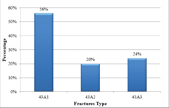
According to the AO/OTA classification, 43A1 fractures were most frequent (56%), followed by 43A3 (24%) and 43A2 (20%) (Fig. 1). These data describe the fracture spectrum included in the study. The study was not powered for reliable subgroup comparisons among AO subtypes, and the source dataset did not contain standardized measurements of distal fragment length, metaphyseal widening, or detailed comminution beyond AO/OTA subtype.
Functional outcomes improved progressively during follow-up. At presentation and 2 weeks, all patients were in the poor AOFAS category. By 6 weeks, most patients had moved into fair or good categories; at 6 months, 60% had excellent and 32% good outcomes (Table 2). Mean AOFAS scores increased from 50.30 ± 1.99 preoperatively to 65.36 ± 6.24 at 6 weeks, 73.12 ± 5.99 at 9 weeks, 81.22 ± 6.81 at 12 weeks, and 87.74 ± 7.85 at 6 months (Table 3). The overall within-patient change across the 6 time points was significant (Friedman χ2 = 239.80, df = 5, P < 0.001; Kendall’s W = 0.959), supporting a strong temporal improvement in the AOFAS score within this cohort.
Functional outcome in patients with extra-articular distal tibia fractures
| Outcome | Preoperative | 2 weeks | 6 weeks | 9 weeks | 12 weeks | 6 months |
|---|---|---|---|---|---|---|
| Excellent (90–100%) | 0 | 0 | 0 | 3 | 14 | 30 |
| Good (80–89%) | 0 | 0 | 5 | 15 | 20 | 16 |
| Fair (70–79%) | 0 | 0 | 25 | 20 | 10 | 2 |
| Poor (<70%) | 50 | 50 | 20 | 12 | 6 | 2 |
| Total | 50 | 50 | 50 | 50 | 50 | 50 |
| P-value | <0.0001 | |||||
Mean AOFAS scores during follow-up
| Time interval | Mean AOFAS score | Standard deviation | Pairwise P-value versus preoperative |
|---|---|---|---|
| Pre-operative | 50.3 | 1.99 | – |
| 2 Weeks | 50.3 | 1.99 | 1 |
| 6 Weeks | 65.36 | 6.24 | <0.0001 |
| 9 Weeks | 73.12 | 5.99 | <0.0001 |
| 12 Weeks | 81.22 | 6.81 | <0.0001 |
| 6 Months | 87.74 | 7.85 | <0.0001 |
Longitudinal analysis: Friedman χ2=239.80, df=5, P<0.001; Kendall’s W=0.959. Pairwise P-values shown in the table are descriptive comparisons with the pre-operative score; the Friedman test is the primary repeated-measures inference. AOFAS: American Orthopaedic Foot and Ankle Society
Functional outcomes improved progressively throughout follow-up. At presentation and during the initial 2 weeks, all patients had poor functional status. By 6 weeks, most patients had fair to good outcomes. At 6 months, excellent outcomes were observed in 60% of patients and good outcomes in 32% of patients. The improvement in functional outcome during follow-up was statistically highly significant (P <0.0001) (Table 2). The mean AOFAS score remained unchanged during the first 2 post-operative weeks but improved significantly from 6 weeks onward. The score increased progressively from 65.36 ± 6.24 at 6 weeks to 87.74 ± 7.85 at 6 months, reflecting substantial functional recovery and improvement in ankle function. This improvement was statistically highly significant (Table 3).
Fig. 2 shows time to radiological union. Among the 48 fractures that united during the 6-month observation period, union was recorded at 14 weeks in 18%, 16 weeks in 22%, 18 weeks in 36%, 20 weeks in 12%, and 24 weeks in 8% of the total cohort; two patients (4%) were classified as non-union in the study dataset. Radiographic follow-up also assessed maintenance of reduction; one patient (2%) was recorded as malunion. Because exact coronal, sagittal, and rotational angles were not prospectively captured, the study cannot provide a quantitative malalignment distribution.
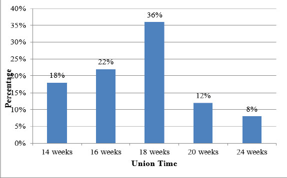
Fig. 2 demonstrates the time to radiological union among study participants. Most fractures achieved union within the expected healing period, suggesting satisfactory fracture stabilization and biological healing following intramedullary interlocking nailing.
Fig. 3 illustrates progression to weight-bearing: 66% of patients achieved weight-bearing at 8–10 weeks, 30% at 10–12 weeks, and two patients (4%) with non-union were not walking at the final recorded assessment. Progression was individualized according to clinical and radiographic healing within the standardized rehabilitation framework.
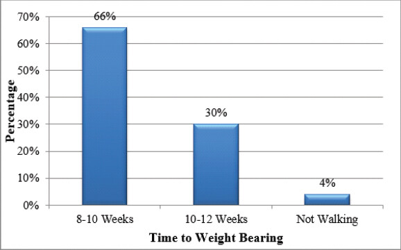
Fig. 3 illustrates the time to weight-bearing among patients. Gradual progression from partial to full weight-bearing was achieved in most cases during follow-up, indicating adequate fracture stability and successful post-operative rehabilitation.
Post-operative complications were relatively low. Superficial infection was the most common complication and was observed in 8% of patients. Delayed union and non-union were noted in 4% of cases each, while malunion occurred in only 2% of patients (Table 4).
Post-operative complications in patients with extra-articular distal tibia fractures
| Complication | No. of Cases | Percentage |
|---|---|---|
| Superficial infection | 4 | 8 |
| Malunion | 1 | 2 |
| Delayed union | 2 | 4 |
| Non-union | 2 | 4 |
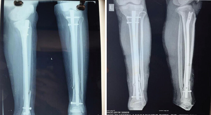
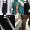
Pre-operative anteroposterior and lateral radiographs demonstrating an extra-articular distal one-third tibial fracture (AO type 43A) before surgical management with closed intramedullary interlocking nailing (Fig. 4).
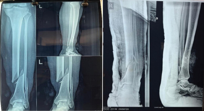
Post-operative anteroposterior and lateral radiographs at follow-up showing satisfactory fracture reduction, appropriate implant positioning, maintained alignment, and radiological union following closed intramedullary interlocking nailing (Fig. 5).
Discussion
In the present study, young adult males formed the majority of the cohort, and road traffic accidents were the most common mode of injury, similar to observations reported by Peng et al. and Daas et al. [13,14]. AOFAS scores improved over serial follow-up, and the Friedman analysis confirmed a significant within-patient temporal change. However, this improvement should not be interpreted as proof that intramedullary nailing is superior to plating, external fixation, or other strategies because the study had no comparison group and treatment selection was not randomized. Bleeker et al. reported favorable outcomes after intramedullary nailing in a broader comparative evidence base [15].
Radiological union was observed in most patients during the 6-month follow-up. Intramedullary interlocking nailing is intended to provide load-sharing fixation while limiting soft-tissue disruption [16], and previous studies have reported high union rates with this approach [15,17]. Nevertheless, the present study’s short follow-up limits assessment of later union events, implant-related problems, persistent symptoms, and post-traumatic ankle degeneration.
Superficial infection was the most frequent recorded complication (8%), while delayed union (4%), non-union (4%), and malunion (2%) were less frequent. Comparative studies by Daolagupu et al. and Nath et al. have evaluated nailing against plating [18,19], but the present single-arm study cannot reproduce such comparative conclusions. In addition, only AO/OTA 43A extra-articular fractures and closed or Gustilo-Anderson Type I open injuries were included, so these complication estimates should not be extrapolated to intra-articular fractures or higher-grade open injuries.
Taken together, the findings describe short-term outcomes after closed intramedullary interlocking nailing in a selected cohort rather than establishing comparative efficacy. Interpretation should account for the observational design, restricted eligibility criteria, absence of a control group, and the 6-month follow-up period.
Overall, the findings of the present study support the use of closed intramedullary interlocking nailing as an effective treatment modality for extra-articular distal tibial fractures with favorable clinical, functional, and radiological outcomes.
Within the limits of this prospective single-center observational study, closed intramedullary interlocking nailing was associated with progressive improvement in AOFAS scores and generally satisfactory short-term fracture healing in selected adults with extra-articular distal one-third tibial fractures. These findings support IMILN as one treatment option in appropriately selected patients but do not establish it as a preferred or superior treatment. Comparative multicenter studies with larger samples and longer follow-up are required to define its relative benefits and long-term outcomes.
This study has several limitations. First, the sample comprised only 50 patients from a single tertiary-care center, limiting statistical power and external validity. Second, the prospective observational, non-randomized design and absence of a control group introduce potential selection and confounding bias and preclude conclusions about superiority over distal tibial plating, external fixation, or other treatments. Third, follow-up was limited to 6 months, which is insufficient to assess long-term function, late implant-related complications, or post-traumatic ankle arthritis. Fourth, eligibility was restricted to patients aged 18–60 years with extra-articular distal one-third fractures and only closed or Gustilo-Anderson Type I open injuries; therefore, the findings do not directly apply to elderly patients, intra-articular fractures, more proximal patterns, or Gustilo-Anderson Type II/III open fractures. Fifth, fracture morphology was recorded principally by AO/OTA subtype, without standardized quantitative recording of comminution, distal fragment length, or metaphyseal widening. Sixth, alignment was assessed radiographically, but exact coronal/sagittal angles and rotational measurements were not prospectively recorded. Seventh, the AOFAS score is partly clinician-rated and may show ceiling effects; contemporary patient-reported outcome measures were not collected. Finally, objective serial measurements of ankle dorsiflexion, plantarflexion, and subtalar motion were not available. Future multicenter comparative studies with larger samples, standardized radiographic alignment measurements, validated patient-reported outcomes, objective range-of-motion assessment, and longer follow-up are needed.
In appropriately selected adults with extra-articular distal tibial fractures, closed intramedullary interlocking nailing can provide stable fixation with progressive short-term functional recovery while preserving soft tissues and fracture biology. Treatment choice should remain individualized according to fracture morphology, soft-tissue condition, patient factors, and surgeon judgment; the present non-comparative study does not establish IMILN as superior or preferred over alternative fixation methods.
Closed intramedullary interlocking nailing is a reliable and effective treatment modality for extra-articular distal tibial fractures. The procedure provides stable fixation with satisfactory functional and radiological outcomes while preserving soft-tissue integrity. Most patients achieved good to excellent functional recovery with minimal post-operative complications. Early mobilization, shorter hospital stay, and favorable union rates support the use of IMILN as an effective treatment option for the management of extra-articular distal tibial fractures.
Conclusion
In this prospective single-center cohort of selected adults with extra-articular distal one-third tibial fractures, closed intramedullary interlocking nailing was associated with progressive improvement in AOFAS scores and generally satisfactory short-term fracture healing. Most patients achieved good-to-excellent functional outcomes by 6 months, with relatively few recorded complications. However, because the study was small, non-randomized, non-comparative, and limited to 6 months of follow-up, these findings should not be interpreted as demonstrating superiority over plating, external fixation, or other treatment options. Larger multicenter comparative studies with longer follow-up are required to define the relative benefits and long-term outcomes of this technique.
Clinical Message
Extra-articular distal tibial fractures can be effectively managed with closed intramedullary interlocking nailing. Preservation of soft tissue and fracture biology, combined with stable fixation and appropriate post-operative rehabilitation, results in predictable fracture union and excellent functional recovery while minimizing complications. This technique should be considered a preferred treatment option for appropriately selected patients.
Conflict of Interest:
Nil
Source of Support:
Nil
Consent:
The authors confirm that informed consent was obtained from the patient for publication of this article
How to Cite this Article
Dashore M, Shah M, Jain A, Bhargava P, Sahu P, Chouhan A. Clinical and Functional Outcomes of Closed Intramedullary Interlocking Nailing in Extra-articular Distal Tibial Fractures: A Prospective Observational Study. Journal of Orthopaedic Case Reports 2026 October;16(10): 376-383.
References
- Wennergren D, Bergdahl C, Ekelund J, Juto H, Sundfeldt M, Möller M. Epidemiology and incidence of tibia fractures in the Swedish Fracture Register. Injury 2018;49:2068-74. [Google Scholar] | [PubMed]
- Vallier HA. Current evidence: Plate versus intramedullary nail for fixation of distal tibia fractures in 2016. J Orthop Trauma 2016;30 Suppl 4:S2-6. [Google Scholar] | [PubMed]
- Guo C, Ma J, Ma X, Wang Y, Sun L, Lu B. Comparing intramedullary nailing and plate fixation for treating distal tibial fractures: A meta-analysis of randomized controlled trials. Int J Surg 2018;53:5-11. [Google Scholar] | [PubMed]
- Liu XK, Xu WN, Xue QY, Liang QW. Intramedullary nailing versus minimally invasive plate osteosynthesis for distal tibial fractures: A systematic review and meta-analysis. Orthop Surg 2019;11:954-65. [Google Scholar] | [PubMed]
- Wang B, Zhao Y, Wang Q, Hu B, Sun L, Ren C. Minimally invasive percutaneous plate osteosynthesis versus intramedullary nail fixation for distal tibial fractures: A systematic review and meta-analysis. J Orthop Surg Res 2019;14:456. [Google Scholar] | [PubMed]
- Kapil Mani KC, Pangeni BR, Marahatta SB, Sigdel A, Amuda KC. Comparative study between intramedullary interlocking nailing and minimally invasive percutaneous plate osteosynthesis for distal tibia extra-articular fractures. Chin J Traumatol 2022;25:90-4. [Google Scholar] | [PubMed]
- Elnewishy A, Elkholy M, Hamada A, Salem M. Comparing minimally invasive percutaneous plate osteosynthesis with interlocking intramedullary nail fixation for the management of adult extra-articular distal tibial fractures: A comprehensive systematic review and meta-analysis. Cureus 2023;15:e49214. [Google Scholar] | [PubMed]
- Sagar BV, Nandi SS, Kulkarni SR, Bagewadi R. Functional outcomes of tibia fractures treated with intramedullary interlocking nails by suprapatellar approach: A prospective study. Cureus 2023;15:e40485. [Google Scholar] | [PubMed]
- Mishra J, Pani S, Das T, Khandelwal C, Mishra S. The lateral para-patellar approach for intramedullary tibia nailing in distal tibia extra-articular fractures: A prospective cohort study. Cureus 2024;16:e62940. [Google Scholar] | [PubMed]
- Liu H, Xu W, Xiong Y, Zhang J, Luo D, Wu J. Distal tibial fractures fixation using retrograde tibial intramedullary nail in high-risk patients: A retrospective study. Acta Orthop Belg 2024;90:559-65. [Google Scholar] | [PubMed]
- Zhou H, Zhang Y, Wei L, Ge X, Liu D, Dong Y. Postoperative functions and complications in minimally invasive percutaneous plate osteosynthesis compared with intramedullary nail for distal tibial fractures: A meta-analysis and systematic review. Int J Surg 2025;111:5582-92. [Google Scholar] | [PubMed]
- Li X, Chen K, Xue H, Cheng J, Yu X. Efficacy comparison between intramedullary nail fixation and plate fixation in distal tibia fractures: A meta-analysis of randomized controlled trials. J Orthop Surg Res 2024;19:403. [Google Scholar] | [PubMed]
- Peng B, Wan T, Tan W, Guo W, He M. Novel retrograde tibial intramedullary nailing for distal tibial fractures. Front Surg 2022;9:899483. [Google Scholar] | [PubMed]
- Daas S, Jlidi M, Baghdadi N, Bouaicha W, Mallek K, Lamouchi M. Risk factors for malunion of distal tibia fractures treated by intramedullary nailing. J Orthop Surg Res 2024;19:5. [Google Scholar] | [PubMed]
- Bleeker NJ, Van De Wall BJ, IJpma FF, Doornberg JN, Kerkhoffs GM, Jaarsma RL. Plate vs. Nail for extra-articular distal tibia fractures: How should we personalize surgical treatment? A meta-analysis of 1332 patients. Injury 2021;52:345-57. [Google Scholar] | [PubMed]
- Padubidri AA, Sorkin AT, Gudeman A, Natoli RM, Gaski GE. Outcomes following intramedullary nailing of select periarticular distal tibia fractures. J Surg Orthop Adv 2023;32:246-51. [Google Scholar] | [PubMed]
- Prasad P, Nemade A, Anjum R, Joshi N. Extra-articular distal tibial fractures, is interlocking nailing an option? A prospective study of 147 cases. Chin J Traumatol 2019;22:103-7. [Google Scholar] | [PubMed]
- Daolagupu AK, Mudgal A, Agarwala V, Dutta KK. A comparative study of intramedullary interlocking nailing and minimally invasive plate osteosynthesis in extra articular distal tibial fractures. Indian J Orthop 2017;51:292-8. [Google Scholar] | [PubMed]
- Nath R, Saxena S, Singh C, Kumar C, Singh SK. Comparative study of the management of distal tibia fractures by nailing versus plating. Cureus 2023;15:e48321. [Google Scholar] | [PubMed]
© 2026 Journal of Orthopaedic Case Reports - Published by Indian Orthopaedic Research Group
About the Authors
-

Mohit Dashore
-

Mrudul Shah
-

Ankit Jain
-

Parv Bhargava
-

Prateek Sahu
-

Abhishek Chouhan
Related Articles in Journal of Orthopaedic Case Reports
 August 1, 2026 A Prospective Analysis of Physeal-Sparing Intramedullary Interlocking Nail for Adolescent Tibial Shaft Fractures
 December 1, 2025 A Prospective Case Series: Functional Outcomes after Calcaneal Tuberosity Partial Resection for Haglund’s Syndrome
 October 1, 2026 A Study of Spinopelvic Relationship and its Impact on Functional Outcome of Total Hip Arthroplasty
 October 1, 2026 Outcomes of Primary Definitive Internal Fixation of Compound Fractures within 72 h of Injury: A Prospective Cohort Study