


Introduction
Bone callus formation genesis represents a cardinal phase in post-fracture osseous regeneration, predominantly orchestrated by immunocytes and immunomodulatory cytokines, synergizing with osteoprogenitor cells and osteoinductive trophic factors such as bone morphogenetic proteins (BMPs), platelet-derived growth factor, and transforming growth factor-beta [1]. The paradigm crystallized within the osteoimmunological domain repudiates the antecedent dogma asserting that callus augmentation is dictated solely by osteoblastic/osteoclastic proliferation and functionality. Immunocompetent mediators including M1/M2 polarized macrophages, polymorphonuclear leukocytes, T lymphocytes, and B lymphocytes infiltrate the fracture hematoma, elaborating receptor activator of nuclear factor kappa-B-ligand (RANKL), interleukin (IL)-6, tumor necrosis factor-alpha (TNF-α), IL-17, IL-10, and interferon-gamma to mediate debris phagocytosis, mesenchymal stromal cell chemotaxis, cartilaginous callus morphogenesis, and equilibrated remodeling dynamics [1]. Proinflammatory M1 macrophages and neutrophils initiate RANKL-driven osteoclastogenesis through T/NK cell interplay at inception, whereas reparative M2 phenotypes, regulatory T cells, and Bregs through IL-10 hegemony induce inflammatory resolution, facilitating endochondral mineralization; dysregulation engenders non-union.
Bone callus is being recently evaluated for its potential role as autograft material [1]. Nakase et al. achieved satisfactory therapeutic outcomes for non-union fractures by performing in situ grafting of excised bone callus followed by external fixation [2]. We have limited knowledge about the immune cells, pro-inflammatory cytokines, and senescent markers (p16 and β-galactosidase) slowing bone regeneration in the elderly. Samakkarnthai et al. and Hambright et al. suggested in their respective studies that modulating the immune microenvironment can promote bone regeneration, but the underlying mechanisms remain unclear [3, 4]. Hence, it is imperative to study the role of bone cells, immune cells, osteoinductive growth factors, pro-inflammatory cytokines, and senescent markers by histological and subsequently by immunohistochemical and micro-computed tomography (µCT) analyses.
In this study, the microscopic structure of bone callus was analyzed with a focus on osteoblasts and osteoconductive structure in fracture patients reporting to the orthopedic outpatient department (OPD). It also assessed changes in callus microstructure across different age groups and time periods after injury. The present article reports the preliminary pilot-phase findings based on the first ten adequately processed samples from the initial 23 recruited participants, laying the methodological framework for the continuation phase of the study.
Materials and Methods
Study design and setting
This cross-sectional observational study of 2-year duration was conducted in the Department of Anatomy in collaboration with the Department of Orthopaedics at the All India Institute of Medical Sciences (AIIMS), Rajkot. AIIMS, Rajkot, is a tertiary-level medical institution recognized as an Institute of National Importance by the Government of India, offering high-quality patient care in the Saurashtra region. The aim of the study was to observe the microscopic structure of bone callus, focusing primarily on the density of osteoblasts and the osteoconductive structure in patients with fractures reporting to the orthopedic OPD.
Ethical approval and trial registration
Ethical clearance was obtained from the Institutional Ethics Committee (IEC) of AIIMS, Rajkot (Protocol ID: D/NF/57/2024; O.W. No.: AIIMS/Rajkot/6th IEC/FB/18 dated February 04th, 2025) (Annexure A). The study was prospectively registered with the Clinical Trials Registry – India (CTRI) with the registration number CTRI/2025/02/080391 on February 12, 2025 (Annexure B).
Participants and sampling
Fifty patients were selected using simple random sampling from among patients with fractures reporting to the orthopedic OPD of AIIMS, Rajkot. Written informed consent was obtained from each study participant. The study conducted by Kaur et al. was taken as the reference study; based on it, a prevalence of self-reported fracture of 2.4% and a precision of 1.2% was considered [5].
P = 2.4, Q = 97.6, Precision (L) = 1.2% (one-half of prevalence) with 95% confidence interval and power of the study: 80.0.
Sample size (n) = Z2 × PQ/L2 = (1.96)2 × 2.4 × 97.6/(1.2)2 = 3.841 × 234.24/1.44
n = 625.3 / 1.44 ≈ 48.65 ≈ 50 (rounded up).
Eligibility criteria
-
Inclusion: Patients aged 18 years and above; any bone fractures treated with any modality of open reduction and internal fixation (ORIF) with any type of fixation device; secondary surgeries for internal fixation failures or hypertrophic non-union
-
Exclusion: Patients under 18 years; fractures with infection, brain injury, bone tumors, or systemic diseases; patients treated with hormones, steroids, Vitamin D, or calcium.
Data collection methods
Patients were selected using the above eligibility criteria from among those reporting to the Department of Orthopaedics and written consent was obtained. Patient demographics and medical/surgical history were collected using a specially designed case report form. Bone callus samples were collected by the orthopedic surgeon (co-investigator) intraoperatively and brought to the histology laboratory. The samples were processed and stained using standard histological methods in the histology laboratory of the anatomy department [6]. The hematoxylin and eosin (H&E)-stained specimens were photographed and analyzed using Image analysis software (NIH, USA) (ImageJ) software to quantify osteoblast density, trabecular thickness, trabecular number, trabecular spacing, and bone volume fraction (bone volume/total volume) [7]. The osteoconductive variables were compared with gender (male and female) and different age groups – younger (15–24 years), middle-aged (25–64 years), and older (≥65 years). The variables assessed in the ten bone callus samples (test samples) were compared with those of a control sample – an adult carpal bone (lunate).
Data analysis methods
Quantitative data were expressed as mean ± standard deviation, and statistical analysis was performed using the Statistical Package for the Social Sciences software, version 16.0 (IBM, Chicago, IL, USA) [8]. Comparison of quantitative data between the different age groups, gender, and varying periods of reporting was done using analysis of variance (ANOVA) and unpaired Student’s t-test, respectively. For the ANOVA test, a P < 0.01 was considered statistically significant. For the t-test, a P < 0.05 was considered statistically significant; P ≥ 0.05 was reported as not significant.
Results
The estimated sample size for this study was calculated as 50 patients; however, in this article, we present the preliminary observations based on the first 10 samples collected from the first ten participants. The study of 2-year duration was divided into a pilot phase (first 6 months) and a continuation phase (remaining 18 months). Twenty-three participants were recruited in the pilot phase, and their respective bone callus samples were analyzed; only 10 samples were found adequate with satisfactory microscopic findings. The remaining 13 samples were excluded from the study due to insufficient callus tissue, inadequate processing quality, or unsatisfactory histological characteristics and were discarded through the institutional biomedical waste management protocol.
Participant demographics
Of the ten adequate samples, seven were from male and three from female patients. Four participants belonged to the young age group (15–24 years) and six to the middle-aged group (25–64 years). No participant from the older age group (≥65 years) was recruited during the pilot phase. The mean reporting period after fracture injury was 6.2 ± 1.5 weeks in the younger group and 8.4 ± 2.1 weeks in the middle-aged group. All patients underwent ORIF. Demographic and clinical details are summarized in Table 1.
Demographic and clinical characteristics of study participants (n=10)
| Parameter | Young age group (15–24 years) | Middle age group (25–64 years) |
|---|---|---|
| n | 4 | 6 |
| Gender (M:F) | 03:01 | 04:02 |
| Fracture type | Long bone (n=3), Short bone (n=1) | Long bone (n=5), Short bone (n=1) |
| Fixation modality | ORIF with plate/screw | ORIF/intramedullary nail |
| Reporting period after injury (mean±SD, weeks) | 6.2±1.5 | 8.4±2.1 |
| Comorbidities | None | Hypertension (n=2), Diabetes (n=1) |
ORIF: Open reduction and internal fixation, SD: Standard deviation. Age groups as per Statistics Canada classification [9]. Comorbidities were recorded at the time of recruitment; patients with active systemic disease or steroid/hormone use were excluded as per eligibility criteria
Gender-wise comparison of osteoconductive variables
The osteoconductive variables – osteoblast density, trabecular number, trabecular thickness, trabecular spacing, and bone volume fraction – were studied and compared by gender. (Table 2 and Fig. 1a, b, c, d, e) display the gender-wise comparison with calculated t and P-values.
Gender-wise comparison of osteoconductive variables (n=10)
| Variables | Gender | Mean±SD | t-value | P-value |
|---|---|---|---|---|
| Osteoblasts number (N/mm) | Male | 426.7±19.0 | 1.57 | 0.15 (NS) |
| Female | 402.5±23.1 | |||
| Trabecular thickness (mm) | Male | 0.46±0.04 | 0.32 | 0.76 (NS) |
| Female | 0.44±0.03 | |||
| Trabecular spacing (mm) | Male | 0.34±0.03 | 0.87 | 0.41 (NS) |
| Female | 0.36±0.02 | |||
| Trabecular number (N/mm) | Male | 1.67±0.06 | 0.94 | 0.37 (NS) |
| Female | 1.61±0.05 | |||
| Bone volume fraction (%) | Male | 54.6±3.0 | 1.48 | 0.17 (NS) |
| Female | 51.5±2.7 |
SD: Standard deviation, NS: Not significant (P>0.05). Statistical comparison by unpaired Student’s t-test. N/mm2 = number per square millimeter; mm: millimeter; N/mm: number per millimeter. Male n=7, Female n=3
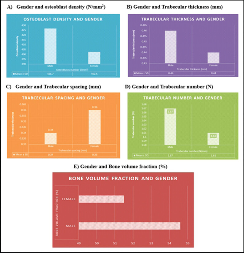
Table 2 shows that male callus samples exhibited slightly higher osteoblast density (426.7 ± 19.0 N/mm2) than female counterparts (402.5 ± 23.1 N/mm2) and higher bone volume fraction in males (54.6 ± 3.0%) than females (51.5 ± 2.7%), indicating more active osteogenesis, possibly due to higher bone turnover. The differences between male and female samples were not statistically significant (P > 0.05), likely due to the small sample size (n = 10). These observations support the general trend that osteoblast density correlates with trabecular density and bone volume fraction.
Age-wise comparison of osteoconductive variables
The osteoconductive variables were studied and compared across different age groups. (Table 3 and Fig. 2a, b, c, d, e) display the age-wise comparison with calculated t and P-values.
Age-wise comparison of osteoconductive variables (n=10)
| Variables | Age group | Mean±SD | t-value and P-value |
|---|---|---|---|
| Osteoblast number (/mm2) | 15–24 years (n=4, young) | 431.3±8.5 | t=2.18, P=0.18 (NS) |
| 25–64 years (n=6, middle) | 395.0±20.4 | ||
| Trabecular thickness (mm) | 15–24 years (young) | 0.49±0.03 | t=1.76, P=0.22 (NS) |
| 25–64 years (middle) | 0.43±0.04 | ||
| Trabecular spacing (mm) | 15–24 years (young) | 0.33±0.01 | t=1.95, P=0.19 (NS) |
| 25–64 years (middle) | 0.36±0.03 | ||
| Trabecular number (N/mm) | 15–24 years (young) | 1.74±0.06 | t=2.10, P=0.17 (NS) |
| 25–64 years (middle) | 1.58±0.09 | ||
| Bone volume fraction (%) | 15–24 years (young) | 57.3±2.5 | t=2.26, P=0.16 (NS) |
| 25–64 years (middle) | 50.9±3.8 |
SD: Standard deviation, NS: Not significant (P>0.05). Statistical comparison by unpaired Student’s t-test. Older age group (≥65 years) absent in pilot sample. All P>0.05 attributed to small pilot sample size; continuation phase (n=50) expected to demonstrate statistical significance
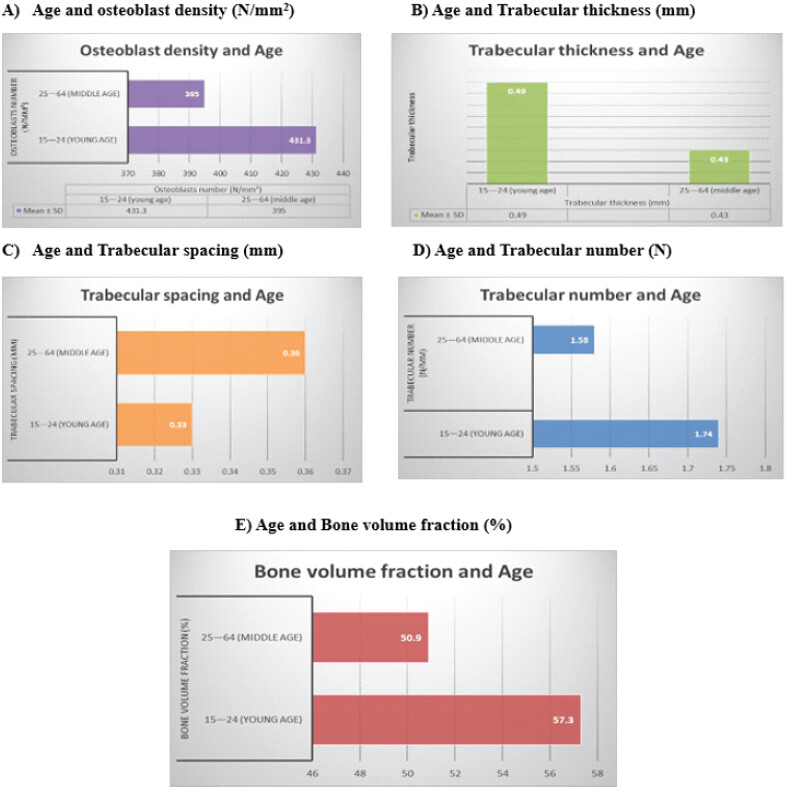
Table 3 shows that the younger age group (15–24 years) displays higher mean values of trabecular thickness, trabecular number, bone volume fraction, and osteoblast count, consistent with superior bone remodeling and callus quality. The middle-aged group (25–64 years) has slightly reduced microarchitectural parameters, suggesting a normal age-related decline. These differences were not statistically significant (P > 0.05), again likely due to the small sample size.
Comparison of test samples with control adult bone
Osteoconductive variables in the ten test samples were compared with the adult bone control (lunate). Fig. 3 displays the callus microscopic features in the younger, middle-aged, and control samples. A quantitative comparison of osteoconductive variables between test samples and control is presented in Table 4.
![Figure 3: Microscopic comparison of bone callus specimens with control adult bone (hematoxylin and eosin [H&E], ×10 and ×40). Photomicrographs illustrating the histological features of bone callus and control bone stained with H&E: (a) younger age group (15–24 years); (b) middle-aged group (25–64 years); (c) control adult carpal bone (lunate). Panel A (young callus): Active bone remodeling with abundant osteoblasts lining the trabeculae, a prominent osteoconductive framework, dense trabecular network, and minimal intertrabecular spacing, reflecting vigorous bone regeneration. Panel B (middle-aged callus): Reduced osteoblast density along trabecular surfaces with comparatively wider intertrabecular spacing and lower trabecular number, consistent with an expected age-related decline in osteogenic activity. Panel C (control adult lunate): Mature lamellar bone architecture with osteocytes housed in lacunae, regular Haversian systems, and a quiescent periosteal surface; serves as the structural reference for adult cortical bone. All sections were stained with hematoxylin and eosin. Photomicrographs were captured using a binocular light microscope and quantified with ImageJ software. Objective magnification as indicated within each panel (×10 and ×40); no calibrated scale bar was superimposed on the photomicrographs. ImageJ: Image analysis software (NIH, USA), OB: Osteoblast, OC: Osteocyte, T: Trabecula, M: Marrow space, H: Haversian canal. Control: Adult carpal bone (lunate) obtained from an age-matched cadaveric specimen. A and B: Bone callus test samples, C: Control adult bone.](https://jocr.co.in/wp/wp-content/uploads/455FF3-converted.jpg)
Comparison of osteoconductive variables: Test samples versus control adult bone (lunate)
| Variables | Young callus (15–24 years) | Middle-aged callus (25–64 years) | Control (adult lunate) | Trend |
|---|---|---|---|---|
| Osteoblasts number (N/mm2) | 431.3±8.5 | 395.0±20.4 | 310.2±15.6 | Young>Middle>Control |
| Trabecular thickness (mm) | 0.49±0.03 | 0.43±0.04 | 0.39±0.02 | Progressive decrease with age |
| Trabecular spacing (mm) | 0.33±0.01 | 0.36±0.03 | 0.41±0.03 | Progressive increase with age |
| Trabecular number (N/mm) | 1.74±0.06 | 1.58±0.09 | 1.42±0.07 | Young>Middle>Control |
| Bone volume fraction (%) | 57.3±2.5 | 50.9±3.8 | 44.7±3.1 | Active callus>quiescent bone |
Control=Adult carpal bone (lunate) from age-matched cadaveric specimen. Values expressed as mean±standard deviation. Both young and middle-aged callus samples demonstrated higher osteoblast density, trabecular thickness, trabecular number, and bone volume fraction compared to adult control bone, consistent with the active osteogenic phase of fracture healing. Bone volume fraction expressed as percentage
Both young and middle-aged bone callus groups demonstrated consistently higher osteoconductive parameters compared to the quiescent adult control bone (lunate), confirming that bone callus – irrespective of age – retains significantly greater osteogenic activity than mature cortical bone. Young callus showed the highest values across all parameters, underscoring the age-dependent gradient in regenerative potential (Table 4 and Fig. 3).
Discussion
There is a paucity of human studies examining the microscopic structure of bone callus, as most callus studies have been conducted in animal models. In addition, there is a dearth of studies focusing on the osteogenic potential of bone callus in different age groups and at varying reporting periods after fracture injury. Han et al. studied the osteogenic potential of bone callus in a nude mouse model and reported that early callus showed regular trabecular morphology, whereas middle- and late-stage callus showed mature lamellar bone. They observed that osteoblast density was highest in early bone callus compared to the late stage; trabecular number, spacing, and thickness decreased with time from early to late callus [1]. Our study findings were consistent with those of Han et al.
Cohn Yakubovich et al. in their study on immunosuppressed rats reported that intermittent parathyroid hormone (PTH) therapy promotes migration of systemically administered hematopoietic mesenchymal stem cells and induces osteogenic differentiation, resulting in rib fracture repair. Ribs healed through PTH therapy were found to be stiffer than intact non-fractured ribs [10].
Wen et al. studied the microstructure of non-unions of human humerus shaft fractures using scanning and transmission electron microscopy and observed that the non-union fracture had a trabecular structural framework similar to that of woven bone. They concluded that non-crystalline calcium phosphate deposition and inadequate mineralization of collagen fibers are the two characteristic features seen in non-unions of human humerus shaft fractures, distinct from normally repaired bone callus [11]. Our study observations are consistent with this theory.
Yang et al. studied 36 male Sprague-Dawley rats and observed that application of recombinant BMP-2 at the completion of the rapid distraction period significantly increased the osteogenic process of bone callus [12]. Cattaneo et al. studied approximately 6 samples of fracture bones from cadavers with known time of survival between injury and death and reported that the presence of blood clots and red blood cell residues on the margins of fractured fragments strongly indicates vital reaction and active bone healing [13]. Cui et al. confirmed that sclerostin antibody (Scl-Ab) promotes fracture healing in rodent and non-human primate models [14]. Iwamoto et al. observed that marrow stromal cells at fracture sites of human long bones differentiate into polygonal cells with higher alkaline phosphatase activity and that these polygonal cells secrete calcified matrix in bone callus [15].
The present preliminary findings, although based on a small pilot cohort, provide a histological foundation for the continuation phase of this study, where a complete sample of 50 participants – including the older age group (≥65 years) – will be analyzed. The total planned sample size for this study is 50 participants; 10 have been analyzed and are reported here, and recruitment and analysis of the remaining participants are ongoing, with completion anticipated by March 2027. Furthermore, the identification of osteoblast-rich callus in younger patients supports the future application of immunohistochemical analysis for cytokines (TNF-α, IL-6, IL-10) and senescence markers (p16, β-galactosidase) to delineate the molecular underpinnings of age-dependent differences in fracture healing. µCT analysis is planned to provide three-dimensional quantification of trabecular architecture, corroborating and extending the two-dimensional histomorphometric findings reported herein. The methodological constraints of this pilot-phase analysis – including its limited sample size, single-stain histology, and two-dimensional image analysis – are discussed in detail in Section 5 (limitations).
Limitations
This pilot-phase study is limited primarily by its very small effective sample size. Of the 23 patients recruited during the 6-month pilot phase, 13 (56.5%) were excluded because of inadequate callus tissue quantity, processing artifacts, or unsatisfactory histological quality, leaving only 10 evaluable samples for analysis. This high attrition substantially limits statistical power and the generalizability of the reported findings; the non-significant P-values obtained for most comparisons should therefore not be interpreted as evidence of true equivalence between groups but rather reflect the limited power of this pilot cohort to detect real differences. All between-group comparisons in this report should be regarded as hypothesis-generating observations rather than confirmatory findings, pending validation in the full cohort of 50 participants planned for the continuation phase.
A related concern is potential selection bias: Only patients who underwent ORIF and who yielded a callus specimen of adequate quantity and histological quality were retained in the final analytic sample. Patients with smaller, fragmented, or technically unsuitable callus – who may represent a biologically or clinically distinct subgroup, for example those with more comminuted fractures, poorer healing, or technically difficult surgery – were systematically excluded. This non-random attrition may have biased the study population toward callus samples with more favorable histological characteristics, potentially overestimating the apparent osteogenic quality of bone callus reported here.
The cross-sectional design constitutes a further important limitation. Each participant contributed a single callus specimen obtained at one time point, precluding longitudinal assessment of callus maturation, remodeling, or osteogenic change within the same patient over the course of healing. Serial sampling from the same patient was not ethically or practically feasible within a single surgical procedure; the between-subject comparisons reported here are therefore only an indirect proxy for the temporal trajectory of callus microarchitecture and should not be interpreted as evidence of within-patient change over time.
The demographic composition of the pilot cohort also constrains interpretation. The gender distribution was unequal (7 males and 3 females), limiting the precision and reliability of gender-based comparisons. No participant ≥65 years of age was recruited during the pilot phase, precluding any assessment of age-related senescence, osteoblast exhaustion, or osteogenic decline in elderly individuals – a population of particular clinical relevance given the high incidence of fragility fractures in this age group. Furthermore, the broad categorization of the middle-aged group (25–64 years) may obscure clinically relevant heterogeneity in bone-healing potential within this 40-year span; patients at the younger and older ends of this category may differ considerably in osteogenic capacity, and this categorization should be refined into narrower age bands in the continuation phase.
Variation in the interval between fracture and sample collection (mean 6.2 ± 1.5 weeks in the younger group and 8.4 ± 2.1 weeks in the middle-aged group, with individual variation around these means) may independently have influenced osteoblast density and trabecular parameters, since callus morphology evolves dynamically through the inflammatory, soft-callus, hard-callus, and remodeling phases of healing. This timing variability was not statistically controlled for in the present analysis and may confound the observed age- and gender-based differences. In addition, the study population was heterogeneous with respect to fracture site (long versus short bone) and fixation modality (plate-and-screw versus intramedullary nailing; Table 1), which may have introduced additional biological variability in callus formation, vascularity, and microarchitecture independent of age or gender.
Histological assessment in this study relied exclusively on H&E staining. While H&E permits reliable assessment of general tissue architecture, it does not allow definitive identification or quantification of specific cell populations, inflammatory mediators, or osteogenic markers. In particular, osteoblast density was determined by morphological criteria alone and was not confirmed using immunohistochemical markers such as osteocalcin, RUNX2, or alkaline phosphatase; morphological identification of osteoblasts on H&E sections carries a recognized risk of misclassification (for example, confusion with fibroblasts or pre-osteoblasts), which may have introduced measurement error into the osteoblast density estimates reported here. Immunohistochemical confirmation is planned for the continuation phase of this study.
Trabecular microarchitecture was quantified using two-dimensional ImageJ analysis of single histological sections. This approach cannot fully represent the three-dimensional architecture, connectivity, and spatial organization of trabecular bone, and two-dimensional parameters are known to correlate imperfectly with true three-dimensional bone volume fraction and connectivity density. Three-dimensional µCT, planned for the continuation phase, will be required to validate and extend these findings. In addition, only a single lunate specimen served as the adult control; a single specimen is insufficient to establish a robust reference range for normal mature bone microarchitecture, and inter-individual variability in normal adult trabecular structure could not be characterized. A larger panel of control specimens is planned for subsequent analyses.
Several potential confounding factors were not comprehensively evaluated in this pilot analysis, including fracture severity/comminution, smoking status, nutritional status, metabolic bone health (for example, Vitamin D and calcium status or bone mineral density), concurrent medications, and the mechanical stability achieved by fixation. Any of these factors could independently influence callus osteoblast density and trabecular microarchitecture, and their absence from the current analysis limits the ability to attribute the observed age- and gender-based differences specifically to biological ageing or sex.
Finally, and most importantly, this study characterizes only the histological and histomorphometric features of bone callus; it does not establish the clinical efficacy of bone callus as an autograft material. Although the callus samples examined demonstrated favorable osteogenic histological characteristics relative to mature adult bone, their actual osteogenic and osteoconductive performance when used as a graft has not been tested clinically or experimentally (for example, in an animal implantation model or a clinical case series with union outcomes). The clinical translation of these histological observations therefore remains hypothetical at this stage and should not be inferred from morphology alone.
Conclusion
Younger individuals (15–24 years) showed higher mean values of osteoblast count, trabecular thickness, trabecular number, and bone volume fraction compared to the middle-aged group, consistent with a possible trend toward superior osteogenic potential; however, these pilot-phase differences did not reach statistical significance (Table 3) and require confirmation in a larger cohort. The middle-aged group exhibited an expected age-related decline in osteoconductive parameters. Male callus samples demonstrated higher osteoblast density and bone volume fraction than female samples, though without statistical significance in this pilot cohort. Both callus groups showed greater osteogenic activity compared to the mature adult control bone (lunate), supporting the premise that bone callus retains significant regenerative potential across age groups.
These preliminary findings provide histological rationale for exploring bone callus as a potential autograft material – particularly in younger patients – and provide the histomorphometric baseline for the subsequent immunohistochemical and µCT phases of this study. However, the actual osteogenic and osteoconductive performance of bone callus as a graft has not been tested clinically or experimentally in this study, and this histological characterization should not be taken to establish clinical graft efficacy. Confirmation of these findings in the full cohort (n = 50) is anticipated to identify statistically significant age- and gender-based differences in callus microstructure, informing future targeted strategies for fracture management, pending dedicated clinical/experimental graft-efficacy studies.
Clinical Message
For orthopedic surgeons managing fracture patients, this preliminary histomorphometric analysis indicates that bone callus generated during normal fracture healing is biologically active and osteoblast-rich, particularly in younger patients, lending rational support to its consideration as a locally available autograft substitute during ORIF and related reconstructive procedures. Because callus microarchitecture varies with patient age, gender, and the interval since injury, callus intended for grafting should ideally be harvested from actively remodeling regions in younger patients wherever surgically feasible, rather than assumed to be uniformly osteogenic across all patients. However, these observations remain histological rather than functional: The actual bone-forming and graft-incorporation performance of harvested callus has not yet been tested clinically or experimentally, and clinicians should not substitute callus for established autograft sources such as iliac crest bone until this is confirmed in the larger, ongoing continuation phase of this study.
Conflict of Interest:
Nil
Source of Support:
Nil
Consent:
The authors confirm that informed consent was obtained from the patient for publication of this article
How to Cite this Article
Charmode SH, Mishra AK, Mehra S. Osteogenic Potential of Bone Callus: A Cross-sectional Study on Microscopic Structure and Healing Stages. Journal of Orthopaedic Case Reports 2026 October;16(10): 455-463.
References
- Han W, He W, Yang W, Li J, Yang Z, Lu X. The osteogenic potential of human bone callus. Sci Rep 2016;6:36330. [Google Scholar] | [PubMed]
- Nakase T, Kawai H, Yoshikawa H. In situ grafting of excised fracture callus followed by ilizarov external fixation for treatment of nonunion after open fracture of tibia. J Trauma 2009;66:550-2; discussion 552-3. [Google Scholar] | [PubMed]
- Samakkarnthai P, Saul D, Zbinden A. In vitro and in vivo determinants of senescent osteoblast function in bone. Aging Cell 2023;22:e13761. [Google Scholar] | [PubMed]
- Hambright WS, Niedernhofer LJ, Huard J, Robbins PD. Murine models of accelerated cellular senescence in skeletal maintenance and repair. Mech Ageing Dev 2023;210:111786. [Google Scholar] | [PubMed]
- Kaur P, Anjana RM, Tandon N, Singh MK, Mohan V, Mithal A. Increased prevalence of self-reported fractures in Asian Indians with diabetes: Results from the ICMR-INDIAB population based cross-sectional study. Bone 2020;135:115323. [Google Scholar] | [PubMed]
- Singh DR. Principles and Techniques in Histology, Microscopy and Photomicrography. 2nd ed. New Delhi: CBS Publishers and Distributors; 2018. [Google Scholar] | [PubMed]
- Rosadi I, Karina K, Rosliana I, Sobariah S, Afini I, Widyastuti T. In vitro study of cartilage tissue engineering using human adipose-derived stem cells induced by platelet-rich plasma and cultured on silk fibroin scaffold. Stem Cell Res Ther 2019;10:369. [Google Scholar] | [PubMed]
- IBM Corporation. IBM SPSS Statistics for Windows. Ver. 16.0. Armonk, NY: International Business Machines Corporation; 2007. [Google Scholar] | [PubMed]
- Age Categories, Life Cycle Groupings. Statistics Canada: Canada's National Statistical Agency. Available from: https://www.statcan.gc.ca/en/start. [Last accessed on 2025 Jun 15]. [Google Scholar] | [PubMed]
- Cohn Yakubovich D, Sheyn D, Bez M, Schary Y, Yalon E, Sirhan A. Systemic administration of mesenchymal stem cells combined with parathyroid hormone therapy synergistically regenerates multiple rib fractures. Stem Cell Res Ther 2017;8:51. [Google Scholar] | [PubMed]
- Wen HB, Cui FZ, Zhu XD. Microstructural features of non-union of human humeral shaft fracture. J Struct Biol 1997;119:239-46. [Google Scholar] | [PubMed]
- Yang JH, Kim HJ, Kim SE, Yun YP, Bae JH, Kim SJ. The effect of bone morphogenic protein-2-coated tri-calcium phosphate/hydroxyapatite on new bone formation in a rat model of femoral distraction osteogenesis. Cytotherapy 2012;14:315-26. [Google Scholar] | [PubMed]
- Cattaneo C, Andreola S, Marinelli E, Poppa P, Porta D, Grandi M. The detection of microscopic markers of hemorrhaging and wound age on dry bone: A pilot study. Am J Forensic Med Pathol 2010;31:22-6. [Google Scholar] | [PubMed]
- Cui L, Cheng H, Song C, Li C, Simonet WS, Ke HZ. Time-dependent effects of sclerostin antibody on a mouse fracture healing model. J Musculoskelet Neuronal Interact 2013;13:178-84. [Google Scholar] | [PubMed]
- Iwamoto M, Shibano K, Watanabe J, Asada-Kubota M, Ogawa R, Kanamura S. Culture of marrow stromal cells derived from bone marrow specimens formed at fracture site of human long bone. Bone 1993;14:799-805. [Google Scholar] | [PubMed]
© 2026 Journal of Orthopaedic Case Reports - Published by Indian Orthopaedic Research Group
About the Authors
-

Sundip Hemant Charmode
-

Abhishek Kumar Mishra
-

Simmi Mehra
Related Articles in Journal of Orthopaedic Case Reports
 August 1, 2026 Platelet-Rich Plasma Augmentation of Osteosynthesis in Femoral Neck Fractures: A Prospective Comparative Study Demonstrating Accelerated Union and Improved Function
 May 1, 2026 Assessment of Teriparatide’s Effect on Post-operative Functional Outcome and Fracture Healing
 May 1, 2026 Effect of Vitamin D Supplementation Regimens on Fracture Healing and Serum Biomarker Profile in Long-Bone Fractures: A Prospective Randomized Study
 May 1, 2026 The Impact of Medial Cortical Reduction on the Outcomes of Fixation in Unstable Intertrochanteric Fractures