
Displaced capitellum fractures may be associated with underlying DRUJ instability, mimicking Essex-Lopresti injuries, therefore careful wrist and forearm clinical and radiological assessment with appropriate management is essential in such injury patterns.
Dr. Aqil Ahamed Mohideen Ahamed Sha, Princess Royal University Hospital, Farnborough Common, Orpington, United Kingdom. E-mail: aqil.sha@nhs.net
Abstract
Introduction: The Essex-Lopresti injury, as defined in the literature, is characterised by a distinct triad: radial head (RH) fracture, tearing of the interosseous membrane and disruption of the distal radioulnar joint (DRUJ). We present an atypical case of DRUJ disruption resulting from a displaced capitellum fracture rather than an RH fracture.
Case Report: A 35-year-old female sustained a distal humerus capitellum fracture with associated DRUJ instability following a high-energy mechanical fall. Surgical management involved open reduction and internal fixation (ORIF) of the capitellum fracture using headless compression screws, along with closed reduction and plaster cast immobilisation of the DRUJ. Post-operative radiographs confirmed satisfactory position and healing of the capitellum fracture and restoration of DRUJ stability. Clinically, the patient demonstrated a stable full range of motion at both the wrist and elbow with good function.
Conclusion: Due to the potential dysfunction of these complex injury patterns, we feel displaced capitellum fractures should prompt a higher index of suspicion for associated DRUJ injury, warranting thorough examination and appropriate imaging of the forearm and wrist also. Capitellum ORIF, along with reduction and plaster cast stabilisation of the DRUJ, followed by appropriate rehabilitation, can lead to successful outcomes in such cases.
Keywords: Capitellum fracture, distal radioulnar joint instability, Essex-Lopresti injury.
In 1951, Peter Essex-Lopresti described this complex injury pattern consisting typically of a radial head (RH) or neck fracture with disruption of the intraosseous membrane (IOM) leading to distal radioulnar joint (DRUJ) instability [1]. The Essex-Lopresti injury (ELI) involves loss of structural integrity of the lateral column of the elbow, specifically an RH fracture, with injury to the IOM resulting in proximal migration of the radial shaft and consequent disruption or dislocation of the DRUJ [1]. The associated longitudinal instability and impaired load transmission across the forearm lead to significant pain and dysfunction. This pattern of injury to the IOM and DRUJ rarely occurs in the presence of an isolated displaced capitellum fracture instead of that of the RH and has only been described twice in the available literature [2,3]. This includes one case of chronic injury and one acute with management incorporating surgical stabilisation of the DRUJ. We present an atypical injury pattern of acute forearm longitudinal instability with subsequent DRUJ disruption accompanied by a displaced capitellum fracture. This was treated successfully with surgical fixation of the capitellum and non-operative management of the DRUJ complex. We describe our surgical technique for the capitellum and the treatment protocol for the DRUJ injury.
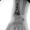
A 35-year-old female sustained a high-energy injury to her left arm following a fall down the stairs onto her outstretched hand. After presenting to the emergency department with elbow pain and swelling, plain elbow radiographs (Fig. 1) demonstrated a displaced capitellum fracture, and subsequent computed tomography (Fig. 2) confirmed a modified Dubberley type 4A fracture [4]. An above-elbow backslab was applied for initial immobilisation before a pre-operative outpatient review by our local upper limb specialist (lead author, KG) found tenderness around the wrist as well as the elbow. A plan was made for open reduction and internal fixation of the capitellar fracture, with examination under anaesthetic of the wrist and forearm to assess for associated longitudinal instability. Under general anaesthesia, a positive DRUJ ballottement test confirmed DRUJ instability, and intraoperative fluoroscopy (Fig. 3a and b) of the wrist demonstrated diastasis. With a tourniquet, an anterior “lazy-S” longitudinal incision was made over the radio-capitellum joint, spanning the anterior transverse elbow crease. Adequate exposure of the capitellum was achieved through an anterior approach, as described by Yang et al., [5] identifying multiple capitellum and lateral trochlear fragments. These were anatomically reduced, and definitive fixation was achieved using five 2.5 mm headless compression screws (HCS) (Acumed Acutrak), aimed anteroposteriorly perpendicular to the vertical shear fracture plane, apart from one aimed distal to proximal for a distal articular fragment. Intraoperative imaging confirmed anatomical reduction of the fragments (Fig. 3c), a congruent articular surface and satisfactory implant placement. The lateral ulnar collateral ligament was visualised and found to be intact. Following fixation of the capitellum, fluoroscopy demonstrated ongoing DRUJ widening and incongruency. With regard to the surgical decision-making, it was concluded that the use of Kirchner wires (K-wires) from the distal ulna to the distal radius to hold the reduction would anyway need to be protected with an elbow-spanning plaster in neutral forearm rotation. As we were able to achieve a satisfactory reduction of the distal joint in neutral forearm rotation and maintain this position without K-wires [6,7], we merely stabilised the acute wrist injury with an above-elbow plaster, maintaining this forearm position (Fig. 3d and e). At the 2-week post-operative follow-up (Fig. 4a), an undisplaced fracture of the scaphoid tubercle became apparent on radiology review. The patient was changed into a below-elbow sugar-tong cast to allow elbow flexion and extension, safeguard the DRUJ by preventing forearm pronosupination and immobilise the scaphoid fracture [6,7]. At 4 weeks following surgery, she was converted to a below-elbow cast to protect the scaphoid fracture, and at the 8-week follow-up, the cast was removed. At this time point, clinically and radiographically, the DRUJ remained reduced and stable (Fig. 4b, c), and both scaphoid and capitellum fractures appeared healed adequately (Fig. 4b, c, d, e). Accompanying physiotherapy allowed good pain-free elbow range of motion (ROM) from 30° to 150° of flexion with full pronation and supination and pain-free wrist ROM of flexion to 85° and extension to 80°. At 12 months the patient was recalled for review and reported mild intermittent lateral elbow pain, particularly in cold weather, but full restoration of ROM (Fig. 5a, b, c, d, e). The disabilities of the arm, shoulder, and hand score were 8.3, and the Mayo Elbow Performance Score was 85.
The ELI is classically characterised by an RH fracture, disruption of the IOM and dislocation of the DRUJ [1]. The responsible mechanism involves direct impact on an outstretched hand with the elbow extended. The force is transmitted proximally predominantly through the long axis of the radius, causing the RH to fracture upon impact with the capitellum. Concurrent forced rotation of the forearm can lead to tearing of the interosseous membrane and subsequent disruption of the DRUJ [1]. These injuries compromise the longitudinal stability of the forearm, allowing proximal migration of the radius, particularly if the RH fragments are markedly displaced. In contrast to the typical ELI pattern, we report a comminuted and displaced capitellum fracture accompanied by DRUJ dislocation, but the mechanism and injury pattern appear to be similar. The capitellum and RH together form the lateral column of the elbow and contribute significantly to its stability, with the radiocapitellar joint being described as an important secondary stabiliser in O’Driscoll’s fortress [4]. Loss of the capitellum’s structural integrity can mimic the destabilising effect of an RH fracture by disrupting the lateral column and potentially leading to longitudinal forearm instability from tearing of the IOM and proximal translation of the radius, a key component in the ELI mechanism [1]. In our case, the comminuted anterior capitellum and lateral trochlear fragments were reduced under direct vision and fixed with HCS to achieve a stable and congruent joint line [5,8,9,10]. The use of an anterior incision and approach ensured adequate exposure, even of the medial fragments, and allowed the desired implant orientation, which is not always easily achievable through a lateral approach [8,9,10]. In this approach, care must be taken to identify the radial nerve laterally and its superficial branch more distally under brachioradialis, along with protecting the medial neurovascular structures by careful retraction. On restoring the lateral column of the elbow and restoring longitudinal stability, intraoperative closed reduction of the DRUJ was performed under imaging guidance and, in the acute setting, attained without significant difficulty [6,7]. We opted to avoid k-wiring, as this was seen to be unnecessary in this case, where reduction was achievable and maintainable with neutral forearm rotation. K-wire fixation of the DRUJ would mandate plaster stabilisation with the elbow and forearm immobilised in the reduced position. In our patient, K-wire fixation was seen to be an unnecessary adjunct with risks of wire movement, breakage, and pin-site infection [7,11]. In a recent published report of a similarly high-energy injury treated acutely, the authors described addressing the longitudinal radioulnar dissociation with an open TFCC suture-anchor repair and K-wire stabilisation from the distal ulna through to radius [2]. This was protected with initial immobilisation and subsequent rehab, avoiding forearm rotation until the wire was removed at 6 weeks postoperatively. In our comparable post-operative regime, we attained a functional range without the need for further wrist surgery. However, Takahashi et al. discussed that the case was a high-energy, more complex B-type modified Dubberley injury pattern with posterior capitellar comminution requiring iliac crest bone grafting and a lateral condyle fracture treated with a combined posterior plate and tension-band wire configuration [2]. In a report from 2014, which was the earliest and only other available publication describing this type of injury complex, Hernández-Cortés et al. warned of the possibility of a longitudinal dissociation following a displaced capitellum injury [3]. The potential perils are discussed with a 32-year-old man who presented with a 5-month chronic injury following a neglected displaced capitellum fracture, proximal migration of the radius and DRUJ dislocation. This was salvaged with a simultaneous Sauvé-Kapandji procedure at the wrist and reverse Sauvé-Kapandji at the elbow [3]. In addition to the described benefits of an anterior approach for a vertical shear anterior capitellum fracture, particularly if the configuration extends medially into the trochlear [5,8,9,10], the key distinction and clinical significance of our report lie in the successful non-operative treatment of the DRUJ with appropriate rehabilitation. We feel that in the relevant injury pattern, acute fixation for the displaced capitellum fracture with plaster immobilisation alone of an associated DRUJ disruption, along with appropriate subsequent physiotherapy, restores timely elbow and forearm ROM, stability and function. At 1 year following surgery, the patient reported mild intermittent lateral pain, which in the presence of a persistently congruent joint line and stable implants on radiographs and an absence of any signs of infection, was put down to either post-traumatic cartilage wear from the complex intra-articular nature of the fracture or well-recognised implant changes in differing temperatures.
This case highlights the importance of maintaining a high index of clinical suspicion for concomitant DRUJ injuries when a capitellum fracture is identified and the need for comprehensive pre-operative and intra-operative assessment with clinical examination and imaging. In the acute setting, DRUJ instability can be successfully managed non-operatively following simultaneous surgical fixation of a concomitant displaced capitellum fracture, leading to favorable outcomes.
Displaced capitellum fractures may be associated with longitudinal forearm instability and DRUJ disruption; a high index of suspicion must be had and consideration made to examine and image the forearm and wrist in such injuries. In the acute setting, it is recommended that fixation of capitellum fractures be coupled with intraoperative evaluation of DRUJ stability. With this injury pattern, DRUJ instability can be successfully treated with closed reduction and immobilisation, without the need for surgical fixation and subsequent early rehabilitation to restore elbow and wrist function.
References
- 1. </p> [Google Scholar] [PubMed]
- 2. <ol> [Google Scholar] [PubMed]
- 3. <li>Essex-Lopresti P. Fractures of the radial head with distal radio-ulnar dislocation; report of two cases. J Bone Joint Surg Br 1951;33B:244-7.</li> [Google Scholar] [PubMed]
- 4. <li>Takahashi D, Yano K, Shibata S, Kaneshiro Y, Yokoi T, Sakanaka H. Surgical treatment for acute longitudinal radioulnar dissociation with humeral capitellar fracture: A case report. J Hand Microsurg 2024;16:100052.</li> [Google Scholar] [PubMed]
- 5. <li>Hernández-Cortés P, Gómez-Sánchez R, Pajares-López M, O’Valle-Ravassa F. Sauvé-Kapandji and reverse Sauvé-Kapandji procedures for treating chronic longitudinal radioulnar dissociation with capitellum fracture. Acta Orthop Traumatol Turc 2014;48:593-7.</li> [Google Scholar] [PubMed]
- 6. <li>Watson JJ, Bellringer S, Phadnis J. Coronal shear fractures of the distal humerus: Current concepts and surgical techniques. Shoulder Elbow 2020;12:124-35.</li> [Google Scholar] [PubMed]
- 7. <li>Yang X, Chang W, Chen W, Liu S, Zhu Y, Zhang Y. A novel anterior approach for the fixation of ulnar coronoid process fractures. Orthop Traumatol Surg Res 2017;103:899-904.</li> [Google Scholar] [PubMed]
- 8. <li>Zampetakis K, Stavrakakis IM, Alpantaki K, Kastanis G, Ktistakis I, Tsioupros A, <em>et al</em>. Systematic review of acute isolated distal radioulnar joint dislocation: Treatment options. J Clin Med 2024;13:7817.</li> [Google Scholar] [PubMed]
- 9. <li>Köroglu M, Özdeş HU, Taşkıran G, Aslantürk O. Acute isolated volar distal radioulnar joint dislocation: First surgery or conservative? Trauma Case Rep 2023;48:100952.</li> [Google Scholar] [PubMed]
- 10. <li>Yu T, Tao H, Xu F, Hu Y, Zhang C, Zhou G. Comparison of lateral approach versus anterolateral approach with Herbert screw fixation for isolated coronal shear fractures of humeral capitellum. J Orthop Surg Res 2019;14:230.</li> [Google Scholar] [PubMed]
- 11. <li>Sidhdhapuria P, Pathan S, Chaudhary V. Anterior approach of the elbow for fixation of capitulum fractures. Int J Orthop Sci 2019;5:583-6.</li> [Google Scholar] [PubMed]
- 12. <li>Ballesteros-Betancourt JR, Fernández-Valencia JA, García-Tarriño R, Domingo-Trepat A, Sastre-Solsona S, Combalia-Aleu A, Llusá-Pérez M. The limited anterior approach of the elbow for open reduction and internal fixation of capitellum fractures. Surgical technique and clinical experience in 2 cases with more than 2 years follow-up. Rev Esp Cir Ortop Traumatol 2017;61:176-84.</li> [Google Scholar] [PubMed]
- 13. <li>Murphy S, Fahey E, Fleming P. Isolated distal radioulnar joint dislocation in an adolescent: A case report and systematic review. J Orthop Case Rep 2025;15:20-4.</li> [Google Scholar] [PubMed]
- 14. </ol> [Google Scholar] [PubMed]
- 15. <p> [Google Scholar] [PubMed]


Related Articles in Journal of Orthopaedic Case Reports
 June 1, 2026 Case Report of Simultaneous Galeazzi Fracture Dislocation and Ipsilateral Scaphoid
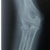 May 1, 2026 Operative Management of a Late-Presenting McKee Type IV Capitellum Coronal Shear Fracture via an Anterolateral Approach: A Rare Case Report
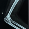 April 1, 2026 Functional and radiological outcome of capitellum fracture fixation with headless screws through lateral approach to elbow, a prospective observational study
 July 10, 2024 A Unique “Galeazzi-Like” Fracture through a Vacated External-Fixator Pin Track: A Case Report