

A double-layered antibiotic-loaded cement spacer may provide both effective infection control and mechanical stability during two-stage reconstruction for hip infections.
Hiroki Kobayashi, Department of Orthopedic Surgery, National Defense Medical College, Tokorozawa, Japan. E-mail: supercova0205@gmail.com
Abstract
Introduction: Septic arthritis of the hip and periprosthetic joint infection (PJI) are serious conditions that can cause joint destruction and severe functional impairment. In cases with advanced joint destruction or implant-related infection, two-stage reconstruction using an antibiotic-loaded cement spacer is widely performed. However, conventional polymethylmethacrylate (PMMA) spacers have limitations, including a short duration of antibiotic release and potential antibiotic inactivation caused by polymerisation heat. A double-layered antibiotic-loaded cement spacer consisting of an antibiotic-loaded calcium phosphate paste core covered with PMMA cement has been developed to combine sustained antibiotic release with mechanical stability.
Case Report: We report two infected hip cases treated using this novel spacer: One patient with PJI following hemiarthroplasty and one patient with septic arthritis of the hip. In both patients, extensive debridement and implantation of a double-layered antibiotic-loaded cement spacer were performed as the first stage of treatment. Inflammatory markers improved promptly after surgery, and no mechanical complications, including spacer fracture or dislocation, occurred during the interval period. Second-stage total hip arthroplasty was successfully performed in both cases. At the final follow-up, both patients were ambulatory without evidence of recurrent infection.
Conclusion: A double-layered antibiotic-loaded cement spacer may provide both effective infection control and mechanical stability during two-stage reconstruction for hip infections. This technique may represent a useful treatment option for the management of septic arthritis of the hip and PJI.
Keywords: Double-layered antibiotic-loaded cement spacer, periprosthetic joint infection, septic arthritis of the hip, calcium phosphate, drug delivery system.

Septic arthritis of the hip and periprosthetic joint infection (PJI) are infections that can result in joint destruction and severe functional impairment, and early diagnosis and appropriate treatment are essential [1,2]. In cases in which infection cannot be controlled by conservative treatment or in which advanced joint destruction is present, surgical treatment based on irrigation and debridement is required. In particular, for PJI, two-stage reconstruction, consisting of debridement of the infected tissue and implant removal, followed by temporary placement of an antibiotic-loaded spacer and subsequent reimplantation, is widely performed as the standard treatment strategy [3,4,5]. Antibiotic-loaded bone cement spacers have the advantage of delivering high concentrations of antibiotics locally; however, it has been reported that antibiotic release is concentrated in the early postoperative period and subsequently decreases rapidly [6,7]. In addition, the possibility of antibiotic inactivation due to the exothermic reaction during polymerization has also been pointed out [6]. In re[3,4,5].rs, antibiotic-loaded calcium phosphate materials have attracted attention as drug delivery systems (DDS). These materials do not generate substantial heat during setting and have the advantage of sustaipolymerisation release [8,9]. However, because of their insufficient mechanical strength, their use alone in weight-bearing joints is limited. To overcome these limitations, a double-layered antibiotic-loaded cement spacer has been reported, consisting of an antibiotic-loaded calcium phosphate bone paste core covered with an outer layer of bone cement (Fig. 1) [10].
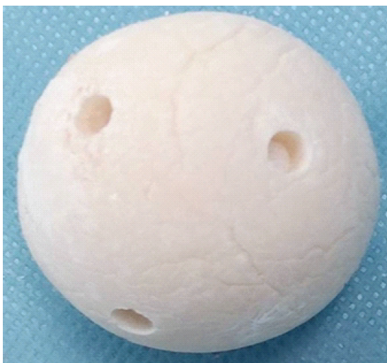
Figure 1: Double-layered antibiotic-loaded cement spacer consisting of an antibiotic-loaded calcium phosphate paste core covered with polymethylmethacrylate cement.
We report two cases in which this spacer was used for the treatment of one patient with PJI and one patient with septic arthritis of the hip, both of whom achieved satisfactory[8,9].tion control.
Case 1:
A 70-year-old woman presented with right hip pain. She had no remarkable medical history and no history of diabetes mellitus or immunosuppressive therapy. Nine years before presentation, she underwent bipolar hemiarthroplasty for a right femoral neck fracture at another hospital. Three years before presentation, she developed right hip pain while working on a farm. Because elevated inflammatory markers recurred repeatedly, infection was suspected, and intermittent antibiotic treatment was administered at the previous hospital. In March of year X, her pain worsened, and she was referred to our department in April of the same year with suspected PJI. At the initial visit, she had a limp and experienced pain during movement. Radiographs demonstrated a radiolucent area around the femoral stem in the femoral shaft, as well as osteolysis around the greater trochanter (Fig. 2a).
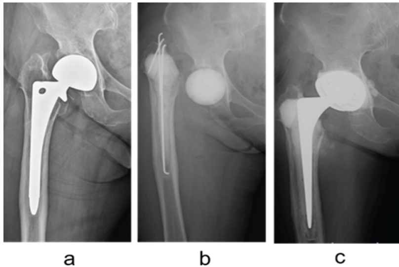
Figure 2: (a) Radiographs obtained at the initial visit. A radiolucent area is observed, and osteolysis is also present around the greater trochanter. (b) Post-operative radiograph showing implantation of the double-layered antibiotic-loaded cement spacer within the hip joint. The bone defect around the greater trochanter was filled with hydroxyapatite paste. (c) Radiograph obtained after revision total hip arthroplasty.
Laboratory examination revealed a white blood cell count of 5,000/μL and a C-reactive protein (CRP) level of 0.4 mg/dL, showing no elevation of inflammatory markers. Joint fluid culture was negative. However, the aspirated fluid was purulent, and glucose analysis showed a value of <5 mg/dL, strongly suggesting infection. Magnetic resonance imaging (MRI) demonstrated findings suggestive of abscess formation around the right hip joint (Fig. 3).
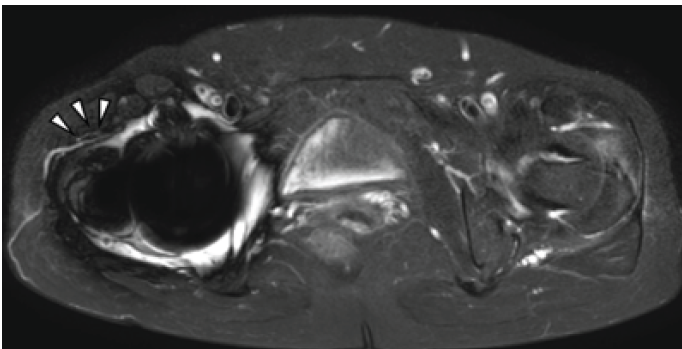
Figure 3: Magnetic resonance imaging obtained at the initial visit. A high-signal intensity area distinct from metal artifact is observed around the right hip joint (arrowhead), suggesting abscess formation.
Based on these findings, the patient was diagnosed with PJI. Because the infection had a chronic course, abscess formation was present on imaging studies, and the causative organism could not be identified due to negative culture results, one-stage reconstruction was considered high risk, and a two-stage reconstruction strategy was selected. In June of year X, implant removal, irrigation, and debridement were performed under general anaesthesia, and a double-layered antibiotic-loaded cement spacer was fabricated and implanted (Fig. 2b). The spacer consisted of an antibiotic-loaded calcium phosphate bone paste core covered with bone cement, with multiple communicating holes connecting the inner core to the external environment. Weight-bearing ambulation with crutch assistance was permitted immediately after surgery. Because the patient had a cefazolin allergy, clindamycin (2,400 mg/day) was initiated on the day of surgery. Rifampicin (450 mg/day) and levofloxacin (500 mg/day) were added on post-operative day 1. Clindamycin was discontinued on post-operative day 9, and CRP normalised on post-operative day 16. On post-operative day 39, the antibiotic regimen was changed to minocycline (200 mg/day) and levofloxacin (500 mg/day). After infection was considered to be controlled, revision total hip arthroplasty was performed 3 months after the initial surgery (Fig. 2c). Antibiotic therapy was discontinued 4 months after reimplantation. At the final follow-up, 1 year and 5 months after surgery, the patient was able to walk independently, and no recurrence of infection was observed.
Case 2:
A 68-year-old man presented with left hip pain. His medical history included psoriasis vulgaris, tinea pedis, and onychomycosis. In mid-October of year X, he fell from a bed and developed left hip pain. Because the pain gradually worsened, he visited a previous hospital and was referred to our department with a suspected subchondral insufficiency fracture of the femoral head. At presentation, he was unable to walk because of pain. Plain radiographs demonstrated collapse of the left femoral head (Fig. 4a). MRI revealed joint effusion in the left hip joint (Fig. 5).
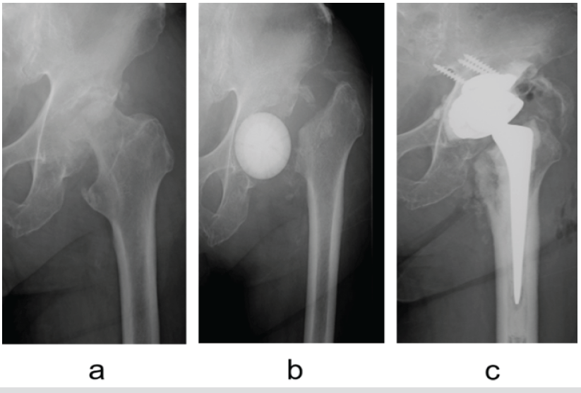
Figure 4. (a) Radiograph obtained at the initial visit showing collapse of the left femoral head and an acetabular bone defect. (b) Post-operative radiograph showing implantation of the double-layered antibiotic-loaded cement spacer within the hip joint. (c) Post-operative radiograph showing implantation of the double-layered antibiotic-loaded cement spacer within the hip joint.
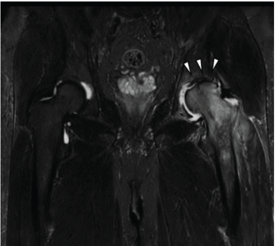
Figure 5: Magnetic resonance imaging obtained at the initial visit demonstrating collapse of the femoral head and fluid collection around the hip joint (arrowhead).
Laboratory examination showed a white blood cell count of 6,600/μL and a CRP level of 3.0 mg/dL, indicating mild elevation of inflammatory markers. Culture of aspirated joint fluid yielded Staphylococcus hominis subsp. Hominis, and septic arthritis of the hip was diagnosed. Because severe skin lesions associated with tinea pedis were present and primary total hip arthroplasty was considered to carry a high risk of implant-related infection, a two-stage reconstruction strategy was selected. After admission, irrigation, debridement, and implantation of a double-layered antibiotic-loaded cement spacer were performed (Fig. 4b). Cefazolin (6 g/day) was administered until post-operative day 8, after which minocycline (200 mg/day) and trimethoprim-sulfamethoxazole (2 tablets/day) were administered. Inflammatory markers normalised on postoperative day 33. Total hip arthroplasty was performed 4 months after the initial surgery (Fig. 4c). At the final follow-up, 1 year after surgery, the patient was able to walk independently, and no recurrence of infection was observed.
Septic arthritis and PJI are among the most challenging infectious conditions encountered in orthopaedic surgery. These severe diseases can cause rapid destruction of articular cartilage and bone, leading to irreversible functional impairment within a short period of time [1,2]. The hip joint, in particular, is a deep-seated joint in which diagnosis is often delayed, and extensive bone loss and soft-tissue damage are frequently present at the time infection is identified. Furthermore, in PJI, eradication of infection becomes extremely difficult because of bacterial adherence to implant surfaces and subsequent biofilm formation [2,11]. In addition to these factors, treatment outcomes are greatly influenced by patient-related factors, such as age, underlying diseases, and immune status, as well as by the virulence of the causative organism. Consequently, these conditions are associated with high recurrence rates and are recognised as refractory diseases that often require multiple surgical interventions [12,13]. Therefore, multidisciplinary treatment consisting of surgical debridement, implant management, systemic antibiotic therapy, and local antibiotic strategies is essential. One of the most important factors contributing to the refractory nature of PJI is bacterial biofilm formation. After adhering to the implant surface, bacteria produce a matrix composed of extracellular polymeric substances and proliferate as microcolonies. Within this structure, bacterial metabolic activity is reduced, and susceptibility to antimicrobial agents is markedly decreased compared with planktonic bacteria [2,12]. In addition, extracellular polymeric substances act as a barrier to antibiotic penetration and serve as a defence mechanism against host immune responses [12]. Furthermore, alterations in gene expression within biofilms induce antimicrobial resistance-related mechanisms, and dormant cells known as persister cells have been reported to contribute to chronic infection and recurrence [12,13]. Given these pathological characteristics, complete eradication of biofilm-associated bacteria is difficult to achieve with a single course or short-term administration of antibiotics alone. Therefore, maintaining a high local concentration of antimicrobial agents over a prolonged period is crucial for infection control. However, antibiotic release from conventional polymethylmethacrylate (PMMA) spacers reaches a peak during the early postoperative period and then declines rapidly, which may be insufficient to provide a sustained antimicrobial environment against biofilm-associated infections [6,7]. This limitation may contribute to persistent infection and recurrence following conventional treatment. Considering the characteristics of biofilm-related infections, the importance of local drug delivery systems capable of providing high and sustained concentrations of antimicrobial agents at the site of infection has gained increasing attention. In addition to delivering antibiotic concentrations that are difficult to achieve through systemic administration, local DDS can minimise systemic toxicity while providing potent local antimicrobial effects [9,14,15]. Moreover, by filling the dead space created after debridement while simultaneously releasing antimicrobial agents, local DDS can contribute to both infection control and tissue reconstruction [15,16]. At present, PMMA bone cement remains the most widely used local DDS carrier. However, antibiotic release from PMMA is largely dependent on an initial burst release during the early postoperative period, which limits its ability to maintain effective antibiotic concentrations over an extended period [6,7]. In contrast, bioabsorbable materials such as calcium sulphate and calcium phosphate have recently attracted attention as local DDS carriers because their porous structures allow relatively sustained antibiotic release [8,9,15]. Calcium phosphate, in particular, offers excellent biocompatibility and does not generate substantial heat during the setting process. On the other hand, these materials possess limited mechanical strength and may fail mechanically when used alone in weight-bearing joints such as the hip. Furthermore, calcium sulphate has been associated with complications including persistent wound drainage and hypercalcemia due to its rapid resorption [9]. Thus, an ideal local DDS should provide both sustained antibiotic release and mechanical support; however, conventional materials have been unable to adequately satisfy both requirements simultaneously. To address this issue, Ikeda et al. reported a double-layered antibiotic-loaded cement spacer that combines the sustained-release properties of calcium phosphate with the mechanical stability of PMMA. In addition, the spacer is designed with communicating holes that facilitate the continuous release of antibiotics from the inner core [10]. In vitro studies have demonstrated improved durability of antibiotic release as well as enhanced mechanical properties with this design [10]. Therefore, this structure appears to represent a rational local DDS strategy for the management of biofilm-associated infections. In the present cases, inflammatory markers improved promptly after spacer implantation, and second-stage reconstruction was successfully performed without mechanical complications such as spacer fracture or dislocation during the interval period. These clinical outcomes are consistent with previously reported results of two-stage reconstruction procedures [3,4] and suggest that the spacer functioned effectively in terms of both infection control and mechanical stability. Achieving both infection control and structural stability during the spacer period is a major challenge in the treatment of hip joint infections, and the clinical significance of this technique may therefore be considerable. Several limitations of this report should be acknowledged. First, only two cases were included, and caution is required when generalising the findings. Second, antibiotic selection and the timing of reconstruction were determined individually for each patient, making it difficult to clearly evaluate the independent effect of the spacer itself. Finally, long-term reinfection rates and functional outcomes have not yet been sufficiently assessed. Further accumulation of cases and additional investigation will therefore be necessary.
We experienced two cases treated with a double-layered antibiotic-loaded cement spacer, including one case of PJI and one case of septic arthritis of the hip. This technique may provide both effective infection control and mechanical stability and may represent a useful treatment option for two-stage total hip reconstruction.
In hip infections requiring two-stage reconstruction, a double-layered antibiotic-loaded cement spacer may overcome the limitations of conventional PMMA spacers by combining prolonged antibiotic release with adequate mechanical strength.
References
- 1. Li C, Renz N, Trampuz A. Management of periprosthetic joint infection. Hip Pelvis 2018;30:138-46. [Google Scholar] [PubMed]
- 2. Zimmerli W, Trampuz A, Ochsner PE. Prosthetic-joint infections. N Engl J Med 2004;351:1645-54. [Google Scholar] [PubMed]
- 3. Kunutsor SK, Whitehouse MR, Blom AW, Beswick AD, INFORM Team. Re-infection outcomes following one- and two-stage surgical revision of infected hip prosthesis: A systematic review and meta-analysis. PLoS One 2015;10:e0139166. [Google Scholar] [PubMed]
- 4. Kini SG, Gabr A, Das R, Sukeik M, Haddad FS. Two-stage revision for periprosthetic hip and knee joint infections. Open Orthop J 2016;10:579-88. [Google Scholar] [PubMed]
- 5. Depypere M, Kuehl R, Metsemakers WJ, Senneville E, McNally MA, Obremskey WT, et al. Recommendations for systemic antimicrobial therapy in fracture-related infection: A consensus from an international expert group. J Orthop Trauma 2020;34:30-41. [Google Scholar] [PubMed]
- 6. Slane J, Vivanco J, Rose W, Ploeg HL, Squire M. Mechanical, material, and antimicrobial properties of acrylic bone cement impregnated with silver nanoparticles. Mater Sci Eng C Mater Biol Appl 2015;48:188-96. [Google Scholar] [PubMed]
- 7. Goltzer O, McLaren A, Overstreet D, Galli C, McLemore R. Antimicrobial release from prefabricated spacers is variable and the dose is low. Clin Orthop Relat Res 2015;473:2253-61. [Google Scholar] [PubMed]
- 8. Steadman W, Chapman PR, Schuetz M, Schmutz B, Trampuz A, Tetsworth K. Local antibiotic delivery options in prosthetic joint infection. Antibiotics (Basel) 2023;12:752. [Google Scholar] [PubMed]
- 9. Kallala R, Haddad FS. Hypercalcaemia following the use of antibiotic-eluting absorbable calcium sulphate beads in revision arthroplasty for infection. Bone Joint J 2015;97-B:1237-41. [Google Scholar] [PubMed]
- 10. Ikeda S, Uchiyama K, Minegishi Y, Ohno K, Nakamura M, Yoshida K, et al. Double-layered antibiotic-loaded cement spacer as a novel alternative for managing periprosthetic joint infection: An in vitro study. J Orthop Surg Res 2018;13:322. [Google Scholar] [PubMed]
- 11. Romanò CL, Scarponi S, Gallazzi E, Romanò D, Drago L. Antibacterial coating of implants in orthopaedics and trauma: A classification proposal in an evolving panorama. J Orthop Surg Res 2015;10:157. [Google Scholar] [PubMed]
- 12. Costerton JW, Stewart PS, Greenberg EP. Bacterial biofilms: A common cause of persistent infections. Science 1999;284:1318-22. [Google Scholar] [PubMed]
- 13. Arciola CR, Campoccia D, Montanaro L. Implant infections: Adhesion, biofilm formation and immune evasion. Nat Rev Microbiol 2018;16:397-409. [Google Scholar] [PubMed]
- 14. Parvizi J, Gehrke T, International Consensus Group on Periprosthetic Joint Infection. Definition of periprosthetic joint infection. J Arthroplasty 2014;29:1331. [Google Scholar] [PubMed]
- 15. Metsemakers WJ, Fragomen AT, Moriarty TF, Morgenstern M, Egol KA, Zalavras C, et al. Evidence-based recommendations for local antimicrobial strategies and dead space management in fracture-related infection. J Orthop Trauma 2020;34:18-29. [Google Scholar] [PubMed]
- 16. Tetsworth K, Cierny G 3rd. Osteomyelitis debridement techniques. Clin Orthop Relat Res 1999;360:87-96. [Google Scholar] [PubMed]


Related Articles in Journal of Orthopaedic Case Reports
 September 1, 2026 Iliopsoas Abscess as the Initial Manifestation of Periprosthetic Hip Joint Infection after Total Hip Arthroplasty: A Case Report and Literature Review
 July 1, 2026 Bone Morphogenetic Proteins-2–Augmented Acetabular Bone Stock Restoration During Two-stage Revision for Periprosthetic Joint Infection: A Case Report
 June 1, 2026 Antiphospholipid Syndrome–Associated Livedoid Vasculopathy Mimicking Recurrent Periprosthetic Joint Infection after Total Knee Arthroplasty: A Case Report
 June 1, 2026 Sequential Multi-organism Periprosthetic Joint Infection after Total Hip Arthroplasty: A Case Report