

A combination of bicruciate and meniscus osseous root avulsions is extremely rare.
Dr. Raghavendra Kembhavi, Department of Orthopedics, Sakra IKOC Multispeciality Hospital, Bengaluru, Karnataka, India. E-mail: kembhavi.raghavendra@gmail.com
Abstract
Introduction: Bicruciate bony avulsion fracture with osseous avulsions of dual root lateral meniscus (LM) in combination with posterior horn LM meniscocapsular injury, causing a floating meniscus in lateral tibial plateau depressed fracture, is a very rare complex injury that was managed carefully with a proper workup and surgical planning to get an excellent functional outcome. A few times innocuous-looking tibial plateau fractures can reveal severe hidden injuries that have potentials of disastrous complications later in life, and therefore high degree of suspicion is required for accurate diagnosis.
Case Report: A 22-year-old female patient injured her knee while playing kabaddi due to a severe hyperextension injury. Based on clinicoradiological and intraoperative findings, she was diagnosed as having both Anterior cruciate ligament (ACL) and posterior cruciate ligament (PCL) avulsion fractures with LM anterior and posterior root avulsion, which we coined the term as “ Terrible Avulsion Tetrad” along with posterior horn of LM menisco-capsular tear with depressed lateral tibial plateau. The patient was operated next day of the injury with both arthroscopic and open procedures. Arthroscopically, ACL and both root tears of LM were fixed with the tunnel pull-out suture technique using No. 2 FibreWire and fixing with the endobutton and suture disk. Posterior horn LM menisco-capsular junction tear was repaired with two all-inside meniscal sutures. Lateral tibial plateau was fixed with two CC screws after elevating depressed fragments. Posteriorly PCL avulsion fragment was fixed with an open procedure with an inverted L incision and two cannulated cancellous screws. The patient was put on a knee brace for 6 weeks and was allowed knee range of movements at 3 weeks. The patient was kept on non-weight-bearing for 3 months. At 24 months follow-up, radiology showed complete union of fractures. Functional assessment using the Lysholm knee score showed an excellent outcome with a score of 96, and she was back to her routine activities.
Conclusion: A combination of bicruciate and meniscus osseous root avulsions is extremely rare. Further rare injury combinations can include a floating meniscus as a result meniscocapsular junction tear, along with osseous root avulsion of the meniscus. All these injuries can be accompanied in subtle looking proximal tibia plateau fracture, which needs be properly worked up.
Keywords: Osteoarthritis, effusion, deformity, gastrocnemius, debridement.

Anterior cruciate ligament (ACL) and posterior cruciate ligament (PCL) midsubstance tear are commonly torn ligaments seen in orthopedic practice though ACL injuries are more common than PCL. Combined injuries of ACL and PCL are rare, which are ideally seen in high-energy trauma like knee dislocations. Avulsion fractures of cruciate ligaments are a relatively uncommon category compared to mid-substance injuries. ACL avulsion fractures are seen in around 2% of ACL injuries, whereas PCL avulsion injuries are seen in 4% of PCL injuries [1,2]. A similar injury pattern applies for meniscal injuries as well. Root avulsions of the medial meniscus (MM) are generally seen in elderly patients as degenerative tears accompanied by trivial trauma, whereas avulsions of the lateral meniscus (LM) are generally associated with traumatic ACL tears [3]. On the other hand, bony avulsions of the meniscus are extremely rare [4]. They are generally seen in high velocity trauma and more seen in the posterior horn than the anterior horn of both menisci.
Proximal tibia fractures are many times innocuous-looking, and often a subtle avulsion of ligaments or menisci are missed out. In this case, we highlight on what initially appeared to be a manageable depressed fracture of the lateral tibial plateau showed multiple injuries of bony avulsion contributing to catastrophic global instability, that is, a simultaneous ACL and PCL avulsion fracture, dual root avulsion of LM with meniscocapsular disruption leading to floating LM.
A 22-year-old professional kabaddi player presented to our casualty with injury to the right knee while playing kabaddi. She sustained a twisting injury to right knee and presented to us with complaints of pain, swelling, and deformity over knee. Players from the opponent team fell on her in a group when the patient landed on her knee in hyperextension while kabaddi riding, causing severe injury. No other obvious injuries were seen. Patient was conscious, oriented, and hemodynamically stable at the time of presentation. On clinical examination, there was severe tenderness, swelling and deformity over the right knee with massive knee effusion. No external wounds were seen. There was significant tenderness over the knee and proximal tibia. Both Lachman’s and posterior drawer’s tests were positive. There was no varus or valgus laxity over knee. There were no distal neurovascular deficits. There was no stretch pain over toes.
Plain radiography showed a displaced proximal tibia fracture with minimal depression over the lateral and posterolateral tibial plateau with PCL avulsed fragment with displacement (Fig. 1).
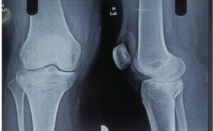
Figure 1: Plain radiograph of knee R showing depressed lateral and posterolateral tibial plateau fracture with a large posterior cruciate ligament avulsion fragment.
Although radiography showed innocuous looking fracture, we decided to work up further with computed tomography (CT) scan and magnetic resonance imaging (MRI) scan in view of significant anterior and posterior knee laxity. CT and MRI scans showed ACL avulsion injury at the tibial side, PCL avulsion injury with a large bony fragment and LM anterior with posterior root avulsion injuries (Fig. 2). There were severe fluid signals due to significant hemarthrosis.
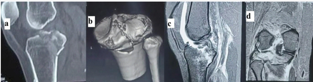
Figure 2: (a and b) Computed tomography and (c and d) magnetic resonance imaging showing anterior cruciate ligament avulsion injury at the tibial side, posterior cruciate ligament avulsion injury with a large bony fragment and lateral meniscus anterior and posterior root avulsion injuries with severe hemarthrosis suggested by large fluid signals.
Meniscocapsular junction of the LM was not visualized clearly due to excessive fluid signals.
Treatment considered was arthroscopic management of the knee injury. First, we decided for initial diagnostic approach with knee arthroscopy to confirm MRI/CT findings and to look for other injuries which were missed on radiology. With the knee in 90° flexion and under tourniquet, 30° arthroscope was used, and an initial diagnostic examination of the knee was made using anterolateral (AL) and anteromedial (AM) working portal. Thorough joint lavage, debridement and hematoma evacuation were done. Avulsed ACL fragment was noted (Fig. 3). In the lateral compartment, both anterior and posterior LM root avulsions were noted (Fig. 4). In addition, we noticed there was a complete tear of menisco-capsular junction of the posterior horn of LM, which was not seen on imaging (Fig. 5).
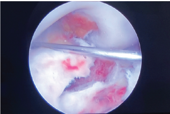
Figure 3: Intraoperative arthroscopic picture showing a large avulsed anterior cruciate ligament fragment.
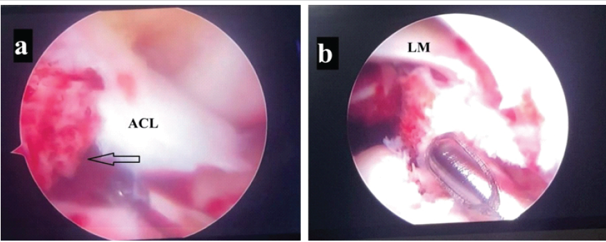
Figure 4: Intraoperative arthroscopic picture showing osseous avulsion of the anterior horn of the lateral meniscus (LM) (a with arrow) and osseous avulsion of posterior horn of LM (b).
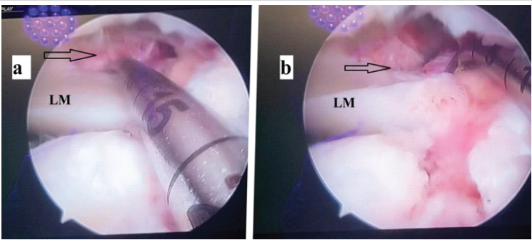
Figure 5: (a and b) Intraoperative arthroscopic picture showing a tear at menisco-capsular junction of the posterior horn of the lateral meniscus (arrows).
Furthermore, depression in the lateral tibial plateau was noted. Posteriorly, avulsion of the PCL fragment was noted. After assessing all the injuries, we planned for systemic approach to address each injuries step wise. First, we passed two fibre wires No. 2 with scorpion wire passer through ACL avulsion at ligament bone junction from AM portal through cannula. With the knee in lazy figure of four position, cinch sutures were placed on both LM meniscal roots with one fibre wire No. 2 on each root avulsion. Using the ACL tibial jig, the first tibial tunnel was made at the footprint of the posterior horn of the LM root with an arthroscope in the AM portal and jig holding from the AL portal with exit at the lateral tibial cortex. 2.9 mm guide pin was used, and it was over reamed with 4 mm cannulated drill bit. Another tibial tunnel was made for the ACL avulsion fragment with an arthroscope from the AL portal and a holding jig from AM portal, with exit at the medial tibial cortex, using 2.9 mm guide pin. It was over reamed with 4 mm cannulated drill bit. No. 2 fibre wire for LM posterior horn avulsion fracture was pulled through the tunnel at the lateral tibial cortex using crescent shaped small hook. Fibre wires holding ACL avulsion fragment and anterior horn of LM avulsion fragment were pulled through the same single tibial tunnel made at the medial tibial cortex using a crescent hook. Holding all the sutures under tension, menisco-capsular junction of the posterior horn of the LM was repaired with two all-inside meniscal repair devices (Sironix SureStich). Now the focus is shifted to depressed lateral tibial plateau fracture. Under arthroscopic guidance and fluoroscopic guidance bone impactor inserted through a small window made over the lateral tibial cortex away from the lateral tibial plateau. Once a satisfactory reduction achieved and to maintain the reduction, two 4 mm cannulated cancellous screws with a washer (4 mm × 45 mm, 4 mm × 50 mm) were passed from the lateral side of the tibial plateau directing slightly posteriorly in the subchondral bone. Cannulated drill bits were placed in already existing tibial tunnels for LM posterior root and ACL avulsion to prevent the cannulated screws entering the tunnels. Once plateau was fixed, LM posterior horn avulsion suture was fixed with endobutton on outer surface of lateral tibial cortex, and stability was checked in an arthroscopic view. ACL avulsion and LM anterior horn sutures were fixed with suture disk on the tibial surface of medial tibial cortex, and again stability assessed arthroscopically (Fig. 6).
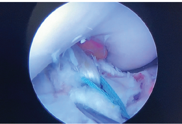
Figure 6: Intraoperative arthroscopic picture showing stable fixation of osseous avulsion of the posterior horn of the lateral meniscus with No. 2 fibre wire.
Now the focus shifted to PCL avulsion fragment. The patient was turned to the prone position. Inverted L-shaped incision was made over the posteromedial corner of the knee with a vertical limb along the medial edge of the gastrocnemius muscle and a horizontal limb on just distal to the joint line. Plane between semimembranosus and gastrocnemius was made, and the avulsed fracture fragment was isolated after making a vertical incision on the posterior capsule. Fracture fixation was done with two 4 mm cannulated cancellous screws (4 mm × 30 mm and 4 mm × 35 mm), directing away from the lateral tibial plateau screws and making sure not to pass through tunnels made for ACL and LM. Fluoroscopy showed satisfactory reduction of all the fractures.
Patient was advised on a knee brace for 6 weeks with isometric quadriceps exercises started on day 1. Knee range of movements (ROM) until 90° was started at 3 weeks, with complete ROM at 6 weeks. She was kept on non-weight-bearing for 12 weeks and was allowed full weight bearing at 16 weeks.
Patient was followed up for 24 months postoperatively, and at the latest follow-up she was back to her regular activities of running and playing her professional sports of Kabaddi. She had an excellent functional score (Lysholm Knee Score −96) (Fig. 7). Plain radiography of the knee complete radiological union of ACL, PCL avulsion with complete union of tibial plateau fracture (Fig. 8).
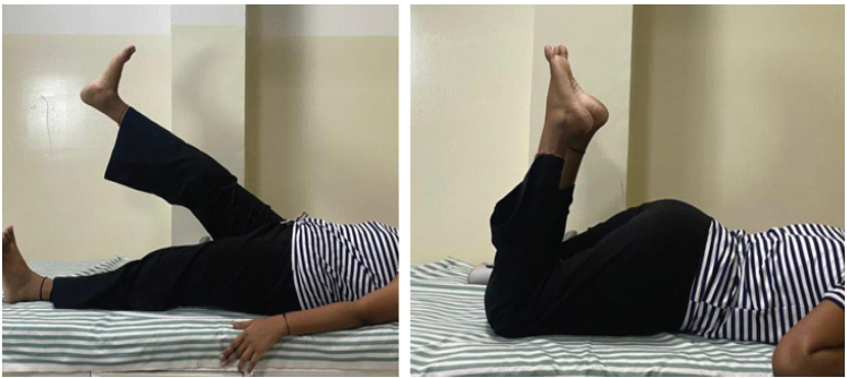
Figure 7: Clinical images of functional outcome at 2-year follow-up.
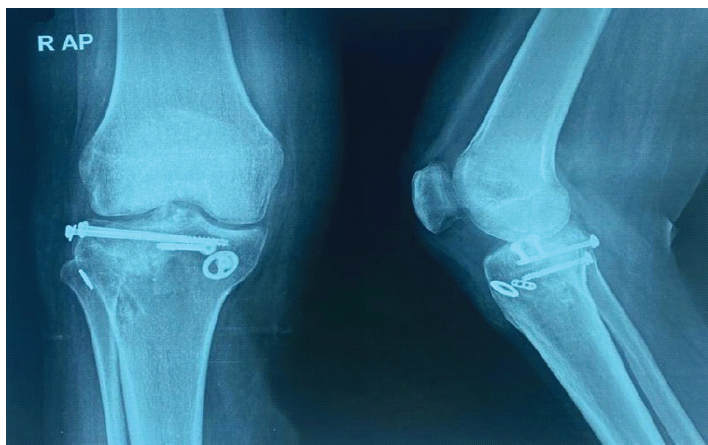
Figure 8: Knee radiograph at 2 years showing complete union of the fracture with well-maintained articular congruity.
This is probably the first case in our knowledge reporting extensive avulsion fractures of ACL, PCL, and dual root of LM with menisco-capsular junction of posterior horn of LM leading to a floating meniscus in a lateral tibial depressed fracture. Previously, many case reports on avulsion injuries of ligaments and meniscus have been reported in various permutation and combination. Mariani et al. reported complete ACL rupture with both MM and LM avulsion injuries in a professional football player [5]. Dartus et al., in their arthroscopic technique, described an injury naming “Tibial Avulsion Triad” which involved ACL tear of the posterolateral bundle, PCL avulsion fracture, with LM posterior horn root avulsion seen in dashboard injury [6]. Therefore, we too named an injury in our patient as “Tibial Avulsion Tetrad” comprising avulsion fractures of ACL, PCL and dual roots of LM. In another interesting case report of a similar complex injury, White et al. described ACL, PCL avulsion fractures with posterior horn MM root avulsion injury in a sleepwalking patient by an unknown injury mechanism [7]. However, closest to our case report was reported by Van der List et al. [8]. In their case report injury described was ACL, PCL avulsion with posterior root osseous avulsions of both meniscus. Therefore, described in our patient is slightly different from all the injuries described above, making it unique in addition to the depressed lateral tibial plateau fracture.
PCL avulsion injuries are commonly associated with other intra-articular injuries, and therefore we need to keep a close look into concurrent injuries, ideally with the direct visualization through arthroscope since MRI too can miss out in the first place due to existing fluid signals secondary to trauma. Pardiwala et al. showed nearly 28% of coexistent pathologies in PCL avulsion fractures in their comparative study of arthroscopic and open fixation of PCL avulsion injuries [9]. PCL avulsions too are commonly associated with bony avulsions injuries of the MM posterior root due to the anatomical close relation to PCL tibial facet [10].
Avulsion injuries of these cruciate ligaments are classically seen in dashboard injuries wherein a posteriorly directed force applied on the proximal tibia with the knee in a flexed position, which is seen in high-energy road traffic accident. Other mechanisms include hyperextension injury, which is ideally seen in sports injuries, and may have concomitant meniscal injuries or other ligamentous injuries [11]. In our patient, hyperextension could be the mechanism of injury since the patient will on the ground with the knee in hyperextension, followed by a group of players from the opposite team falling on the knee causing significant injury.
Floating meniscus is an interesting entity generally seen in high-impact injuries. It results from tear of both root attachments from the tibial surface, with a tear in coronary ligaments of the meniscus, resulting in meniscal complete displacement. Pardiwala et al. reported seven cases of such floating meniscus, where 5 were operated on an emergency basis and two on an elective basis [12]. They reported all their injuries in MM. On the contrary, in our patient, it was an LM floating meniscus with disruption of both anterior and posterior roots, with bony avulsions with tear in the posterior horn menisco-capsular junction of the LM, causing floating meniscus.
Management of avulsion injuries of cruciates or menisci are generally operative, which yields excellent outcomes. ACL avulsion fractures showed favorable outcome in terms of improving Lachmans test and patient-related outcomes following surgical [13]. PCL avulsion fractures too, though rare greater fracture union rates and improved side-to-side posterior translation of tibia following operative management, either open or arthroscopic compared to conservative management [14]. Bony avulsion injuries of MM are far lesser variants of degenerative root tears offer greater functions following arthroscopic repair than non-operative or following meniscectomy [15]. Among all these avulsions, LM bony avulsions are the rarest avulsion injuries and again operative management helps in achieving AL rotational stabilities and prevents long-term complications like osteoarthritis [16].
Bicruciate bony avulsion fracture with osseous avulsions of dual root LM in combination with posterior horn LM meniscocapsular injury, causing “floating meniscus” in lateral tibial plateau depressed fracture, is a very rare complex injury. Such a complex injury needs a careful proper workup and surgical planning to get an excellent functional outcome. A few times innocuous-looking tibial plateau fractures can reveal severe hidden injuries which have potentials of disastrous complications later in the life, and therefore high degree of suspicion is required for accurate diagnosis.
Bicruciate avulsion fractures with meniscal dual root avulsion injuries are extremely rare and never been reported in the literature, and therefore we coined a new terminology- “terrible avulsion tetrad” for such injury. “Floating meniscus” due to both root injuries and meniscocapsular tear can also happen, making it an even rarer injuries. All such complex injuries can happen in innocuous looking proximal tibia fracture. Therefore, even in such fractures, we should work up accordingly with proper investigations like CT or MRI to look out for missed out injuries when in doubt. Arthroscopy too is a great tool in finding out missed injuries like disrupt meniscocapsular junction, as in this case, which was missed in MRI due to excessive fluid signals. Early and aggressive treatment approach, including arthroscopic and open approaches helps in an excellent functional outcome. This case report highlights on such a rare complex injury of the knee, which was managed both arthroscopically as well as with an open procedure, yielding an excellent outcome in a young active patient.
References
- 1. Van Der List JP, Mintz DN, DiFelice GS. The location of anterior cruciate ligament tears: A prevalence study using magnetic resonance imaging. Orthop J Sports Med 2017;5:2325967117709966. [Google Scholar] [PubMed]
- 2. Vermeijden HD, Jonkergouw A, Van Der List JP, DiFelice GS. The multiple ligament-injured knee: When is primary repair an option? Knee 2020;27:173-82. [Google Scholar] [PubMed]
- 3. Krych AJ, Bernard CD, Kennedy NI, Tagliero AJ, Camp CL, Levy BA, et al. Medial versus lateral meniscus root tears: Is there a difference in injury presentation, treatment decisions, and surgical repair outcomes? Arthroscopy 2020;36:1135-41. [Google Scholar] [PubMed]
- 4. LaPrade CM, James EW, Cram TR, Feagin JA, Engebretsen L, LaPrade RF. Meniscal root tears: A classification system based on tear morphology. Am J Sports Med 2015;43:363-9. [Google Scholar] [PubMed]
- 5. Mariani PP, Iannella G, Cerullo G, Giacobbe M. Avulsion of both posterior meniscal roots associated with acute rupture of the anterior cruciate ligament. J Orthop Traumatol 2015;16:259-62. [Google Scholar] [PubMed]
- 6. Dartus J, Kahhaleh E, Belzile EL, Matache BA. Arthroscopic repair of the “Tibial Avulsion Triad”-ACL posterolateral bundle, PCL, and lateral meniscus posterior root: The triple tunnel technique. Arthrosc Tech 2022;11:e2383-8. [Google Scholar] [PubMed]
- 7. White CC, Powell CW, Langley C, Bruce JR. A sleepwalking patient presenting with concomitant ACL, PCL, and meniscal root avulsion fractures: A case report. J Orthop Case Rep 2023;13:54-8. [Google Scholar] [PubMed]
- 8. Van Der List JP, Waterman BR. Arthroscopic repair of bony avulsions of both cruciate ligaments and both posterior meniscal roots. Video J Sports Med 2026;6:26350254251369542. [Google Scholar] [PubMed]
- 9. Pardiwala DN, Agrawal D, Patil V, Saini U, Dhawal P. Comparison of open versus arthroscopic fixation for isolated PCL tibial bony avulsions: A prospective randomized study with minimum 2 year follow-up. Arthroscopy 2012;28:e413-4. [Google Scholar] [PubMed]
- 10. Katsman A, Strauss EJ, Campbell KA, Alaia MJ. Posterior cruciate ligament avulsion fractures. Curr Rev Musculoskelet Med 2018;11:503-9. [Google Scholar] [PubMed]
- 11. Hooper PO 3rd, Silko C, Malcolm TL, Farrow LD. Management of posterior cruciate ligament tibial avulsion injuries: A systematic review. Am J Sports Med 2018;46:734-42. [Google Scholar] [PubMed]
- 12. Pardiwala DN, Desouza C, Jesia A. The ‘Floating Medial Meniscus’ lesion in KD3M knee dislocations. Indian J Orthop 2024;59:494-500. [Google Scholar] [PubMed]
- 13. Chang CJ, Huang TC, Hoshino Y, Wang CH, Kuan FC, Su WR, et al. Functional outcomes and subsequent surgical procedures after arthroscopic suture versus screw fixation for ACL tibial avulsion fractures: A systematic review and meta-analysis. Orthop J Sports Med 2022;10:23259671221085945. [Google Scholar] [PubMed]
- 14. Gopinatth V, Mameri ES, Casanova FJ, Khan ZA, Jackson GR, McCormick JR, et al. Systematic review and meta-analysis of clinical outcomes after management of posterior cruciate ligament tibial avulsion fractures. Orthop J Sports Med 2023;11:23259671231188383. [Google Scholar] [PubMed]
- 15. Lee DR, Lu Y, Reinholz AK, Till SE, Lamba A, Saris DB, et al. Root repair has superior radiological and clinical outcomes than partial meniscectomy and nonoperative treatment in the management of meniscus root tears: A systematic review. Arthroscopy 2025;41:390-417. [Google Scholar] [PubMed]
- 16. Minami T, Muneta T, Sekiya I, Watanabe T, Mochizuki T, Horie M, et al. Lateral meniscus posterior root tear contributes to anterolateral rotational instability and meniscus extrusion in anterior cruciate ligament-injured patients. Knee Surg Sports Traumatol Arthrosc 2018;26:1174-81. [Google Scholar] [PubMed]



Related Articles in Journal of Orthopaedic Case Reports
 October 1, 2026 Revision Total Knee Arthroplasty in a Case of Operated Right Total Knee Arthroplasty with Implant Loosening and Pigmented Villonodular Synovitis
 July 1, 2026 Extradigital Glomus Tumor of the Knee Mimicking Osteoarthritis: A Case Report
 July 1, 2026 Atypical Presentation of MRSA Septic Arthritis with Concurrent Pyomyositis of the Calf Muscles in an Immunocompetent Adult
 July 1, 2026 Stepwise Surgical Correction of Fixed Flexion Deformity in Total Knee Arthroplasty: Functional Outcomes