


Introduction
Achondroplasia is the most common skeletal dysplasia. It presents unique challenges in fracture management due to rhizomelic limb shortening, metaphyseal deformities, and abnormal bone geometry [1,2,3]. This report details the surgical management of a distal humerus fracture in a 28-year-old achondroplastic male treated with reconstruction plating.
Case Report
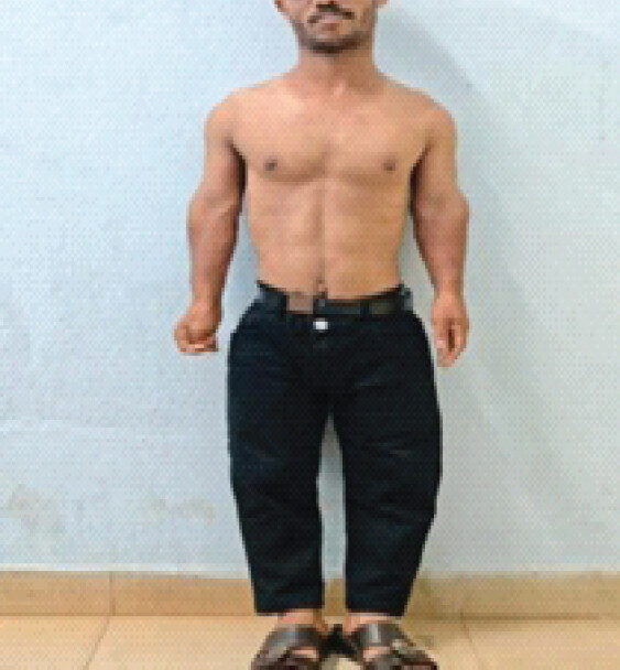
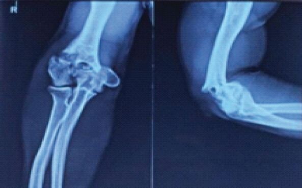
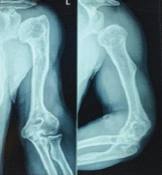
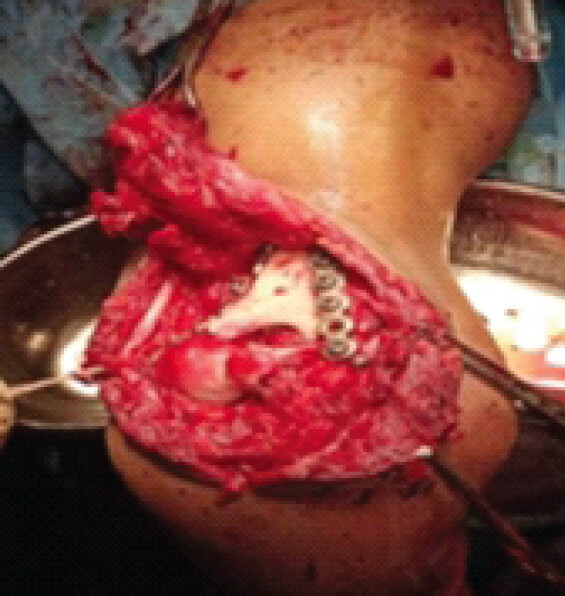
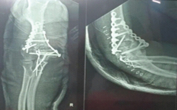
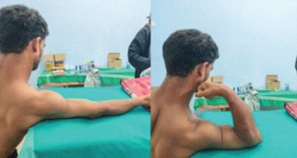
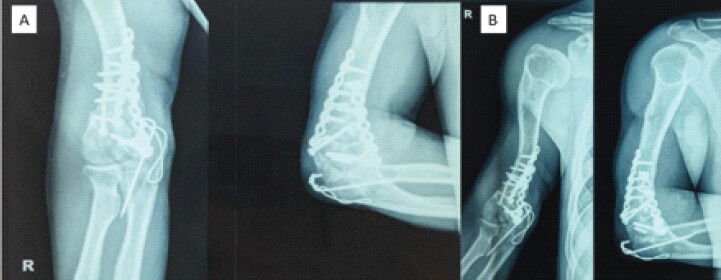
A 28-year-old right-hand-dominant male with achondroplasia sustained a closed, comminuted intra-articular distal humerus fracture (AO/OTA type C3) following a motor vehicle accident. Physical examination revealed rhizomelic limb shortening (Fig. 1), elbow swelling, tenderness, and severely restricted range of motion and no neurovascular deficits. X-ray elbow Anteroposterior and Lateral confirmed articular comminution and metaphyseal involvement (Fig. 2), consistent with the characteristic distal humeral deformities seen in achondroplasia [1, 2, 3] (Fig. 3). Under general anesthesia, a posterior approach via chevron olecranon osteotomy was utilized. Key steps included identification of the ulnar nerve (Fig. 4) and mobilization to prevent iatrogenic injury. Articular fragments were reduced under direct visualization. Fixation was done with dual 3.5-mm reconstruction plates contoured to match the shortened, angulated distal humerus anatomy (Fig. 4). Orthogonal plating (medial and posterolateral) achieved stable fixation across articular fragments. Locking screws secured the metaphyseal segment, compensating for osteopenic bone, and reduction confirmed with fluoroscopy [1,4,5]. The olecranon osteotomy was repaired with tension-band wiring (Fig. 5). Early Post-operative period showed no neurovascular complications. Mobilization began 72 h postoperatively with passive range of motion exercises. 6-month postoperatively, the patient achieved an acceptable range of motion, 5–110° flexion and extension, 70° pronation/supination (Fig. 6). Mayo Elbow performance score (MEPS): 85 (“good”). Radiographic union achieved at 14 weeks postoperatively (Fig. 7 A&B) [1,5]. Prominent K-wire was removed at 14 weeks due to skin irritation. Potential long-term complications, such as implant prominence and heterotopic ossification, could not be fully assessed due to short follow-up.
Discussion
Anatomic and pathophysiological considerations in achondroplasia
Achondroplasia, caused by Fibroblast growth factor 3 gain-of-function mutations, results in impaired endochondral ossification, metaphyseal flaring, and osteopenia [3,4]. These factors complicate distal humerus fracture fixation due to:
-
Abnormal bone geometry: Shortened limbs with exaggerated metaphyseal curvature necessitate extensive plate contouring [1,6]
-
Reduced bone mineral density: Osteopenia increases risks of screw loosening and cut-out, mitigated here by locking screws [3,7]
-
Concomitant deformities: Pre-existing radial head dislocation (reported in 33% of achondroplastic patients) and elbow contractures may compromise functional outcomes [6,8].
Comparative fixation strategies
While pre-contoured plates are standard for distal humerus fractures, reconstruction plates offer critical advantages in achondroplasia:
-
Adaptability: Superior contouring capacity accommodates metaphyseal angulation and shortened segments [1,6]
-
Biomechanical stability: Dual orthogonal plating resists torsional forces better than unilateral methods in osteopenic bone [1,2]
-
Compatibility with deformity: Unlike monolateral external fixators (used in lengthening), recon plates avoid pin-site complications and permit immediate mobilization [6,8,9]
-
Delayed union: This case’s 14-week consolidation aligns with achondroplastic humerus, where healing indices average 25–32 days/cm (vs. 20 days/cm in non-achondroplastic bone) [6,7,8].
Complication profile
-
Neurovascular injury: Ulnar nerve neuropraxia occurred in 9% of achondroplastic elbow surgeries according to existing literature; proactive mobilization prevented this in our case [1,8]
-
Post-fixation fracture: Reported in 13% of humeral lengthening cases, avoided here through metaphyseal locking screws [6,7]
-
Cubitus valgus: A documented sequela of distal humerus fractures, prevented by anatomical articular reduction [2].
Functional outcomes in context
The achieved MEPS of 85 (“good”) compares favorably with:
-
Lengthening studies: Post-lengthening MEPS averages 80–89 despite elbow stiffness in 30% of cases [6,8], but may not show subtle deficits in strength and endurance
-
Non-achondroplastic fractures: Standard fixation yields MEPS >85 in 70–80% of cases, suggesting comparable efficacy when accommodating dysplasia [1,2].
Objective muscle strength measurements using dynamometry were not performed during follow-up as the patient was back to his work and activities of daily living, and to avoid unnecessary expenses for the patient. Advanced post-operative imaging, such as computed tomography scan, was not utilized to confirm articular congruity and union, and rehabilitation compliance and physiotherapy intensity were not objectively quantified in detail for the same reason.
Technical recommendations
-
Nerve management: Routine ulnar nerve transposition reduces neuropathy risk [1,8]
-
Osteosynthesis enhancement: Locking screws in metaphyseal segments and supplemental bone grafting for comminuted zones [10,11]
-
Rehabilitation protocol: Hinged bracing (0°–30° initially) prevents stiffness without compromising stability [1,6].
The presence of an underlying achondroplastic bone deformity may have influenced surgical complexity. Pre-existing elbow deformities and altered biomechanics associated with achondroplasia may act as confounding factors in post-operative functional recovery. The use of reconstruction plates required extensive intraoperative contouring, which may reduce reproducibility across surgeons with varying experience.
Conclusion
Reconstruction plating provides anatomically adaptable fixation for complex distal humerus fractures in achondroplasia, balancing biomechanical demands with pathological bone constraints. This report represents a single case, limiting the generalizability of the findings to the broader achondroplastic population. The rarity of distal humerus fractures in patients with achondroplasia restricts opportunities for larger cohort analysis and statistical validation. The follow-up duration of 6 months may not adequately assess long-term functional outcomes, implant survival, or the development of post-traumatic arthritis. This case demonstrates that meticulous technique and implant selection can achieve functional outcomes comparable to non-dysplastic populations, though longer healing times should be anticipated. Future studies should establish standardized protocols for fracture management in skeletal dysplasias.
Clinical Message
Accurate anatomical reconstruction and fixation with specially contoured dual reconstruction plates provide positive outcomes for distal humerus fractures in adults with achondroplasia, mitigating challenges of dysplastic bone architecture and density.
Conflict of Interest:
Nil
Source of Support:
Nil
Consent:
The authors confirm that informed consent was obtained from the patient for publication of this article
References
- Horton WA, Hall JG, Hecht JT. Achondroplasia. Lancet 2007;370:162-72. Google Scholar | PubMed
- Kim SJ, Agashe MV, Song SH, Choi HJ, Lee H, Song HR. Comparison between Upper and lower limb lengthening in patients with achondroplasia: A retrospective study. J Bone Joint Surg Br 2012;94:128-33. Google Scholar | PubMed
- Ain MC, Shirley ED. Spinal manifestations of achondroplasia. J Bone Joint Surg Am 2009;91 Suppl 4:16-25. Google Scholar | PubMed
- Shadi M, Musielak B, Koczewski P, Janusz P. Humeral lengthening in patients with achondroplasia and in patients with post-septic shortening: Comparison of procedure efficiency and safety. Int Orthop 2017;42:419-26. Google Scholar | PubMed
- Sanchez-Sotelo J, Torchia ME, O'Driscoll SW. Complex distal humeral fractures: Internal fixation with a principle-based parallel-plate technique. J Bone Joint Surg Am 2007;89:961-9. Google Scholar | PubMed
- Kim SJ, Balce GC, Agashe MV, Song SH, Song HR. Is bilateral lower limb lengthening appropriate for achondroplasia?: Midterm analysis of the complications and quality of life. Clin Orthop Relat Res 2012;470:616-21. Google Scholar | PubMed
- Paley D. Extensive limb lengthening for achondroplasia and hypochondroplasia. Children (Basel) 2021;8:540. Google Scholar | PubMed
- Venkatesh KP, Modi HN, Devmurari K, Yoon JY, Anupama BR, Song HR. Femoral lengthening in achondroplasia: Magnitude of lengthening in relation to patterns of callus, stiffness of adjacent joints and fracture. J Bone Joint Surg Br 2009;91-B:1612-7. Google Scholar | PubMed
- Mishima K, Kitoh H, Iwata K, Matsushita M, Nishida Y, Hattori T. Clinical results and complications of lower limb lengthening for fibular Hemimelia: A report of eight cases. Medicine (Baltimore) 2016;95:e3787. Google Scholar | PubMed
- Nashi N, Kagda FH. Current concepts of bone grafting in trauma surgery. J Clin Orthop Trauma 2023;43:102231. Google Scholar | PubMed
- Grzeskowiak RM, Freeman LR, Harper DP, Anderson DE, Mulon P. Effect of cyclic loading on the stability of screws placed in the locking plates used to bridge segmental bone defects. J Orthop Res 2020;39:516-24. Google Scholar | PubMed
© 2026 Journal of Orthopaedic Case Reports - Published by Indian Orthopaedic Research Group
About the Authors
-

Anand Raj Rajendrababu
-

Arun Nirmal
-

Harikumar Kurumalikkal Sukumaran
-

Anjali Sudha Vijay
Related Articles in Journal of Orthopaedic Case Reports
 August 1, 2026 A Bilobed Lipoma Presenting in the Post-Excision Space of the Radial Head 5 Years after Radial Head Excision: A Case Report
 August 1, 2026 Dual Fixation in Complex Long Bone Fractures: A Case Series of 30 Patients
 June 1, 2026 Role of Tranexamic Acid in Orthopedic Trauma: Current Evidence, Clinical Applications, and Ongoing Controversies
 May 1, 2026 Effect of Intrawound Vancomycin Powder as an Adjunct to SystemicAntibiotic Prophylaxis on Surgical Site Infection Following Open TibialFracture Fixation: A Randomized Prospective Study