

Titanium and cobalt-chromium rods provide comparable deformity correction and complication rates in adolescent idiopathic scoliosis; therefore, rod selection should be individualized according to curve characteristics, surgical requirements, and surgeon preference.
Dr. Sitanshu Barik, Department of Orthopedics, All India Institute of Medical Sciences, Nagpur, Maharashtra, India. E-mail: sitanshubarik@gmail.com
Abstract
Introduction: As biomaterial technology advances, rods made of cobalt-chromium appear to be an option for correcting deformities in adolescent idiopathic scoliosis (AIS). This study aimed to compare the efficacy of titanium and cobalt-chrome rods in correcting the deformity and to compare the clinical outcomes between the two groups.
Methods: Two independent reviewers performed a literature search using PubMed, EMBASE, Scopus, and Google Scholar databases. The search focused on identifying articles that compared the surgical treatment of AIS using titanium and cobalt-chromium rods.
Results: Six studies were included for qualitative and quantitative analysis, along with interpretation, of which one was a randomized controlled trial, while the rest of the five were cohort studies. Three hundred thirty-two subjects were part of the studies. There was no significant difference noted in either Cobb’s angle of the primary curve at the final follow-up (standardized mean difference [SMD] – 0.14, 95% confidence interval [CI] (−0.23)–0.51) or the correction rate (SMD – (−0.01), 95% CI (−0.67)–0.36). A trend toward lower final kyphosis was noted in the titanium group, although it was statistically not significant (SMD – (−0.21), 95% CI (−0.43)–0.01).
Conclusion: No significant difference in outcomes or complication rates was noted following the use of either the titanium or cobalt-chromium rods. The advantages and disadvantages of both of these biomaterials should be considered before routine usage in AIS surgeries. Further studies with high power and validity using randomization, blinding, and higher sample size, preferably multicentric with longer follow-ups, are recommended. Level of Evidence – III
Keywords: Adolescent idiopathic scoliosis, titanium, cobalt-chromium, Cobb’s angle.

Adolescent idiopathic scoliosis (AIS) is a three-dimensional spine deformity with an estimated prevalence of 0.5–5.2% [1]. When the AIS deformity exceeds 50° in growing children, surgical intervention generally warrants correcting the deformity and preventing further progression [2,3]. Surgery in AIS aims to achieve tri-planar deformity correction and a balanced spine over the pelvis [4]. The correction of deformity depends on many factors, such as curve flexibility and pattern, the type of anchors used (pedicle screws, hooks), the type of rods used, and the technique used for correction [4]. While the curve patterns and flexibility are surgeon-independent, the spine anchors and the rods used are surgeon-dependent. The pedicle screw and rod system have stood the test of time in correcting the deformity [5]. The design of the pedicle screws has mostly remained unchanged.
In contrast, the material used in the rods for the deformity correction has changed from stainless steel to titanium and, recently, cobalt-chrome (CC) rods [6]. Titanium rods (TR) have the advantages of biocompatibility, inert nature, more flexibility, less corrosion, and fewer radiologic artifacts, while the inherent flexibility of these rods is the disadvantage [7,8]. On the other hand, the CC has similar advantages to TR rods except that they are stiffer than the TR rods [6]. The evidence from the literature provides mixed conclusions regarding outcomes after use of TR and CC rods [9,10,11,12]. However, the existing literature presents a conflicting picture. While some studies suggest that stiffer CC rods provide superior mechanical advantages and potentially better correction, others report equivalent clinical and radiographic outcomes between CC and TR alloys. While the TR rods get weakened during intraoperative corrective forces due to pliability, the CC rods exert significant force on the screw rod interface, which may lead to screw pull-out [8]. The correction rates with cobalt chrome rods are also reported to be better when compared to TI and stainless steel rods [11].
These contradictory findings from individual, often underpowered studies highlight the need for a higher level of evidence. A systematic review and meta-analysis that quantitatively synthesizes the available data is crucial to resolve this ambiguity. By pooling the results of all comparative studies, this review aims to provide a definitive conclusion on whether CC rods offer a significant advantage over TR rods in achieving deformity correction in AIS surgery, thereby offering evidence-based guidance to spinal deformity surgeons.
Literature search
Two independent reviewers (SI and SB) performed a comprehensive yet exhaustive literature search using PubMed, EMBASE, Scopus, and Google Scholar databases. The study protocol was registered in PROSPERO (CRD42023387740). There were no protocol changes between registration and completion of the manuscript. The search focused on identifying the articles that compared the surgical treatment of AIS using TR and CC rods. The articles published between January 2000 and November 2023 were scanned for relevance to our study. Medical subject heading (MeSH) words used for the literature search were “scoliosis” and “Scoliosis/surgery,” and non-MeSH headings used were “adolescent idiopathic scoliosis,” “TI rods,” “CC rods,” “Lenke curves” and “deformity.” The keywords were used in various combinations with Boolean operators to retrieve the maximum possible relevant results. Unpublished data were excluded. Conflicts between reviewers were resolved by mutual discussion, and the final decision in case of sustained conflict was taken by the senior research member in the team (VK). Duplicates were eliminated by the authors using Zotero’s deduplication function, followed by manual elimination after merging all the references.
Study selection
The PICOS format was used to define the inclusion criteria for study selection. The following strategy was used: Patients – The studies describe AIS patients treated with TR and CC rods, , Outcomes – studies reporting the effectiveness of TR and CC rods in the maintenance of deformity correction, effect on kyphosis and lordosis, number of segments fused, implant fractures, screw pull-out, and corrosion were included. Study design – randomized controlled trials (RCTs) or case–control studies comparing the two rods were included. Biomechanical studies (TR vs. CC). Exclusion criteria – Studies that compared the rods in adult scoliosis, Single-arm studies that do not compare TR and CC rods, Case reports, and reviews were excluded.
Quality assessment
Two independent reviewers assessed the quality of the included studies using the NIH quality assessment tool [13]. Two tools, namely, one for observational and cohort studies and a second for RCTs, were used. Both tools contain 14 questions that need to be answered concerning the studies. They may be answered as “yes,” “no,” “cannot determine,” “not reported,” and “not applicable.” Any reviewer dispute was settled after discussing with the senior researcher (VK). The detailed quality assessment is elaborated in a table.
Data extraction
The data were extracted after two authors analyzed the selected articles (SI, SB). The data extracted included the type of study, demographic data, and outcomes. The study characteristics included the author’s name, title, study design, information on the type of curve (Lenke), journal, and year of publication, evidence level, and study quality. The extracted demographic data included the number of patients, mean age, and the number of males and females. Curve characteristics such as pre-and post-operative Cobb’s angle of major and minor curves, correction percentages, number of segments fused, SRS-22 score with its components, and complications like rod fractures, corrosion, and screw pull-out constituted the measured outcome variables. Once the data were extracted, the studies were classified into two groups for analysis – (1) with TR and (2) with CC rod (CR). In the case of data being incomplete or unclear, the study’s corresponding author was contacted for additional information.
Statistical analysis
Mean, standard deviation, and range were used to describe the data collected. Open Meta Analyst (Brown University, USA version 13) with a random-effect model was used for meta-analysis. Comparative studies included in the review were assessed using a formal meta-analysis using the Der Simonian and Laird method, and the outcome was reported as a standardized mean difference (SMD). The correction of coronal Cobb angle, pelvic obliquity, operative time, and blood loss was used for comparison. The I2 statistic was used to assess the heterogeneity. A p-value of < 0.05 was considered statistically significant for the overall effect of the Z test. Low, moderate, and high heterogeneity levels were denoted as I2 values of 25%, 50%, and 75%, respectively. A sensitivity analysis of the meta-analysis was done to identify any study contributing to heterogeneity. A narrative review of the data that cannot be statistically analyzed was done.
Literature search
The initial search of all the databases yielded 1069 results, additional records through cross references and citation searching yielded 35 more results (Fig. 1). After removing the duplicate studies, an initial abstract and title screening were performed for 684 studies. Six hundred and fifty-two articles were excluded as they were not defining the aim of the study after initial title and abstract screening. The full text of 32 studies was analyzed. Single arm studies (n = 13), biomechanical studies (n = 6), adult deformity studies (n = 2), and other reasons (n = 4) were excluded. Finally, six studies were included for qualitative and quantitative analysis [8,10,11,14,15 16].
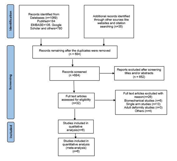
Figure 1: Preferred reporting items for systematic reviews and meta-analyses flowchart depicting the study methodology.
Study characteristics
Of the six studies, one was an RCT, and five were cohort studies [8,10,11,14,16]. Among the cohort studies, three were retrospective [10,11,14]. The lone randomized trial followed the principles of randomization and blinding and had an adequate sample size with 80% power [15]. None of the cohort studies provided any sample size calculation or power description (Table 1). All the forest plots showed high heterogeneity (I2 – 62.4% to 91.7%) except for the one for final kyphosis (I2 – 0%, P = 0.02). Sensitivity analysis showed that all the studies contributed to high heterogeneity.
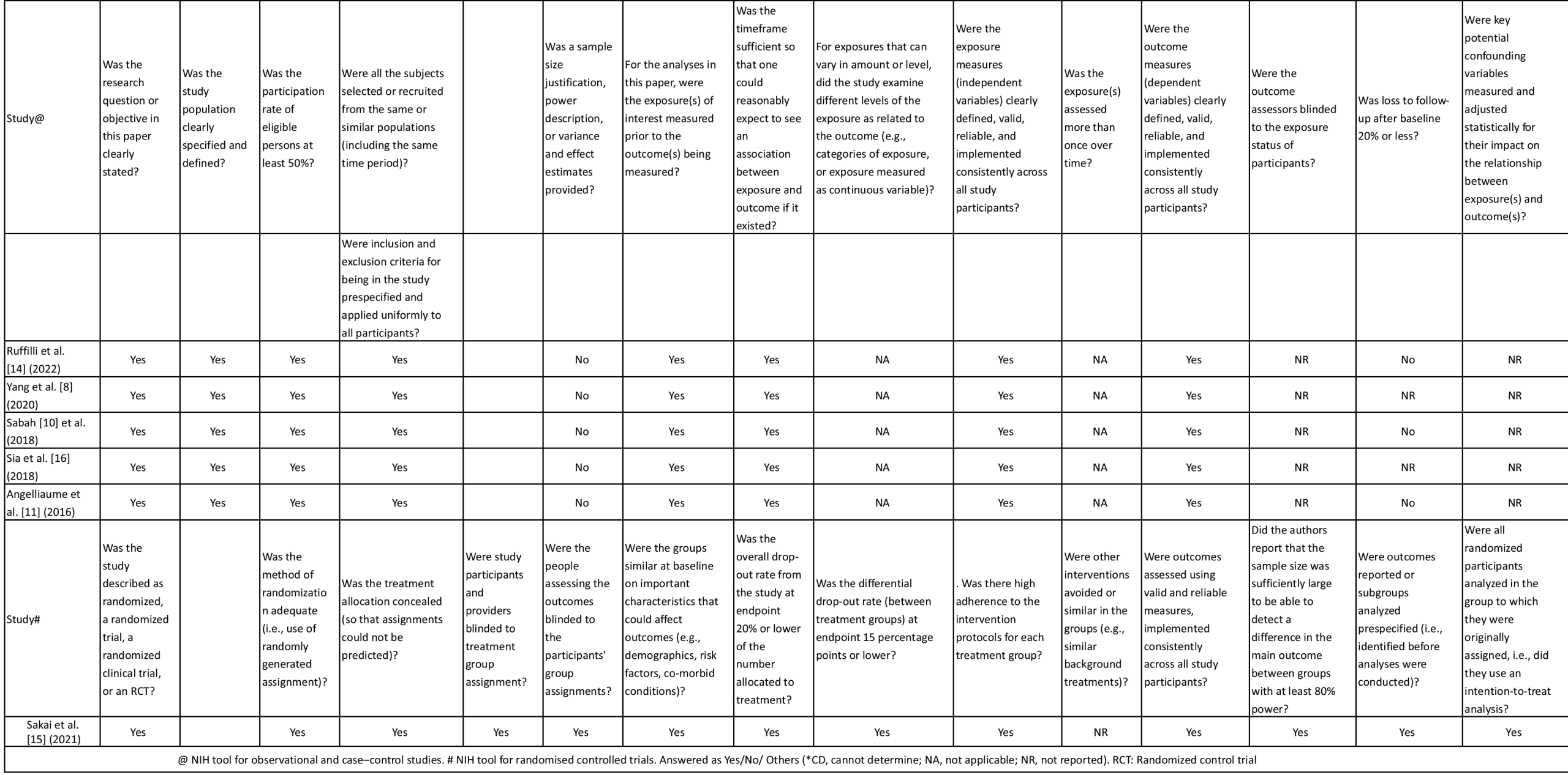
Table 1: Quality assessment of the studies included using NIH tool
Demographic data
The study population was broadly divided into two groups, namely TR and CC (CC). Three hundred thirty-two subjects were included in the six studies (TR – 178, CC – 154) (Table 2). No significant difference was observed in the age groups of both groups (TR – 15 mean, CC – 14.9 mean, SMD – 0.19). There was no significant difference in the gender distribution in both groups (Males – 21/131, 14.3%). Lenke Type 1 curve was the most common curve type in the studies. There was no significant difference in the follow-up period in either group (SMD – 0.14, 95% CI (−0.16)–0.45). Similarly, there was no significant difference in the number of fused levels in both groups (SMD – (−0.32), 95%CI (−1)–0.44).
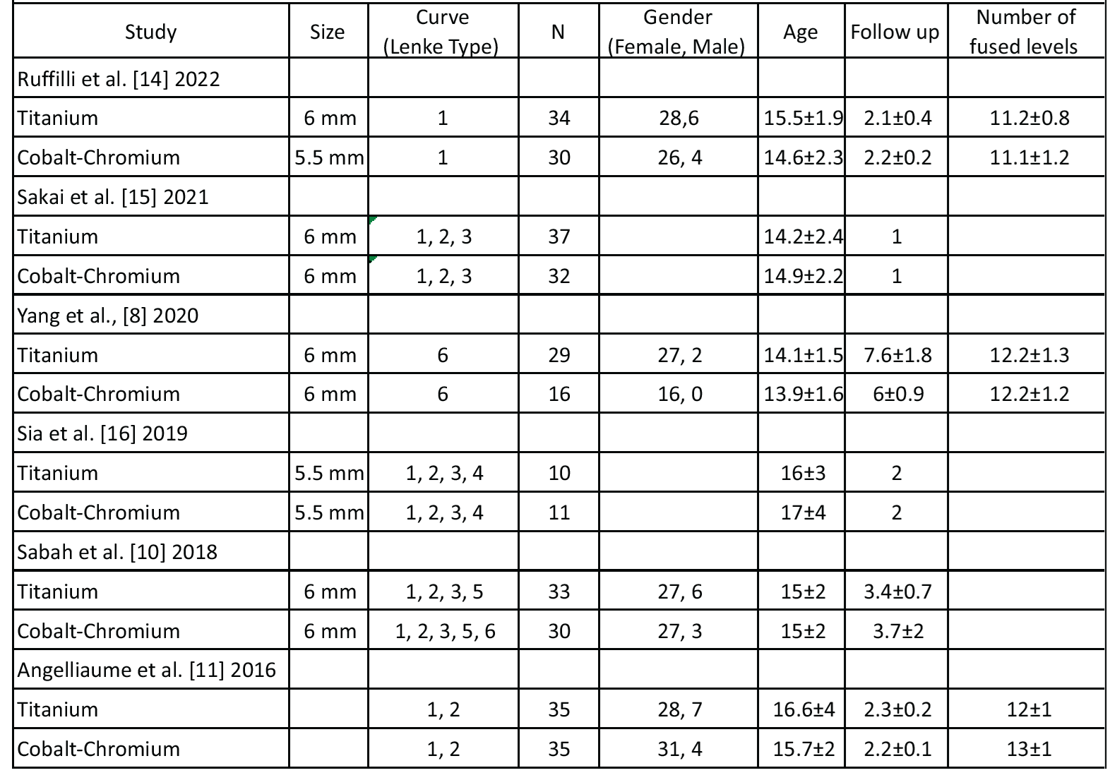
Table 2: Demographic data of the studies included in the review
Outcomes
There was no significant difference in the pre-operative Cobb’s angle of the primary curve in both groups (SMD – 0.18, 95% CI (−0.09)–0.33) (Table 3). There was no significant difference noted in either Cobb’s angle of the primary curve at the final follow-up (SMD – 0.14, 95% CI (−0.23)–0.51) or the correction rate (SMD – (−0.01), 95% CI (−0.67)–0.36) (Fig. 2a and b). A trend toward lower final kyphosis was noted in the TR group, although it was statistically not significant (SMD – (−0.21), 95% CI (−0.43)–0.01) (Fig. 2c).
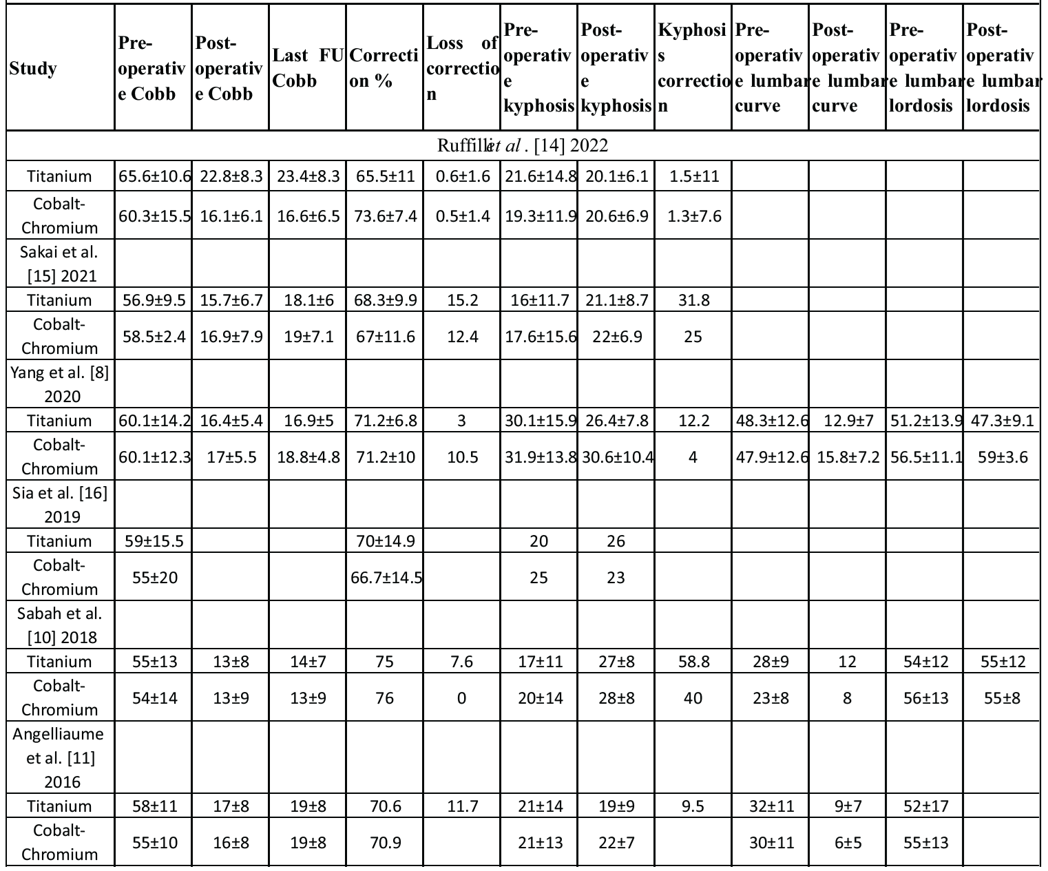
Table 3: Surgical outcomes in the two groups analysed in this review
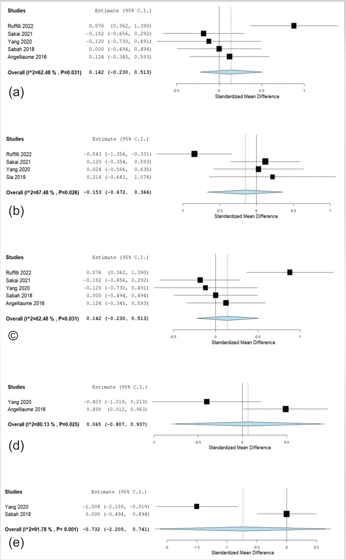
Figure 2: (a) Forest plot depicting the comparison of results in both groups concerning final primary curve, (b) correction rate, (c) final kyphosis, (d) lumbar curve, and (e) lumbar lordosis.
Sakai et al., reported the blood loss (TR – 791 mL, CC – 735 mL) and surgery time (TR – 351.6 min, CC – 342 min) [15]. Two studies noted the final correction in the lumbar curve, which was not statistically significant between both the groups (SMD – 0.06, 95% CI (−0.08)–0.93) (Fig. 2d) [8,11]. Yang et al., and Sabah et al., noted the correction in lumbar lordosis, which was statistically not significant among both the groups (SMD – (−0.73), 95% CI (−2.2)–0.74) (Fig. 2e) [8,10].
Complications
TI group
Yang et al., reported three early complications of pulmonary origin (n = 2) and wound dehiscence (n = 1), and three late complications of delayed infection (n = 1), metal breakage (n = 1), and curve decompensation (n = 1) [8]. Sabah et al., and Angelliaume et al., noted two cases each of proximal junctional kyphosis [10,11].
CC group
Yang et al., reported two early complications of pulmonary origin (n = 1) and wound dehiscence (n = 1) [8]. Sabah et al., reported one case of proximal junctional kyphosis and infection [12]. Angelliaume et al., [10] noted two proximal junctional kyphosis [11] cases.
The challenge in AIS deformity correction is to obtain and secure a balanced spine with stable fusion, which brings into play the role of rods because, apart from the initial correction, they provide stability until fusion [17]. The commonly used biomaterials in AIS as rods are TR alloy and CC alloy.
With the availability of both TR alloy and CC alloy, it is the surgeon’s choice to use the implant that works best for them. When higher corrective forces are anticipated in a patient, CC alloy rods would be the choice as TR rods are 5 times less stiffer than CC alloy rods [8]. Further, in dysplastic spine deformities, the chances of pull-out are high due to narrow and dysplastic pedicles. The use of CC alloy rods in such cases may cause the pullout of screws on the table, and surgeons may choose alloys with lower Young’s modulus, like TI alloys. Many studies in the literature conclude similar findings that thicker rods and CC alloy rods effect better correction [8,18]. The role of restoration of the sagittal profile in post-operative well-being cannot be underestimated. Few studies like Urbanski et al., and Ruffilli et al., have shown a better improvement in thoracic sagittal profile with TI alloy rods compared to stiffer rods [14,19]. Sabah et al., hypothesized that before fusion of the instrumented curves, there may be an elastic pullback of the less stiffer rods, leading to increased kyphosis, which alters the sagittal balance [10]. The results from our meta-analysis also state that the correction rates are similar between CC and TR. To summarize, the factors influencing deformity surgeons to choose CC alloy rods are severe and rigid curves, whereas the factors like more petite curves, less severe deformities, dysplastic curves, and better thoracic sagittal alignment influence the surgeons to choose TR.
The rod derotation maneuver is commonly used to correct the deformity in AIS [20]. It is done by using pre-contoured rods to the concave and convex sides of the curve, followed by rotation of the rods in a counterclockwise direction. The more the rod stiffness, the more its ability to hold the correction after the surgery until the fusion is complete. However, few in vitro studies like Serhan et al., on biomechanical forces required for correction have proved the superiority of corrective forces exerted by CC alloy rods [6]. Intraoperatively, TR is easy to apply because of their flexibility. The elasticity of TR decreases the efficiency of in-situ bending during the correction. Postoperatively, they have been associated with better imaging and low infection rates compared to stainless steel [21]. CC rods come with the advantages of TR and some mechanical advantages of stainless steel, which have been confirmed in vitro. They provide 42% better correction forces than TR rods in vitro [6]. The first in vivo study of a CC rod was compared with a stainless steel rod of the same diameter, and it showed an improved frontal correction rate and stability [12].
Besides the coronal correction, the CC rod provides better sagittal correction by posteromedial translation [11]. Due to its stiffness, better correction in the sagittal profile can be obtained without lamina breakage while using sublaminar bands. This review shows more hypokyphosis in the TR as compared to the CC group. Though the hypokyphosis is more in the TR group, which the pliable nature of TI may explain, the difference in resultant kyphosis between the two groups did not affect the ultimate deformity correction and patient outcomes. The use of multiple in a stiffer curve can lead to a better correction of the sagittal profile as there is more effective force transfer from the rod to the bone via the screws. Another factor influencing the sagittal correction is the interplay of thoracic kyphosis and lumbar lordosis. The stiffer rod can change lumbar lordosis more effectively, as the lumbar segment is more flexible when compared to the thoracic segment [8]. However, this change in lumbar lordosis also affects post-operative thoracic kyphosis.
The increased stiffness of the CC rods comes with a few disadvantages, along with the advantages mentioned above. There is always a concern about screw pull-out during maneuvering due to the stiffness of cobalt chrome rods. If not for the frank screw pull-out, micro-loosening at the screw-bone interface takes place during the correction maneuver. To prevent this complication, a few authors recommend an increased implant density when CC rods are used. As TR is more flexible than CC and is stronger than the purchasing power of the screws, it can provide the same, if not better, correction compared to CC rods [8]. Theoretically, TR rods being more flexible should have a lower rate of screw pull-out, but in practice, this risk of complication may be overrated, as noted in this review, with no incidence of screw pull-out reported in both the study groups.
Although there are subtle differences between the two types of rod used in the various studies, the difference was not big enough to cause a significant difference in correction or complication rates. This lack of discernible difference may be explained by the influence of different variables in the deformity correction surgery, like the hold of pedicle screws, the morphology of pedicles, the amount of soft tissue and bony releases, the type of correction maneuvers, etc. The actual difference due to the biomaterial of the rod may be slight in the presence of the above-described factors.
This is the first analytical review that compared the surgical outcomes of deformity correction in AIS using TR and CC rods. Only comparative studies were considered in this review to minimize the heterogeneity. About its limitations, all the studies were limited by their small sample size, thus reducing the power of the study. The types of curves included, sizes of the rods used, and follow-up period were heterogeneous across the groups, which might explain the high I2 values noted in this analysis. Sub-group analysis could not be performed due to the lesser number of studies. The studies included did not provide information on the usage of polyaxial or monoaxial screws.
This meta-analysis found no high-quality evidence to support the superiority of CC rods over TR in achieving major coronal or sagittal correction in AIS surgery. The choice of rod material may be less critical than surgical technique and patient-specific factors. Surgeons can therefore select rod material based on other criteria, such as cost, familiarity, or desired intraoperative flexibility, without expecting a significant difference in radiographic outcomes. Future high-powered RCTs must control for rod diameter and curve rigidity to definitively answer this question.
Titanium and cobalt-chromium rods achieve comparable radiographic correction and complication rates in adolescent idiopathic scoliosis. Rod selection should therefore be guided by curve rigidity, bone and pedicle morphology, desired intraoperative flexibility, implant characteristics, cost, and surgeon experience rather than an expectation of superior clinical outcomes with either material.
References
- 1. Konieczny MR, Senyurt H, Krauspe R. Epidemiology of adolescent idiopathic scoliosis. J Child Orthop 2013;7:3-9. [Google Scholar] [PubMed]
- 2. Crawford AH, Lykissas MG, Gao X, Eismann E, Anadio J. All-pedicle screw versus hybrid instrumentation in adolescent idiopathic scoliosis surgery: A comparative radiographical study with a minimum 2-Year follow-up. Spine (Phila Pa 1976) 2013;38:1199-208. [Google Scholar] [PubMed]
- 3. Ahuja K, Ifthekar S, Mittal S, Bali SK, Yadav G, Goyal N, et al. Is final fusion necessary for growing-rod graduates: A systematic review and meta-analysis. Glob Spine J 2023;13:209-18. [Google Scholar] [PubMed]
- 4. Ifthekar S, Ahuja K, Sudhakar PV, Mittal S, Yadav G, Kandwal P, et al. Is it safe to save levels and choose the lowest instrumented vertebra as touched vertebra while selectively fusing lenke 1/2 curves? A proportional meta-analysis of existing evidence. Glob Spine J 2023;13:219-26. [Google Scholar] [PubMed]
- 5. Jaquith BP, Chase A, Flinn P, Sawyer JR, Warner WC, Freeman BL, et al. Screws versus hooks: Implant cost and deformity correction in adolescent idiopathic scoliosis. J Child Orthop 2012;6:137-43. [Google Scholar] [PubMed]
- 6. Serhan H, Mhatre D, Newton P, Giorgio P, Sturm P. Would CoCr rods provide better correctional forces than stainless steel or titanium for rigid scoliosis curves? J Spinal Disord Tech 2013;26:E70-4. [Google Scholar] [PubMed]
- 7. Binyamin G, Shafi BM, Mery CM. Biomaterials: A primer for surgeons. Semin Pediatr Surg 2006;15:276-83. [Google Scholar] [PubMed]
- 8. Yang JH, Suh SW, Chang DG. Comparison of surgical correction rates between titanium and cobalt-chrome-alloy as rod materials in adolescent idiopathic scoliosis. Sci Rep 2020;10:10053. [Google Scholar] [PubMed]
- 9. Etemadifar MR, Andalib A, Rahimian A, Nodushan SM. Cobalt chromium-titanium rods versus titanium-titanium rods for treatment of adolescent idiopathic scoliosis; which type of rod has better postoperative outcomes? Rev Assoc Med Bras (1992) 2018;64:1085-90. [Google Scholar] [PubMed]
- 10. Sabah Y, Clément JL, Solla F, Rosello O, Rampal V. Cobalt-chrome and titanium alloy rods provide similar coronal and sagittal correction in adolescent idiopathic scoliosis. Orthop Traumatol Surg Res 2018;104:1073-7. [Google Scholar] [PubMed]
- 11. Angelliaume A, Ferrero E, Mazda K, Le Hanneur M, Accabled F, De Gauzy JS, et al. Titanium vs cobalt chromium: What is the best rod material to enhance adolescent idiopathic scoliosis correction with sublaminar bands? Eur Spine J 2017;26:1732-8. [Google Scholar] [PubMed]
- 12. Lamerain M, Bachy M, Delpont M, Kabbaj R, Mary P, Vialle R. CoCr rods provide better frontal correction of adolescent idiopathic scoliosis treated by all-pedicle screw fixation. Eur Spine J 2014;23:1190-6. [Google Scholar] [PubMed]
- 13. Study Quality Assessment Tools. NHLBI, NIH; (n.d.). Available from: https://www.nhlbi.nih.gov/health-topics/study-quality-assessment-tools [Last accessed on 2023 Jan 13]. [Google Scholar] [PubMed]
- 14. Ruffilli A, Fiore M, Viroli G, Barile F, Manzetti M, Martikos K, et al. 5.5-mm cobalt-chrome vs 6-mm Titanium alloy rods in surgical treatment of lenke 1 adolescent idiopathic scoliosis with high-density pedicle screws and direct vertebral rotation on differently shaped rods: A retrospective comparative cohort study. Int J Spine Surg 2022;17:25-31. [Google Scholar] [PubMed]
- 15. Sakai D, Tanaka M, Takahashi J, Taniguchi Y, Schol J, Hiyama A, et al. Cobalt-chromium versus titanium alloy rods for correction of adolescent idiopathic scoliosis based on 1-year follow-up: A multicenter randomized controlled clinical trial. J Neurosurg Spine 2021;34:897-906. [Google Scholar] [PubMed]
- 16. Sia U, Tan BB, Teo YY, Wong CC. Post-implantation deformation of titanium rod and cobalt chrome rod in adolescent idiopathic scoliosis. Malays Orthop J 2019;13:14-9. [Google Scholar] [PubMed]
- 17. Yagi M, Rahm M, Gaines R, Maziad A, Ross T, Kim HJ, et al. Characterization and surgical outcomes of proximal junctional failure in surgically treated patients with adult spinal deformity. Spine (Phila Pa 1976) 2014;39:E607-14. [Google Scholar] [PubMed]
- 18. Lamartina C, Petruzzi M, Macchia M, Stradiotti P, Zerbi A. Role of rod diameter in comparison between only screws versus hooks and screws in posterior instrumentation of thoracic curve in idiopathic scoliosis. Eur Spine J 2011;20 Suppl 1:S85-9. [Google Scholar] [PubMed]
- 19. Urbanski W, Wolanczyk MJ, Jurasz W, Kulej M, Morasiewicz P, Dragan SL, et al. The impact of direct vertebral rotation (DVR) on radiographic outcome in surgical correction of idiopathic scoliosis. Arch Orthop Trauma Surg 2017;137:879-85. [Google Scholar] [PubMed]
- 20. Weinstein SL, Dolan LA, Cheng JC, Danielsson A, Morcuende JA. Adolescent idiopathic scoliosis. Lancet 2008;371:1527-37. [Google Scholar] [PubMed]
- 21. Glotzbecker MP, Riedel MD, Vitale MG, Matsumoto H, Roye DP, Erickson M, et al. What’s the evidence? Systematic literature review of risk factors and preventive strategies for surgical site infection following pediatric spine surgery. J Pediatr Orthop 2013;33:479-87. [Google Scholar] [PubMed]





Related Articles in Journal of Orthopaedic Case Reports
 April 1, 2026 Total Talus Replacement using a Custom 3D-Printed Titanium Implant for Post-Traumatic Avascular Necrosis of Talus following Failed Internal fixation: A Case Report
 November 1, 2025 Outcome of Pediatric Femoral Shaft Fractures Treated with Titanium Elastic Nailing: A Prospective Study
 October 1, 2025 Alterartions of Cervical Lordosis in Neck Pain Patients
 July 1, 2025 Sequential Rod Rolling for Surgical Correction of Lenke Type 2 Adolescent Idiopathic Scoliosis: A 3D Analysis