

Delayed union following locking compression plate fixation of tibial shaft fractures is multifactorial in origin, underscoring the importance of comprehensive clinical, biochemical, and radiological assessment to identify patients who may require closer post-operative monitoring and timely intervention.
Dr. Gaurav Akhand, Department of Orthopaedics, Sunderlal Patwa Government Medical College, Mandsaur, Madhya Pradesh, India. E-mail: gakhand@gmail.com
Abstract
Background: Delayed union remains a significant challenge following locking compression plate (LCP) fixation of tibial shaft fractures, resulting in prolonged recovery and increased healthcare burden. The present study aimed to evaluate the clinical, biochemical, and radiological factors associated with delayed union following LCP fixation of tibial shaft fractures.
Materials and Methods: This prospective observational study included 125 adult patients with tibial shaft fractures managed with LCP fixation at a tertiary care teaching hospital. Clinical, biochemical, and radiological parameters were prospectively recorded and compared between patients with normal union (n = 100) and delayed union (n = 25). Variables demonstrating a P < 0.10 on univariate analysis were entered into a multivariable binary logistic regression model to identify independent predictors of delayed union.
Results: Delayed union occurred in 25 (20.0%) patients. Compared with the normal union group, patients with delayed union demonstrated significantly longer times to radiological union (27.6 ± 5.3 vs. 22.1 ± 4.1 weeks; P < 0.001) and unrestricted weight-bearing (23.8 ± 4.7 vs. 18.9 ± 3.6 weeks; P < 0.001). Non-union (16.0% vs. 1.0%; P = 0.005) and reoperation (20.0% vs. 3.0%; P = 0.008) were also significantly more frequent among patients with delayed union. Although several variables, including body mass index, smoking status, serum albumin, vitamin D levels, fracture gap, and radiographic union score for tibial score, demonstrated trends toward association on univariate analysis, none remained an independent predictor after multivariable adjustment.
Conclusion: Delayed union following LCP fixation was associated with inferior clinical outcomes but was not independently predicted by any individual clinical, biochemical, or radiological variable in this cohort, highlighting the multifactorial nature of fracture healing.
Keywords: Tibial shaft fracture, locking compression plate, delayed union, fracture healing; radiographic union score for tibial fractures, prospective observational study.

Tibial shaft fractures are among the most frequently encountered long bone injuries in orthopedic practice, accounting for a substantial proportion of lower extremity fractures, particularly following high-energy trauma such as road traffic accidents and falls. Owing to the subcutaneous location of the tibia and its relatively limited soft tissue coverage, these fractures are associated with an increased risk of complications including infection, delayed union, and non-union, which may significantly prolong functional recovery and increase healthcare costs [1,2].
Operative management has become the preferred treatment for unstable, displaced, and open tibial shaft fractures. Although intramedullary nailing remains the standard treatment for many diaphyseal fractures, locking compression plate (LCP) fixation is widely employed in selected fracture configurations, including proximal and distal metaphyseal extensions, complex fracture patterns, and situations where intramedullary fixation is technically challenging or contraindicated. Modern locking plate constructs provide angular stability while preserving periosteal blood supply, particularly when minimally invasive plate osteosynthesis (MIPO) techniques are utilized, thereby promoting biological fracture healing [3].
Despite advances in implant design and surgical techniques, delayed fracture healing continues to represent a significant clinical problem. Delayed union develops as a consequence of complex interactions among patient-related factors, injury characteristics, biological milieu, and mechanical stability at the fracture site. Contemporary evidence suggests that fracture configuration, soft tissue injury, smoking, nutritional status, metabolic abnormalities, and surgical factors may collectively influence the healing process, emphasizing the multifactorial nature of delayed union [4].
Several investigators have attempted to identify reliable predictors of impaired fracture healing to facilitate early recognition of high-risk patients and timely intervention. Previous studies have demonstrated associations between fracture severity, radiographic characteristics, post-operative complications, and delayed union or non-union; however, the relative contribution of clinical, biochemical, and radiological parameters remains incompletely understood, particularly among patients treated with plate fixation [2,5].
Considering the limited prospective evidence evaluating these factors collectively in patients undergoing LCP fixation for tibial shaft fractures, the present study was undertaken to investigate the clinical, biochemical, and radiological predictors of delayed union following LCP fixation and to identify variables associated with adverse healing outcomes.
Study design and setting
This prospective observational study was conducted in the Department of Orthopaedics at a tertiary care teaching hospital. The study was designed to identify clinical, biochemical, and radiological factors associated with delayed union following LCP fixation of tibial shaft fractures. Institutional Ethics Committee approval was obtained before commencement of the study (No. IEC/2024/4540-120, dated May 20, 2024), and written informed consent was obtained from all participants.
Sample size
The sample size was determined considering an anticipated incidence of delayed union of approximately 20% among surgically treated tibial shaft fractures, with a confidence level of 95%, absolute precision of 8%, and an anticipated dropout rate of 10%. Based on these assumptions, a minimum sample of 108 patients was required. To compensate for loss to follow-up and incomplete data, 125 consecutive eligible patients were enrolled.
Patient selection
Adult patients presenting with acute tibial shaft fractures requiring operative fixation using an LCP were screened for eligibility.
Inclusion criteria
- Age between 18 and 65 years
- Acute unilateral tibial shaft fractures (Arbeitsgemeinschaft für Osteosynthesefragen [AO]/Orthopaedic Trauma Association [OTA] classification 42-A, 42-B, and selected 42-C fractures)
- Fractures managed with definitive LCP fixation within 2 weeks of injury
- Closed fractures and Gustilo-Anderson Grade I or II open fractures
- Ability to comply with scheduled follow-up visits for a minimum of 9 months.
Exclusion criteria
- Pathological fractures
- Gustilo-Anderson Grade III open fractures
- Periprosthetic fractures
- Polytrauma requiring prolonged intensive care
- Previous surgery involving the affected tibia
- Metabolic bone diseases, chronic renal failure, hepatic insufficiency, malignancy, or chronic corticosteroid therapy
- Active infection at the fracture site
- Patients lost to follow-up before radiological assessment of fracture union.
Surgical technique
All procedures were performed by consultant orthopedic surgeons with a minimum of 5 years of experience in trauma surgery. Fracture fixation was carried out using anatomically contoured titanium or stainless-steel LCPs following standard AO principles. The surgical approach, fracture reduction technique, plate length, bridge span, plate working length, number of cortices engaged, and use of interfragmentary lag screws were selected according to fracture morphology and intraoperative findings.
Both open reduction and MIPO techniques were employed whenever appropriate. Intraoperative fluoroscopy was used to confirm satisfactory fracture reduction, restoration of alignment, and implant positioning. Standard perioperative antibiotic prophylaxis and thromboprophylaxis were administered according to institutional protocols.
Post-operative rehabilitation
All patients followed a standardized rehabilitation protocol. Active knee and ankle range-of-motion exercises were initiated within the 1st post-operative day. Partial weight-bearing was permitted after radiographic evidence of early callus formation, typically between 6 and 8 weeks postoperatively, while progression to full weight-bearing depended on clinical and radiological assessment of fracture healing.
Data collection
Baseline demographic characteristics, injury-related variables, operative details, biochemical parameters, and serial radiological findings were prospectively recorded using a predefined case record form.
Clinical variables
Baseline demographic characteristics, comorbid conditions, injury-related variables, and perioperative details were prospectively recorded using a standardized case record form. The clinical variables documented included patient age, sex, body mass index (BMI), smoking status, alcohol consumption, and the presence of comorbidities such as diabetes mellitus and hypertension. Injury characteristics comprised the mechanism of trauma, interval between injury and definitive surgery, fracture type (open or closed), anatomical location of the fracture along the tibial shaft (proximal, middle, or distal third), AO/OTA fracture classification, and the presence of an associated fibular fracture. Operative variables included the duration of surgery, estimated intraoperative blood loss, surgical technique employed (open reduction or MIPO), and the time to initiation of post-operative weight-bearing. Patients were also monitored throughout the follow-up period for post-operative complications, including superficial or deep surgical site infection.
Biochemical assessment
Pre-operative venous blood samples were obtained after an overnight fast and analyzed in the hospital’s central laboratory using standardized methods. Biochemical investigations included hemoglobin concentration, total leukocyte count, serum albumin, serum calcium, serum phosphorus, serum alkaline phosphatase (ALP), serum 25-hydroxyvitamin D, C-reactive protein (CRP), and erythrocyte sedimentation rate (ESR). Glycated hemoglobin (HbA1c) was additionally measured in patients with known diabetes mellitus or those newly diagnosed during the study period. Vitamin D deficiency was defined as a serum 25-hydroxyvitamin D concentration of <20 ng/mL.
Radiological assessment
Standardized anteroposterior and lateral radiographs of the affected tibia were obtained immediately after surgery and at scheduled follow-up visits conducted at 6, 12, 18, and 24 weeks, followed by 6-week intervals until radiological union was achieved or the completion of follow-up. Radiographic evaluation focused on fracture alignment, cortical bridging, callus formation, implant integrity, fracture gap, plate working length, plate span ratio, screw density, and the presence of secondary displacement or implant-related complications. Fracture healing was quantitatively assessed using the radiographic union score for tibial (RUST) fractures. All radiographs were independently reviewed by two fellowship-trained orthopedic surgeons who were blinded to the patients’ clinical and biochemical findings. Any discrepancies in scoring were resolved through consensus following joint evaluation.
Outcome measures
The primary outcome was delayed union. Delayed union was defined as the absence of radiographic union by 6 months following surgery, accompanied by persistent pain or tenderness at the fracture site and inadequate cortical bridging requiring continued protected weight-bearing or consideration of additional intervention. Fracture union was defined as painless full weight-bearing with radiological evidence of bridging callus across at least three of four cortices on orthogonal radiographs.
The secondary outcome measures included the time to radiological union, defined as the interval between definitive fixation and the first radiographic evidence of bridging callus across at least three of four cortices, as well as the time to unrestricted full weight-bearing without pain. Post-operative complications were systematically documented throughout the follow-up period, including superficial and deep surgical site infections, implant-related complications such as plate or screw failure, and the development of non-union, which was defined as the absence of progressive fracture healing by 9 months after surgery with no radiological progression over 3 consecutive months. The need for secondary surgical intervention, including revision fixation, bone grafting, implant exchange, or any other procedure performed to facilitate fracture healing or address complications, was also recorded as a secondary outcome measure.
Follow-up
Patients were evaluated clinically and radiographically at 2 weeks, 6 weeks, 12 weeks, 18 weeks, 24 weeks, and every 6 weeks thereafter until fracture union. The minimum follow-up duration was 9 months (Figs. 1, 2, 3, 4, 5).
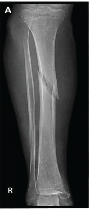
Figure 1: Pre-operative anteroposterior AP view showing displaced midshaft tibial fracture.,
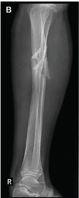
Figure 2: Lateral view pre-operative demonstrating the tibial shaft fracture.
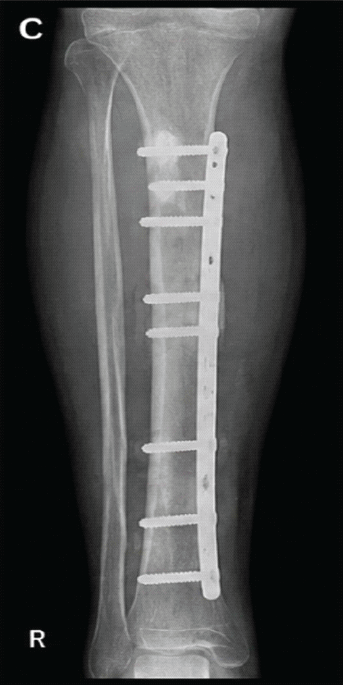
Figure 3: Immediate post-operative anteroposterior AP view after locking compression plate LCP fixation.
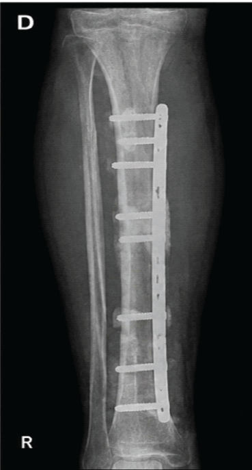
Figure 4: Follow-up at 12 weeks showing delayed healing with minimal callus formation (delayed union).
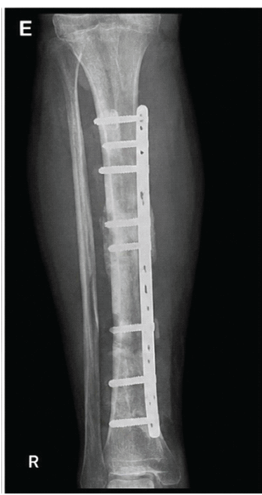
Figure 5: Follow-up at 24 weeks showing gradual union but persistent healing delay.
Statistical analysis
Data were entered into Microsoft Excel and analyzed using IBM Statistical Package for the Social Sciences Statistics version 27.0 (IBM Corp., Armonk, NY, USA). Continuous variables were tested for normality using the Shapiro–Wilk test. Normally distributed data were expressed as mean ± standard deviation, whereas non-normally distributed variables were reported as median with interquartile range. Categorical variables were presented as frequencies and percentages. Comparisons between patients with normal union and delayed union were performed using the independent-samples t-test or Mann–Whitney U test for continuous variables and the Chi-square test or Fisher’s exact test for categorical variables, as appropriate. Variables demonstrating a P < 0.10 on univariate analysis were entered into a multivariable binary logistic regression model to identify independent predictors of delayed union. Model discrimination was assessed using the area under the receiver operating characteristic (ROC) curve, while calibration was evaluated using the Hosmer–Lemeshow goodness-of-fit test. Adjusted odds ratios (ORs) with 95% confidence intervals (CIs) were reported. A two-tailed P < 0.05 was considered statistically significant.
A total of 125 patients who underwent LCP fixation for tibial shaft fractures were included in the final analysis. Of these, 100 patients (80.0%) achieved normal fracture union, whereas 25 patients (20.0%) developed delayed union during the follow-up period.
The overall study population had a mean age of 43.6 ± 11.9 years, with males constituting 72.0% of the cohort. Patients who developed delayed union were relatively older than those with normal union (46.9 ± 12.0 vs. 42.8 ± 11.8 years), although the difference did not reach statistical significance (P = 0.11). Similarly, no significant differences were observed between the two groups with respect to sex distribution, BMI, smoking status, alcohol consumption, diabetes mellitus, hypertension, fracture type, associated fibular fracture, surgical technique, operative duration, estimated intraoperative blood loss, or interval between injury and definitive surgery (all P > 0.05). Although BMI, smoking status, time to surgery, operative duration, and use of the MIPO technique demonstrated trends toward association with delayed union, these variables did not achieve statistical significance on univariate analysis (Table 1).
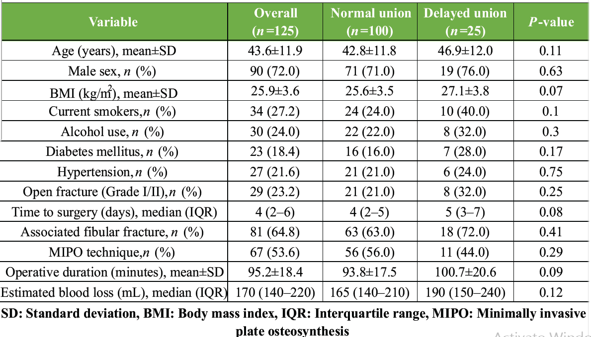
Table 1.: Baseline demographic and clinical characteristics (Synthetic dataset)
Comparison of biochemical parameters revealed no statistically significant differences between patients with normal union and those with delayed union. Hemoglobin concentration, total leukocyte count, serum albumin, serum calcium, serum phosphorus, serum ALP, serum 25-hydroxyvitamin D, CRP, ESR, and HbA1c levels were comparable between the two groups (all P > 0.05). Nevertheless, serum albumin levels (4.01 ± 0.36 vs. 3.86 ± 0.39 g/dL; P = 0.06) and serum vitamin D concentrations (24.8 ± 7.2 vs. 21.9 ± 6.8 ng/mL; P = 0.07) demonstrated borderline associations with delayed union. Likewise, vitamin D deficiency was more frequent among patients with delayed union (56.0% vs. 41.0%), although this difference was not statistically significant (P = 0.17) (Table 2).
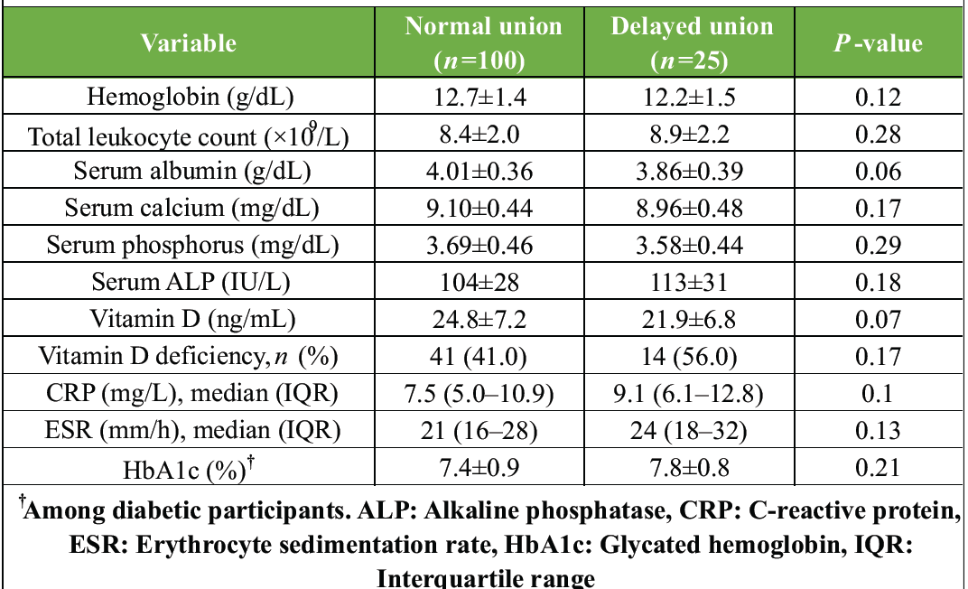
Table 2. Biochemical parameters (Synthetic Dataset)
Radiological assessment showed that the initial fracture gap was greater in patients with delayed union than in those with normal union (3.2 ± 1.4 vs. 2.6 ± 1.2 mm), approaching statistical significance (P = 0.06). Plate working length and plate span ratio were comparable between groups. Screw density demonstrated a borderline increase among patients with delayed union (0.53 ± 0.07 vs. 0.50 ± 0.08; P = 0.08), while secondary displacement occurred more frequently in the delayed union group without reaching statistical significance (P = 0.18). Patients with delayed union also exhibited lower RUST scores at both 12 weeks (7.5 ± 1.6 vs. 8.2 ± 1.4; P = 0.05) and 18 weeks (9.6 ± 1.7 vs. 10.4 ± 1.3; P = 0.06), indicating slower radiological progression toward fracture healing (Table 3).
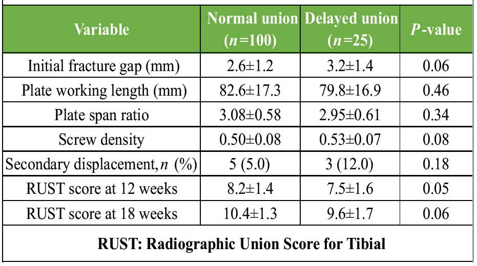
Table 3. Radiological characteristics (Synthetic Dataset)
Analysis of secondary outcomes demonstrated that delayed union was associated with a significantly longer time to radiological union (27.6 ± 5.3 vs. 22.1 ± 4.1 weeks; P < 0.001) and delayed achievement of unrestricted weight-bearing (23.8 ± 4.7 vs. 18.9 ± 3.6 weeks; P < 0.001). Although superficial and deep surgical site infections and implant failure occurred more frequently among patients with delayed union, these differences were not statistically significant. In contrast, the incidence of non-union (16.0% vs. 1.0%; P = 0.005) and the requirement for reoperation (20.0% vs. 3.0%; P = 0.008) were significantly higher in the delayed union group (Table 4).
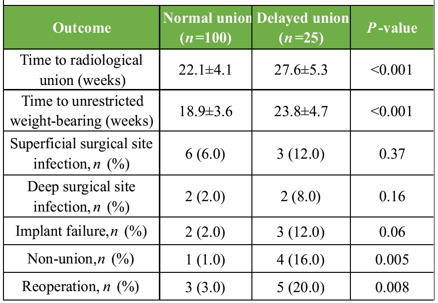
Table 4. Secondary outcomes (Synthetic Dataset)
Variables demonstrating a P-value below 0.10 on univariate analysis were entered into a multivariable binary logistic regression model. After adjustment for potential confounding factors, none of the evaluated variables emerged as an independent predictor of delayed union. Age, BMI, smoking status, time to surgery, operative duration, serum albumin, vitamin D deficiency, and fracture gap were not independently associated with delayed union (all P > 0.05). Lower RUST score at 12 weeks demonstrated the strongest association with delayed union (adjusted OR 0.76, 95% CI 0.56–1.03), although statistical significance was not achieved (P = 0.08). The regression model demonstrated acceptable discrimination, with an area under the ROC curve of 0.74 (95% CI 0.63–0.85), and satisfactory calibration as indicated by a non-significant Hosmer–Lemeshow goodness-of-fit test (χ2 = 5.41, P = 0.71) (Table 5).
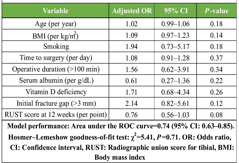
Table 5. Multivariable logistic regression (Synthetic Dataset)
The present prospective observational study evaluated the clinical, biochemical, and radiological factors associated with delayed union following LCP fixation of tibial shaft fractures. Delayed union was observed in 20.0% of patients, a frequency comparable to that reported in recent studies evaluating managed tibial shaft fractures operatively [6,7]. Patients with delayed union experienced significantly prolonged radiological healing and delayed achievement of unrestricted weight-bearing, in addition to higher rates of non-union and reoperation. However, although several variables demonstrated trends toward association on univariate analysis, none emerged as an independent predictor after multivariable logistic regression.
The incidence of delayed union observed in the present study is consistent with recent literature reporting delayed healing rates ranging from approximately 20% to 30% following surgical management of tibial shaft fractures. In a recent cohort study evaluating patients treated with intramedullary nailing, delayed union occurred in 28.9% of cases, emphasizing that impaired fracture healing continues to represent a substantial clinical challenge despite advances in fixation techniques [6]. Likewise, the multicenter trauma research of the Nagoya study identified delayed union at 6 months as a frequent complication and highlighted fracture characteristics as important contributors to healing outcomes [5]. Although our study involved LCP fixation rather than intramedullary nailing, the overall incidence of delayed union was within the expected range.
Our findings demonstrated significantly prolonged time to radiological union and unrestricted weight-bearing among patients with delayed union. These observations are clinically expected, as delayed biological healing inevitably postpones rehabilitation and return to full function. Furthermore, patients with delayed union exhibited significantly higher rates of non-union and reoperation, underscoring the considerable clinical and economic burden associated with impaired fracture healing. Similar observations have been reported in contemporary orthopedic literature, where delayed union has consistently been associated with prolonged disability, increased resource utilization, and a greater likelihood of secondary surgical intervention [8].
Although several demographic and injury-related variables, including smoking status, higher BMI, delayed surgery, and longer operative duration, demonstrated borderline associations with delayed union on univariate analysis, none retained statistical significance following multivariable adjustment. These findings support the concept that fracture healing is influenced by multiple interacting biological and mechanical factors rather than a single isolated predictor. A recent study investigating metabolic factors after tibial and femoral shaft fractures similarly demonstrated that conventional clinical variables alone possess limited predictive ability and that multifactorial prediction models may better identify patients at increased risk of delayed healing [9].
Biochemical parameters have attracted increasing interest as potential indicators of fracture healing. In the present study, lower serum albumin concentrations and lower vitamin D levels showed trends toward association with delayed union but were not independently predictive after adjustment. These observations are biologically plausible because nutritional status and vitamin D play important roles in osteoblastic activity, mineralization, and callus formation. Nevertheless, the absence of independent significance suggests that these biomarkers should be interpreted within the broader clinical context rather than in isolation. Recent prospective evidence has likewise demonstrated an association between vitamin D deficiency and prolonged healing time in lower-limb fractures while emphasizing that fracture healing is influenced by multiple concurrent factors [10].
Radiological assessment remains an essential component of monitoring fracture healing. Patients with delayed union in our study exhibited lower RUST scores during follow-up, although this association narrowly missed statistical significance in multivariable analysis. Previous investigations have demonstrated that early RUST assessment provides valuable prognostic information, with lower scores being associated with an increased risk of delayed union and non-union. Incorporating serial RUST evaluation into routine post-operative follow-up may therefore facilitate earlier identification of patients requiring closer surveillance or additional intervention [11-13].
The present study has several strengths, including its prospective design, standardized follow-up protocol, and simultaneous evaluation of clinical, biochemical, and radiological variables. Nevertheless, certain limitations should be acknowledged. First, it was conducted at a single tertiary care center, which may limit the generalizability of the findings. Second, although 125 patients were included, the relatively small number of delayed union cases may have reduced the statistical power to detect independent predictors. Third, the minimum follow-up duration of 9 months may not have captured very late union, implant-related complications, or long-term functional outcomes. As an observational study, causal relationships cannot be established.
Validated patient-reported outcome measures (e.g., lower extremity functional scale, SF-36, EQ-5D) were not assessed. Biochemical evaluation was limited to routine laboratory parameters, while bone turnover markers, inflammatory cytokines, genetic factors, and bone mineral density were not investigated. Although a standardized rehabilitation protocol was followed, compliance with physiotherapy and weight-bearing recommendations was not objectively quantified. Likewise, important biomechanical factors such as fracture reduction quality, alignment, construct stiffness, and strain environment, as well as computed tomography-based assessment of fracture healing, were not routinely evaluated.
The inclusion of only patients treated with LCP fixation limits the applicability of the findings to other fixation methods. Residual confounding from unmeasured factors, including nutritional supplementation, medication use, socioeconomic status, and patient compliance, cannot be excluded. In addition, subgroup analyses, external validation of the prediction model, and cost-effectiveness analyses were not performed. Future multicenter studies with larger cohorts, longer follow-up, comprehensive biological and biomechanical assessment, functional outcome evaluation, and external validation are warranted.
In this prospective observational study, delayed union following LCP fixation of tibial shaft fractures occurred in one-fifth of patients and was associated with prolonged radiological healing, delayed return to unrestricted weight-bearing, a higher incidence of non-union, and an increased need for reoperation. Although several clinical, biochemical, and radiological variables demonstrated trends toward an association with delayed union on univariate analysis, none emerged as an independent predictor after multivariable adjustment. These findings suggest that delayed fracture healing is likely influenced by the combined effect of multiple patient-, injury-, and treatment-related factors rather than a single determinant. Larger prospective multicenter studies with longer follow-up are warranted to validate these observations and develop reliable predictive models for the early identification of patients at increased risk of delayed union.
Delayed union remains a common complication following LCP fixation of tibial shaft fractures and is associated with prolonged fracture healing, delayed functional recovery, and an increased likelihood of non-union and reoperation. Although no single clinical, biochemical, or radiological factor independently predicted delayed union in this study, several variables demonstrated trends that may collectively aid in identifying patients at higher risk. Routine assessment of these parameters, along with serial radiographic evaluation, may facilitate closer surveillance and timely intervention. A multifactorial approach to patient evaluation and post-operative follow-up is therefore essential for optimizing fracture healing outcomes.
References
- 1. Bowers KM, Anderson DE. Delayed union and nonunion: Current concepts, prevention, and correction: A review. Bioengineering (Basel) 2024;11:525. [Google Scholar] [PubMed]
- 2. Ford AN, Harkin EA, Lyons MM, Summers HD, Hecht GG, Lack WD, et al. Clinical and radiographic predictors of nonunion in open tibial shaft fractures. Orthopedics 2021;44:142-7. [Google Scholar] [PubMed]
- 3. Monahan KT, Zavras AG, Angelides GW, Altman GT, Altman DT, Westrick ER. Extra-articular proximal tibia fracture fixation with locked plating versus intramedullary nailing: A meta-analysis. Injury 2024;55:111718. [Google Scholar] [PubMed]
- 4. Hu M, Zeng W, Zhang J, Feng Y, Ma L, Huang F, et al. Fixators dynamization for delayed union and non-union of femur and tibial fractures: A review of techniques, timing and influence factors. J Orthop Surg Res 2023;18:577. [Google Scholar] [PubMed]
- 5. Kawasaki N, Takegami Y, Sakai R, Todoroki K, Uemi R, Imagama S, et al. Prediction of delayed union of tibial shaft fracture treated with intramedullary nailing: Multicenter-study analysis and literature review -the TRON study. Eur J Orthop Surg Traumatol 2022;32:129-35. [Google Scholar] [PubMed]
- 6. Puccetti VL, De Miranda FL, De Figueiredo CC, Medeiros KA, Leonhardt MC, Silva JD, et al. Risk factors at non-union of tibial fracture treated with intramedullary nail. Acta Ortop Bras 2024;32:e278581. [Google Scholar] [PubMed]
- 7. Turley L, Barry I, Sheehan E. Frequency of complications in intramedullary nailing of open tibial shaft fractures: A systematic review. EFORT Open Rev 2023;8:90-9. [Google Scholar] [PubMed]
- 8. Tamburini L, Zeng F, Neumann D, Jackson C, Mancini M, Block A, et al. A review of tibial shaft fracture fixation methods. Trauma Care 2023;3:202-11. [Google Scholar] [PubMed]
- 9. Ahmed WU, Sen PC, Israk A, Alam MR. Prospective study of tibial shaft fractures: Epidemiology, management and outcome. taj J 2025;38:297-303. [Google Scholar] [PubMed]
- 10. Luo X, Wu N, Wang Z, Lu E, Zhang X. Association between visceral adiposity index and delayed union/nonunion after tibial or femoral shaft fractures: A single-center retrospective cohort study. BMC Musculoskelet Disord 2026 In Press. doi: 10.1186/s12891-026-10074-x. [Google Scholar] [PubMed] [CrossRef]
- 11. Memon R, Raj P, Sheoran M, Singh SK, Kamra P, Bhavadharani P. Correlation between Vitamin D levels and healing time in lower limb fractures. J Orthop Case Rep 2025;15:379-85. [Google Scholar] [PubMed]
- 12. Mundi R, Axelrod D, Chaudhry H, Sahota N, Heels-Ansdell D, Sprague S, et al. Association of three-month radiographic union score for tibia fractures (RUST) with nonunion in tibial shaft fracture patients. Cureus 2020;12:e8314. [Google Scholar] [PubMed]
- 13. Hughes M, Brand J, Edmond T, Zhang T, Raffetto M, Turner KE, et al. Can early postoperative radiographs predict fracture union? A case-control study of femoral shaft nails. Injury 2025;56:112512. [Google Scholar] [PubMed]



Related Articles in Journal of Orthopaedic Case Reports
 October 1, 2026 Pertrochanteric Femur Fracture in a Post-Polio Patient treated with a Pediatric Hip Plate: A Case Report
 August 1, 2026 The “Terrible Tetrad” of the Elbow: A Case Series of Two Patients with a Rare Quadruple-Component Injury and a Proposed New Classification Term
 August 1, 2026 Augmentation Plating with Intramedullary Nailing for Aseptic Long Bone Non-union: A Prospective Observational Study of Clinical, Radiological, and Functional Outcomes
 May 1, 2026 Suprapatellar Tibial Nailing in Skeletally Immature Patients: A Case Series