


Introduction
Patellar instability most commonly occurs in the lateral direction, whereas medial patellar subluxation (MPS) is a rare and poorly recognized clinical entity [1,2]. Most reported cases of medial patellar instability are iatrogenic and occur following excessive lateral retinacular release (LRR) procedures performed for anterior knee pain or patellofemoral instability [1,3]. Betz et al. were among the first to describe MPS as a complication of LRR surgery [4]. First formally described by Hughston and Deese in 1988 as a complication of LRR. MPS has since been recognized across a broader spectrum of etiologies, including iatrogenic over-release, malpositioned medial patellofemoral ligament grafts, tibial tubercle over-medialization, hyperlaxity syndromes, trochlear dysplasia, and trauma [5].
The lateral patellar stabilizing structures include the lateral retinaculum, lateral patellofemoral ligament (LPFL), lateral patellotibial ligament (LPTL), and the vastus lateralis complex, all of which play an important role in preventing excessive medial translation of the patella [3,6]. The lateral retinaculum is composed of superficial oblique fibers and deep transverse fibers, and disruption of these structures can compromise patellar stability [2,7]. Cadaveric and biomechanical studies have demonstrated that the LPFL acts as a significant restraint against medial patellar displacement [2,6].
MPS typically presents with anterior knee pain, painful popping, instability, giving-way episodes, and difficulty during stair climbing or squatting [2,8]. Clinical diagnosis is primarily based on physical examination findings such as excessive medial patellar glide and a positive medial apprehension test [2,8]. Imaging studies, including magnetic resonance imaging (MRI) and dynamic ultrasound assessment, may assist in evaluating patellar tracking abnormalities and associated soft-tissue defects [1,3].
Although the majority of reported cases are associated with prior surgical intervention, isolated or idiopathic MPS without previous trauma or lateral release is exceedingly uncommon [1,9]. Only a limited number of such cases have been described in the literature, making diagnosis and management challenging [2,9]. Due to its rarity, this condition is frequently overlooked or misdiagnosed, often leading to persistent symptoms and functional limitation [9].
Herein, we report a rare case of idiopathic non-traumatic MPS in a patient without previous knee surgery or significant trauma, highlighting the clinical presentation, diagnostic evaluation, and management of this unusual condition.
Case Report
A 44-year-old female presented to the orthopedic outpatient clinic with a chief complaint of persistent left knee pain of approximately 2 years’ duration. She described the pain as predominantly anterior and medial. She reported episodic “giving way” events in which she perceived the knee shifting medially, prompting her to stop and brace herself. This sensation was initially intermittent but had become nearly daily, significantly impairing her ability to perform household tasks and light exercise.
There was no history of acute traumatic knee injury, prior knee surgery, or known connective tissue disorder. She had no family history of patellar instability. She had sought evaluation at two prior facilities. She had received diagnoses of patellofemoral pain syndrome and chondromalacia patella, for which she completed two separate physiotherapy programs over 18 months without sustained improvement. She had also trialled a commercially available patellar stabilizing brace, which she reported provided partial, inconsistent relief. Non-steroidal anti-inflammatory drugs offered minimal symptomatic benefit.
Her general medical history included well-controlled hypothyroidism managed with tablets. She was a non-smoker with a body mass index of 26.4 kg/m2. She was a homemaker and was moderately active before the onset of symptoms.
Physical examination
On examination, there was no effusion, and the passive range of motion of the left knee was from 0° to 100°, after which it was painful. Active and passive motion through the first 30° of knee flexion elicited anteromedial knee pain and apprehension. Peripatellar tenderness was noted predominantly along the medial patellar border. There was no tenderness over the lateral retinaculum.
Patellar mobility assessment revealed markedly increased passive medial patellar translation, exceeding two quadrants, with the knee in full extension. Patellar grind and facet test were positive. A positive medial patellar apprehension test [6] was elicited, with a medially directed force applied to the lateral border of the patella and the knee then progressively flexed, the patient experienced sharp pain and subjective instability as the patella snapped laterally into the trochlear groove at approximately 20–30° of flexion, reproducing her familiar symptom of the knee “shifting.” Lateral patellar mobility was comparatively reduced. The “lateral patellar float” sign, in which the patella translates laterally and rotates upward with medial pressure, was also present. The gravity subluxation test [10] was negative.
The patient reported symptomatic relief when a laterally directed force was applied to the patella during early knee flexion (0–30°), suggesting medial patellar instability.
Quadriceps and hamstring strength were otherwise symmetric at 5/5. The Q-angle was measured at 16°. No palpable defect was identified in the lateral retinaculum – consistent with the absence of prior lateral release. McMurray’s test was positive for the medial meniscus, and the right knee was unremarkable on examination.
Imaging
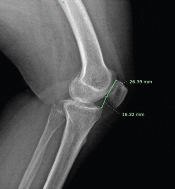
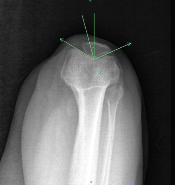
Weight-bearing anteroposterior, lateral, and merchant (axial) radiographs of the left knee were obtained. The lateral view did demonstrate patella baja with the Caton–Deschamps index of 0.62 (Fig. 1). The Merchant view showed a congruence angle of 16°, which was within normal limits, and there was no gross static subluxation (Fig. 2). There was no trochlear dysplasia evident on the lateral radiograph. Medial and lateral femoral condyle morphology was normal.
MRI of the left knee was performed. Axial proton density-weighted sequences demonstrated subtle medial patellar tilt and increased signal within the medial patellar retinaculum consistent with strain or low-grade partial tearing. The lateral retinaculum appeared intact. Patellar articular cartilage demonstrated grade 4 chondromalacia on the medial facet and degenerative changes.
Diagnosis
The constellation of findings, chronic antero-medial knee pain, markedly increased passive medial patellar translation, a strongly positive medial patellar apprehension test reproducing the patient’s cardinal symptom, relieved symptoms on lateral pushing of the patella by the examiner, and an MRI showing evidence of Grade 4 cartilage damage on the medial facet, established the clinical diagnosis of spontaneous MPS.
A trial of a medial-buttressed patellofemoral stabilizing brace was fitted. The patient confirmed significant symptomatic relief while wearing the brace, with a marked reduction in painful episodes. This response to bracing served as additional confirmatory evidence of MPS.
An extended supervised rehabilitation program was then initiated, emphasizing vastus lateralis strengthening, hip abductor and external rotator strengthening, balance and proprioception training, and correction of dynamic valgus patterns. Despite adherence to a structured 5-month physiotherapy program, the patient reported no meaningful reduction in instability episodes or daily functional limitation. Surgical intervention was therefore discussed and consented to.
Surgical management
Given that the patient had intact lateral retinacular tissue (no prior LRR), the operative strategy centered on reducing medial patellar tension. The planned procedure was arthroscopic medial retinacular release, modelled after the technique described by Shannon and Keene in 2007 [11].
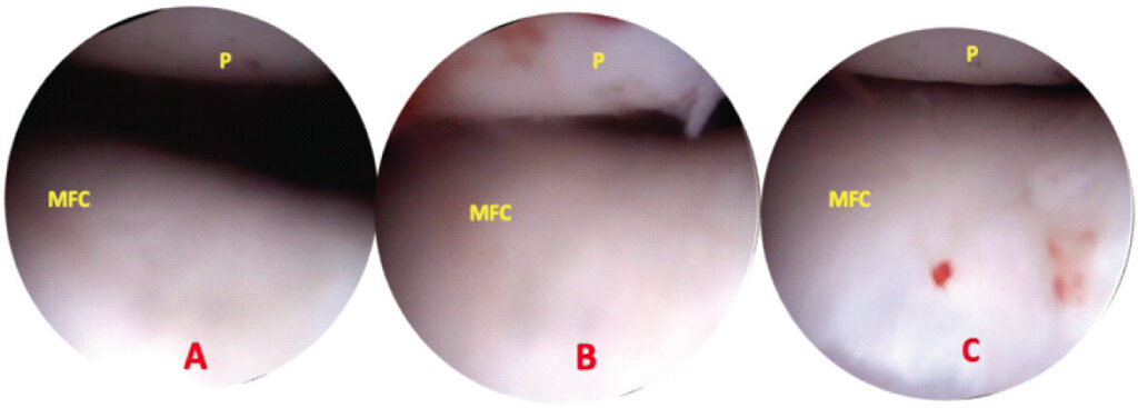
The procedure was performed under spinal anesthesia with the patient supine and a thigh tourniquet applied, but not initially inflated. Examination under anesthesia confirmed medial patellar displacement exceeding two quadrants at full extension, with the patella unable to be displaced laterally. Diagnostic arthroscopy was then performed using standard anterolateral and anteromedial portals (Fig. 3).
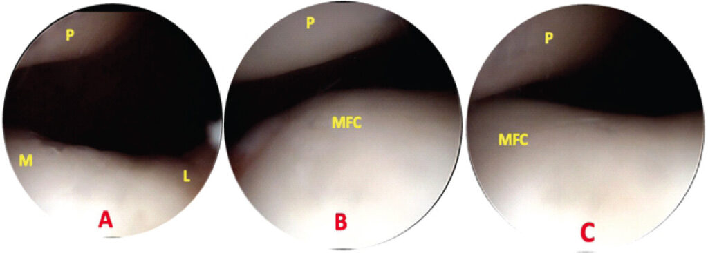
Diagnostic arthroscopy via the anterolateral portal confirmed patellar maltracking and significant medial patellar tilting and laxity in extension, with the patella snapping laterally into the trochlear groove at approximately 25° of flexion. grade IV chondral changes were confirmed on the medial patellar facet. The lateral retinaculum appeared intact; the medial structures appeared thickened and fibrotic.
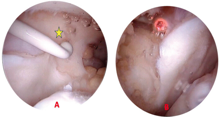
The medial retinaculum was divided arthroscopically beginning at the superior pole of the patella and extending distally to the level of the anteromedial portal – approximately 2 cm medial to the patellar border, taking care to preserve the attachment of the vastus medialis obliquus to the patella to maintain its dynamic medial stabilizing role. Once completed, the patella was grasped and tilted 50–70° on its lateral edge to ensure complete division of all intervening retinacular tissue; the medial undersurface of the patella was palpated to confirm release (Fig. 4).
Following the arthroscopic medial release, patellar tracking was reassessed and demonstrated improved centralization. Patellar tracking with the knee taken through a full arc of motion confirmed stable midline tracking without medial or lateral subluxation. The portals were closed in layers (Fig. 5).
Post-operative course and rehabilitation
The patient was discharged on the same day as a day-care procedure. She was instructed to bear weight as tolerated using crutches for the first 2 weeks. A soft knee brace was worn for comfort during ambulation for 4 weeks. Isometric quadriceps setting exercises and straight leg raises were commenced on post-operative day 1. Active range of motion exercises were initiated within 5 days, and the patient was referred to an outpatient physiotherapy program at 2 weeks.
Rehabilitation focused sequentially on: (1) Restoring full pain-free range of motion, (2) progressive quadriceps strengthening with emphasis on vastus lateralis rehabilitation, (3) hip abductor and external rotator conditioning, (4) proprioceptive and balance training, and (5) return to functional activities. The patient achieved full active range of motion by week 4 and completed her formal physiotherapy program at 3 months, at which point isokinetic testing confirmed symmetric quadriceps and hamstring strength.
At 6 months, the patient reported complete resolution of her instability episodes and a substantial reduction in anterior knee pain. She had returned to her pre-morbid level of daily activity, including regular walking and cycling. At 18-month follow-up, she denied any recurrent subluxation, instability, or giving-way events. Residual mild aching with prolonged stair descent was noted but did not significantly interfere with daily function. Examination confirmed midline patellar tracking, normalized medial patellar mobility (no apprehension), and a negative lateral float sign. The result was graded as excellent by the Merchant and Mercer rating scale criteria [12]. Validated patient-reported outcome measures improved from pre-operative assessment to final 18-month follow-up: Kujala score 45–87, International Knee Documentation Committee-subjective 52–93, Lysholm 61–89, and Visual Analog Scale pain 4–1. The Tegner activity level was unchanged at 3, consistent with her pre-morbid homemaker activity profile.
Discussion
MPS is an uncommon and frequently overlooked cause of anterior knee pain and instability. In contrast to the far more prevalent lateral patellar instability, medial instability remains poorly recognized, largely because the majority of reported cases occur as an iatrogenic complication following excessive LRR [1,5]. Consequently, spontaneous or idiopathic MPS in patients without prior surgery or significant trauma is exceedingly rare, with only isolated case reports available in the literature. The present case highlights the diagnostic challenges associated with this unusual condition and emphasizes the importance of careful clinical evaluation in patients with persistent unexplained patellofemoral symptoms.
The pathophysiology of MPS is closely related to insufficiency of the lateral soft-tissue stabilizers of the patella, including the lateral retinaculum, LPFL, and LPTL. Cadaveric and biomechanical studies have demonstrated that these structures provide significant restraint against excessive medial patellar translation [6]. Excessive release or attenuation of these structures disrupts patellar tracking and predisposes the patella to abnormal medial displacement, particularly during early knee flexion before engagement within the trochlear groove [3,6]. In our case, although there was no history of surgery or trauma, clinical examination demonstrated marked medial patellar translation and a strongly positive medial apprehension test, suggesting functional insufficiency of the lateral restraints.
Idiopathic MPS remains poorly understood. Several authors have suggested that subtle generalized ligamentous laxity, repetitive microtrauma, trochlear morphology abnormalities, muscular imbalance, or chronic maltracking may contribute to spontaneous medial instability [2,9]. However, many patients, including the present case, may not demonstrate obvious predisposing anatomical abnormalities on imaging. The absence of trochlear dysplasia, significant malalignment, or prior surgical intervention in our patient further supports the rarity of true idiopathic medial patellar instability.
Diagnosis of MPS is primarily clinical and requires a high index of suspicion. Patients often report vague anterior knee pain, instability, painful popping, giving-way episodes, and symptoms aggravated during early knee flexion [2,8]. Because these symptoms overlap with more common patellofemoral disorders such as chondromalacia patella or patellofemoral pain syndrome, delayed or missed diagnosis is common. In the present case, the patient had undergone prolonged conservative treatment elsewhere before the diagnosis was established. The medial patellar apprehension test remains one of the most valuable clinical examination findings [8]. Reproduction of symptoms with medially directed patellar translation and relief upon application of a lateral corrective force are considered highly suggestive of MPS [3,8]. Our patient demonstrated both findings consistently.
Imaging findings in MPS are often subtle and may not correlate with symptom severity. Standard radiographs are frequently normal because medial instability is typically dynamic rather than static. MRI may show medial patellar tilt, attenuation of the lateral retinacular structures, or associated chondral injury [1,3]. In the current case, MRI demonstrated medial patellar tilt and Grade IV chondromalacia of the medial patellar facet. This pattern of cartilage degeneration is particularly important because repetitive abnormal medial patellar tracking can increase contact pressure over the medial patellofemoral articulation. Persistent instability may therefore accelerate degenerative cartilage changes and contribute to chronic pain and disability.
Non-operative treatment is generally considered the first-line management strategy for idiopathic MPS. Recommended measures include activity modification, patellar stabilization bracing, quadriceps rehabilitation with emphasis on vastus lateralis strengthening, and hip/core strengthening to improve dynamic limb alignment [2,8]. Although our patient experienced temporary improvement with a patellar stabilizing brace, symptoms persisted despite an extensive supervised physiotherapy program. Failure of prolonged conservative treatment in symptomatic patients may justify surgical intervention.
Surgical management of MPS should be directed by the underlying pathoanatomy. When medial instability arises from insufficiency or attenuation of the lateral restraints, as is typical after excessive lateral release or in constitutional hyperlaxity, restoration of the lateral soft tissues is appropriate, and lateral retinacular imbrication, LPTL augmentation, and LPFL reconstruction have each produced favorable outcomes [3,8]. A distinct subgroup, however, presents with a contracted, fibrotic medial retinaculum that holds the patella in a medially tilted position and prevents its recentralization within the trochlea; in these patients, division of the medial restraint, rather than lateral reconstruction, relieves the primary mechanical block. Shannon and Keene described arthroscopic medial retinacular release for this purpose and reported six excellent and three good results in nine knees, with no complications or subsequent realignment surgery [11]; their series included cases of spontaneous medial subluxation, in which release relieved both instability and pain at a mean follow-up of 2.7 years. The present case is clinically significant because it demonstrates that MPS may occur even in the absence of previous LRR, trauma, or major anatomical abnormality. Awareness of this rare entity is essential to avoid misdiagnosis and prolonged ineffective treatment. Careful clinical examination, particularly the medial patellar apprehension test and symptom relief with lateral patellar support, remains critical for diagnosis. Early recognition may help prevent progressive cartilage degeneration and functional limitation.
Isolated MPS, occurring without prior lateral release or trauma, is exceptionally rare; reviewing the English-language literature, Akşahin et al. identified only five spontaneous cases among 163 reported instances of medial subluxation, the remainder being iatrogenic or post-traumatic [13]. Reported management of these isolated cases has almost uniformly involved restoration of the lateral restraints, as summarized in Table 1. The present case differs from previous reports in both aetiology and surgical technique.
Reported cases of isolated or spontaneous (non-iatrogenic, non-traumatic) medial patellar subluxation
| Study | Year | Age/sex | Category | Surgical treatment | Outcome/Follow-up |
|---|---|---|---|---|---|
| Richman and Scheller [14] | 1998 | 17/M | Isolated | Lateral retinacular imbrication | Tracking centralized, stable |
| Shannon and Keene [11] | 2007 | Adults (2 knees) | Spontaneous | Arthroscopic medial retinacular release | Instability and pain relieved, 2.7 years |
| Akşahin et al. [13] | 2010 | 21/M | Spontaneous | Lateral imbrication+patellotibial ligament augmentation | Corrected |
| Kreinces et al. [15] | 2026 | 32/F | Idiopathic | LPFL reconstruction (semitendinosus allograft) | Improved |
| Present case | 2026 | 44 | Idiopathic | Arthroscopic medial retinacular release | Excellent, 18 months |
M: Male, F: Female, LPFL: Lateral patellofemoral ligament
This report is limited by the inherent constraints of a single-case study design. The diagnosis rested on clinical examination, static radiographs, static MRI, and dynamic intraoperative assessment; stress radiography and dynamic fluoroscopy or MRI, which can objectively quantify medial patellar translation, were not performed and would be valuable adjuncts in future cases. Longer-term follow-up beyond 18 months is required to confirm durability and to monitor progression of the established medial-facet chondral disease.
Conclusion
Idiopathic Medial patellar subluxation is a rare and frequently overlooked cause of anterior knee pain and instability. This case report describes that this clinical entity can occur in the absence of lateral retinacular release, trauma, or other major abnormalities. A high index of suspicion and meticulous clinical examination are more useful for diagnosis than static imaging. Though conservative management remains the gold standard of care, persistent symptoms might require surgical intervention.
Clinical Message
Idiopathic medial patellar subluxation is a rare clinical entity that is often misdiagnosed. Careful clinical examination and early diagnosis will help tailor treatment to prevent patellar chondral damage and enhance functional outcome.
Conflict of Interest:
Nil
Source of Support:
Nil
Consent:
The authors confirm that informed consent was obtained from the patient for publication of this article
References
- Hwang SH, Nam CH, Choi KW, Ahn HS. Medial patella subluxation after minor trauma. Open J Orthop 2017;7:295-300. Google Scholar | PubMed
- Saper MG, Shneider DA. Medial patellar subluxation: Diagnosis and treatment. Am J Orthop (Belle Mead NJ) 2015;44:499-504. Google Scholar | PubMed
- Brinker MR, O'Connor DP, Flandry F, Hughston JC. Diagnosis and surgical correction of medial patellar subluxation. Oper Tech Sports Med 2001;9:183-9. Google Scholar | PubMed
- Betz RR, Lonergan R, Patterson R, Litton J, Yucha T, Boal R. The percutaneous lateral retinacular release. Orthopedics 1982;5:57-62. Google Scholar | PubMed
- Hughston JC, Deese M. Medial subluxation of the patella as a complication of lateral retinacular release. Am J Sports Med 1988;16:383-8. Google Scholar | PubMed
- Huddleston HP, Chahla J, Gursoy S, Williams BT, Dandu N, Malloy P. A comprehensive description of the lateral patellofemoral complex: Anatomy and anisometry. Am J Sports Med 2022;50:984-93. Google Scholar | PubMed
- Hayden DJ, Doshi C, Parikh SN. Lateral patellar retinaculum Z-lengthening. Arthrosc Tech 2021;10:e1883-7. Google Scholar | PubMed
- McCarthy MA, Bollier MJ. Medial patella subluxation: Diagnosis and treatment. Iowa Orthop J 2015;35:26-33. Google Scholar | PubMed
- Saper MG, Shneider DA. Medial patellar subluxation without previous lateral release: A case report. J Pediatr Orthop B 2014;23:350-3. Google Scholar | PubMed
- Nonweiler DE, DeLee JC. The diagnosis and treatment of medial subluxation of the patella after lateral retinacular release. Am J Sports Med 1994;22:680-6. Google Scholar | PubMed
- Shannon BD, Keene JS. Results of arthroscopic medial retinacular release for treatment of medial subluxation of the patella. Am J Sports Med 2007;35:1180-7. Google Scholar | PubMed
- Merchant AC, Mercer RL. Lateral release of the patella. A preliminary report. Clin Orthop Relat Res 1974;103:40-5. Google Scholar | PubMed
- Akşahin E, Yumrukçal F, Yüksel HY, Doğruyol D, Çelebi L. Role of pathophysiology of patellofemoral instability in the treatment of spontaneous medial patellofemoral subluxation: A case report. J Med Case Rep 2010;4:148. Google Scholar | PubMed
- Richman NM, Scheller AD Jr. Medial subluxation of the patella without previous lateral retinacular release. Orthopedics 1998;21:810-3. Google Scholar | PubMed
- Kreinces J, Bedard T, Vredenburgh Z. Idiopathic medial patellar instability treated with lateral patellofemoral ligament reconstruction using semitendinosus allograft. J Orthop Exp Innov 2026;7. https://doi.org/10.60118/001c.156481.. Google Scholar | PubMed | CrossRef
© 2026 Journal of Orthopaedic Case Reports - Published by Indian Orthopaedic Research Group
About the Authors
-

Anurag Bhakhar
-

A Salman Ul Faris
-

B G Ragulajay
-

Bhaskar Sarkar
-

Nirvin Paul
-

Md Quamar Azam
Related Articles in Journal of Orthopaedic Case Reports
 August 1, 2026 Intra-articular Ganglionic Cysts of Knee: A Cause for Anterior Knee Pain
 January 1, 2026 Anteromedial Knee Pain Unmasked: Schwannoma of the Infrapatellar Branch of the Saphenous Nerve – A Rare Case Report
 May 1, 2025 Patellar Osteoid Osteoma: A Rare Cause for Anterior Knee Pain
 April 10, 2024 Simultaneous ACL and Patellofemoral Instability: Surgical Management and Outcomes