


Introduction
Osteosarcoma is the most common primary malignant bone tumor in children and adolescents, typically arising during periods of rapid growth [1]. In contrast, the distal femur and proximal tibia are the most frequent sites. Approximately 10–12% of cases involve the humerus [2], and within this group, long-segment involvement extending from the proximal epiphysis through the diaphysis is relatively rare – reported in <5% of humeral osteosarcomas [3]. Surgical management has shifted from amputation toward limb-salvage strategies, enabled by the success of multi-agent chemotherapy and improved imaging. However, when large portions of the humerus are involved – particularly in skeletally immature patients – the choice of reconstruction becomes challenging.
Endoprosthetic reconstruction allows early mobilization but carries concerns regarding longevity, growth disruption, and functional outcomes in the upper limb [4]. Biological reconstructions, such as autografts, allografts, vascularized fibula, and extracorporeally irradiated autografts, offer long-term solutions with potential for osteointegration but involve longer recovery times and risk of mechanical failure [5, 6].
Extracorporeal radiotherapy (ECRT), in which the tumor-bearing bone is irradiated ex vivo and reimplanted, has emerged as a versatile method that offers precise anatomical fit and avoids immunological complications associated with allografts [7]. Zhao et al. proposed a reconstruction strategy based on tumor location and bone strength score, recommending biological reconstruction for diaphyseal tumors with preserved epiphyses and low strength scores [8]. Their findings indicated that functional outcomes were comparable between biological and prosthetic reconstructions when patients were appropriately selected.
This report describes a unique case of long-segment humeral osteosarcoma managed with a hybrid approach – proximal endoprosthetic reconstruction combined with ECRT-based biological reconstruction of the diaphysis – preserving the distal humerus and elbow function.
Case Report
A 15-year-old right-hand-dominant female presented with insidious onset of pain and swelling in the left upper arm over 2 months. There was no history of trauma, fever, or weight loss. Examination revealed a firm, mildly tender swelling involving the proximal humerus, with restricted shoulder motion but preserved elbow and hand functions. Neurovascular examination was normal.
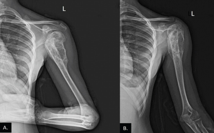
Radiographs demonstrated a long-segment mixed lytic-sclerotic lesion in the proximal epimetaphyseal location of the humerus with extraosseous soft tissue and osteoid matrix (Fig. 1). Magnetic resonance imaging showed that the lesion involved the proximal epiphysis and diaphysis, sparing only the distal 3 cm (Fig. 2). No skip lesions were identified. Computed tomography of the chest and whole-body bone scan were negative for metastases. Core needle biopsy revealed high-grade conventional osteosarcoma.
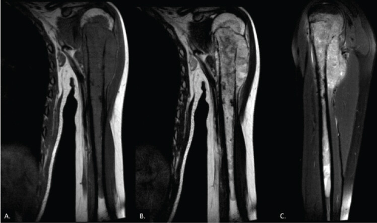
Following diagnosis, the patient received neoadjuvant chemotherapy with the mean arterial pressure protocol (methotrexate, adriamycin, and cisplatin) over 10 weeks. She responded well clinically and radiologically, with reduction in pain and swelling and decreased soft tissue involvement.
Surgical management
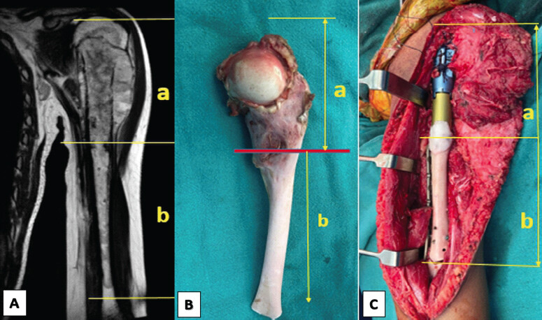
Given the tumor extent and skeletal maturity, a decision was made to perform wide en bloc excision of the tumor followed by limb salvage using a hybrid reconstructive approach. Total humerus replacement was avoided to preserve distal growth and elbow joint function. The surgical plan included wide resection of the proximal and mid-humerus (approximately 17 cm), preservation of the distal 3 cm of the humerus, proximal reconstruction with a cemented modular prosthesis, and mid-diaphyseal reconstruction using ECRT.
Under general anesthesia, the tumor was resected with oncological margins, preserving the distal humerus and elbow joint (Fig. 3). The excised diaphyseal segment was transported for ECRT and irradiated with a single 50 Gy dose. After sterilization, the autograft was reimplanted and fixed to the distal segment using a locking compression plate. The proximal prosthesis was cemented into the reimplanted graft and secured with a plate-screw construct. The rotator cuff was reattached to the prosthesis using heavy sutures.
Histopathology
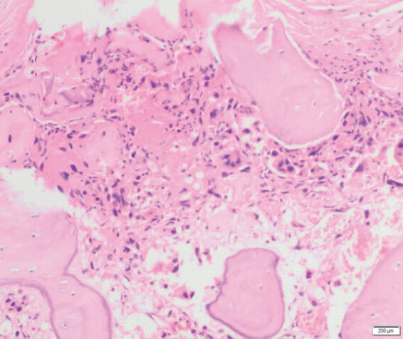
The gross specimen measured 10 × 3.5 × 3 cm. The medullary cavity showed a 6 cm cavity with a 1 cm luminal diameter, and a yellow-gray lesion of 4 × 1 cm located 2 cm from the resection margin. Microscopy revealed scant residual tumor within the medullary cavity. Tumor cells were moderate to markedly pleomorphic, with coarse chromatin and occasional giant cells. Significant chemotherapy-induced changes were observed, including extensive fibrosis, hyalinization, hemorrhage, and lacy osteoid deposition. Mitotic rate was low (2/10 HPF), including atypical forms. There was no lymphovascular invasion identified, and bone resection margins were free of tumor. The case was staged as pT1 (ypT1, AJCC 8th edition). Histologic grade was G3 (poorly differentiated) (Fig. 4).
Outcome
Postoperatively, the arm was immobilized in a sling for 4 weeks to protect the soft-tissue reconstruction, followed by the initiation of cautious passive range-of-motion exercises. X-rays showed a stable fixation of the hybrid composite construct (Fig. 5). Active physiotherapy began at 6 weeks with a focus on maximizing distal joint mobility and strengthening the preserved musculature. Adjuvant chemotherapy was resumed after complete wound healing without any delays. At the 18-month follow-up, the patient demonstrated an excellent functional recovery and reported being entirely pain-free. The functional assessment yielded a Musculoskeletal Tumor Society (MSTS) score of 26 out of 30 and a Disabilities of the Arm, Shoulder, and Hand score of 12, indicating high function with minimal upper extremity disability.
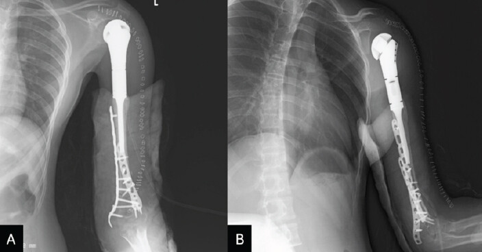
Physical examination revealed a highly functional and stable preserved elbow joint with a range of motion consisting of 130° of flexion, an extension lag of only 5°, and full forearm supination and pronation of 80° each. Shoulder active abduction and forward flexion were limited to 60° and 70°, respectively, an expected consequence of the extensive deltoid and rotator cuff resection; however, the patient successfully returned to routine daily activities and schooling with excellent structural integration of the graft.
Discussion
Limb salvage surgery with joint preservation allows pediatric and adolescent patients with humeral malignancies to retain critical function while maintaining oncologic safety. Zhao et al.’s retrospective series of 28 patients with diaphyseal or metaphyseal humeral malignant tumors offers a valuable framework, using tumor origin, location, and bone strength scoring to guide reconstruction type [8].
Their data suggest that biological reconstruction – such as devitalized autograft replantation – is most suitable when the bone strength score is low (≤10), and the tumor is confined to the diaphysis or metaphysis with an uninvolved epiphysis. In their series, the biological group (mean score 9.7 ± 1.3) achieved excellent MSTS outcomes (27.2) comparable to the prosthetic group (26.1), with no statistically significant difference (P > 0.05) [8].
Extracorporeal irradiation offers several advantages. The technique preserves the natural shape and joint articulation, allows for potential osteointegration, and avoids immunologic or disease transmission risks associated with allografts [7, 9]. Multiple studies report satisfactory mechanical strength in irradiated segments, especially for diaphyseal intercalary reconstructions [10].
In the present case, the hybrid approach balanced the need for early mechanical stability (via prosthesis) and long-term biological durability (via ECRT). The preserved distal segment permitted rigid fixation and avoided a total humeral prosthesis, which often results in poor shoulder and elbow function. The literature suggests that failure rates are significantly lower in ECRT reconstructions involving diaphyseal regions than in joint-involved grafts [11]. Common complications include infection, non-union, and graft fracture; however, these risks are mitigated with precise fixation and aseptic technique [7, 11].
Moreover, ECRT is cost-effective and widely applicable in resource-constrained settings where allografts or custom prosthetics may not be readily available [12]. The irradiated bone, though devitalized, serves as an ideal scaffold for creeping substitution over time, which was observed radiographically in our case.
At 18-month follow-up, the patient had no local recurrence or metastasis, showed evidence of graft union, and retained functional elbow movement. The shoulder abduction was limited to 60°, acceptable given resection of the deltoid and rotator cuff. The MSTS score of 24/30 reflected good functional recovery.
This case supports the decision-making approach advocated by Zhao et al., favoring biological reconstruction when anatomical and mechanical conditions permit. Combining prosthetic and biological methods enables tailored reconstruction, especially in growing children, allowing durable, functional, and oncologically sound outcomes.
Conclusion
In pediatric patients with extensive humeral osteosarcoma, hybrid reconstruction combining a proximal megaprosthesis with ECRT-based autograft reimplantation offers a promising strategy. It ensures wide tumor clearance while preserving limb function and skeletal integrity. When distal joint preservation is possible, this approach avoids total humeral replacement and supports better long-term outcomes. Proper case selection, rigid fixation, and meticulous surgical planning are essential for success. ECRT, particularly in the diaphyseal region, remains a valuable biological option, especially in children where durability, growth potential, and cost are important considerations.
Clinical Message
For pediatric patients presenting with long-segment humeral osteosarcoma, a hybrid reconstruction utilizing a proximal megaprosthesis combined with extracorporeal radiotherapy (ECRT)-sterilized autograft reimplantation represents a highly effective limb-salvage strategy. When tumor anatomy allows for the preservation of the distal humerus, avoiding a total humeral prosthesis through rigid distal plate fixation significantly enhances long-term elbow function, mechanical stability, and skeletal integrity while maintaining oncological safety.
Conflict of Interest:
Nil
Source of Support:
Nil
Consent:
The authors confirm that informed consent was obtained from the patient for publication of this article
References
- Mirabello L, Troisi RJ, Savage SA. International osteosarcoma incidence patterns in children and adolescents, middle ages and elderly persons. Int J Cancer 2009;125:229-34. [Google Scholar] | [PubMed]
- Geller DS, Gorlick R. Osteosarcoma: A review of diagnosis, management, and treatment strategies. Clin Adv Hematol Oncol 2010;8:705-18. [Google Scholar] | [PubMed]
- Aponte-Tinao LA, Ayerza MA, Muscolo DL. Allograft-prosthetic reconstruction of the proximal part of the humerus. J Bone Joint Surg Am 2002;84:718-25. [Google Scholar] | [PubMed]
- Anninga JK, Gelderblom H, Fiocco M, Kroep JR, Taminiau AH, Hogendoorn PC. Prognostic factors in osteosarcoma: A review. Eur J Cancer 2011;47:59-66. [Google Scholar] | [PubMed]
- Biau DJ, Odumala OA, Campion D, Pelet S, Letson GD. Reconstruction after bone tumor resection in children. J Bone Joint Surg Br 2006;88-B:95-100. [Google Scholar] | [PubMed]
- Manfrini M, Gasbarrini A, Malaguti C, Cerisoli M, Bacci G, Mercuri M. Intraoperative extracorporeal irradiation of bone segments for tumor sterilization. J Pediatr Orthop 2004;24:303-8. [Google Scholar] | [PubMed]
- Puri A, Gulia A, Hawaldar R, Rangarajan V, Syed Hasan Q, Desai S. Functional and oncological outcomes after extracorporeal irradiation and re-implantation of tumour-bearing bone segments. Bone Joint J 2014;96-B:1118-21. [Google Scholar] | [PubMed]
- Zhao J, Zhao Z, Li J, Yin J, Wang B. Tumor location and bone strength score are useful in selecting biological and non-biological reconstruction for humeral malignancies. BMC Surg 2019;19:130. [Google Scholar] | [PubMed]
- Krieg AH, Mani M, Speth BM, Stalley PD. Extracorporeal irradiation for local treatment of bone tumors. J Bone Joint Surg Br 2010;92-B:862-9. [Google Scholar] | [PubMed]
- Uyttendaele D, De Schryver A, Claessens H, Roels H, Berkvens P, Mondelaers W. Limb conservation in primary bone tumours by resection, extracorporeal irradiation and re-implantation. J Bone Joint Surg Br 1988;70:348-53. [Google Scholar] | [PubMed]
- Sanjay BK, Moreau PG, Younge DA. Extracorporeal irradiation and reimplantation in limb salvage for bone tumors. Clin Orthop Relat Res 1999;358:111-22. [Google Scholar] | [PubMed]
- Abudu A, Carter SR, Grimer RJ, Tillman RM. Extracorporeal irradiation and reimplantation in primary bone tumours. J Bone Joint Surg Br 1996;78:878-82. [Google Scholar] | [PubMed]
© 2026 Journal of Orthopaedic Case Reports - Published by Indian Orthopaedic Research Group
About the Authors
-

Dr. Prabodh Kantiwal
-

Dr. Syed Mohammed Adnan
-

Dr. Amir Suhail
-

Dr. Manveer Singh
-

Prof. Siddhi Chawla
-

Dr. Sudeep Khera
Related Articles in Journal of Orthopaedic Case Reports
 October 1, 2026 Development of a Non-ossifying Fibroma in the Same Femur 6 Years after Limb-Salvage Surgery for Osteosarcoma: A Case Report
 June 19, 2021 Outcomes of Tumor Resection and Reconstruction by Megaprosthesis around Knee: Our Experience
 October 1, 2026 Soft-Tissue Closure through Acute Deformation and Gradual Correction Using Taylor Spatial Frame in a Gustilo-Anderson Type IIIB Open Tibial Pilon Fracture: A Case Report
 October 1, 2026 Early Surgical Escalation and Structured Multidisciplinary Pathways Improve Limb-Salvage Outcomes in Necrotizing Fasciitis: A Two-Cycle Quality Improvement Project