


Introduction
Periprosthetic joint infection (PJI) is among the most serious complications of hip arthroplasty, occurring after roughly 0.5–2% of primary procedures. Despite better surgical technique and antimicrobial prophylaxis, PJI still carries substantial morbidity, repeated operations, prolonged antibiotic courses, and increased mortality, especially in elderly and frail patients [1,2,3].
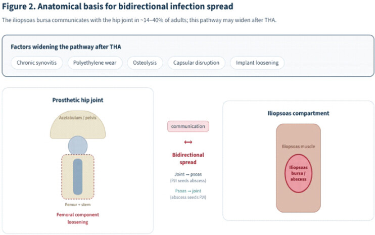
The iliopsoas bursa communicates with the hip joint in up to 40% of individuals, giving infection a route to spread between the prosthetic joint and the iliopsoas compartment. An iliopsoas abscess may therefore be the first sign of a hip PJI or a consequence of one. The presentation is uncommon and easily missed: early symptoms are non-specific, which delays diagnosis and misdirects initial treatment [4,5,6,7,8].
In a patient with an ipsilateral hip prosthesis, a persistent inflammatory syndrome or failed image-guided drainage should raise a strong suspicion of occult PJI. Cross-sectional imaging, microbiological confirmation by image-guided aspiration, and multidisciplinary management establish the diagnosis and define the surgical strategy. We report an elderly patient in whom an iliopsoas abscess revealed a chronic Escherichia coli hip PJI, and we use the case and a review of the literature to outline the diagnostic pitfalls and therapeutic challenges of this rare but important presentation [2,4,6].
Case Report
An 88-year-old man presented with a persistent inflammatory syndrome and abnormal laboratory findings. His anthropometry, functional status, and comorbidities are summarized in Table 1; in brief, he had a well-functioning right total hip arthroplasty (THA) – one of bilateral THAs – multiple cardiovascular and other comorbidities, and long-term oral anticoagulation with apixaban.
| Domain | Details |
|---|---|
| Demographics/ anthropometry | 88-year-old man; 83 kg, 172 cm; BMI 26 kg/m2 |
| Functional status | GIR 3 – moderate dependence (French dependency scale) |
| Cardiovascular | Hypertension; atrial fibrillation on apixaban 2.5 mg twice daily; transcatheter aortic valve implantation (TAVI, 2022) |
| Prior arthroplasty | Bilateral total hip arthroplasty; left total knee arthroplasty |
| Spine/mobility | Cervical and lumbar spinal stenosis; gait impairment |
| Urological | Benign prostatic hyperplasia |
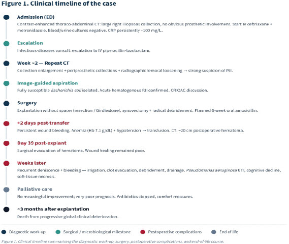
He was admitted through the emergency department. Contrast-enhanced thoraco-abdominal computed tomography (CT) showed a large right iliopsoas abscess with no obvious prosthetic involvement. Empirical intravenous ceftriaxone and metronidazole were started. Blood cultures were negative, and urine cultures showed no significant bacteriuria. Despite broad-spectrum treatment, inflammatory markers stayed high, with C-reactive protein persistently around 100 mg/L.
After an infectious-diseases consultation, treatment was escalated to intravenous piperacillin-tazobactam. Repeat CT 2 weeks later showed that the abscess had enlarged, now with multiple periprosthetic fluid collections and radiographic loosening of the femoral component – findings strongly suggestive of PJI.
Image-guided hip aspiration grew fully susceptible E. coli, confirming chronic PJI. After discussion with the regional referral center for complex bone and joint infection (CRIOAC), the prosthesis was removed without spacer implantation – chosen for the patient’s age, frailty, comorbidities, limited functional reserve, and poor rehabilitation potential. A 6-week course of targeted oral amoxicillin (2 g 3 times daily) was planned postoperatively.
The right THA was explanted with extensive synovectomy, radical debridement of infected and necrotic tissue, and multiple intraoperative specimens for culture. Given the patient’s condition and limited reconstructive options, no antibiotic spacer was placed (resection [Girdlestone] arthroplasty). The early post-operative course was uneventful, and he was transferred to a rehabilitation unit to continue targeted therapy.
Two days after transfer, the surgical wound began to bleed persistently. Progressive anemia (hemoglobin 7.1 g/dL) with hypotension required fluid resuscitation and transfusion. Emergency CT showed a large post-operative hematoma of about 20 cm involving the intramuscular and subcutaneous compartments around the right hip.
He was readmitted to orthopedics, and the hematoma was evacuated 39 days after prosthesis removal. Wound healing remained poor. Several weeks later, recurrent dehiscence with persistent bleeding required a further procedure: irrigation, clot evacuation, debridement, and drainage.
Over the following weeks, his condition declined. Severe cognitive impairment, complete dependence for activities of daily living, recurrent Pseudomonas aeruginosa urinary tract infection, a persistent inflammatory syndrome despite successive antibiotic regimens, and progressive soft-tissue necrosis around the wound prompted multidisciplinary reassessment. With no meaningful improvement and a very poor prognosis, palliative care was started after discussion with the family. Antibiotics were stopped, and comfort measures provided. He died approximately 3 months after prosthesis removal (Fig. 1).
Discussion
PJI remains one of the most serious complications of hip arthroplasty, following approximately 0.5–2% of primary implantations. Chronic PJI in particular is associated with prolonged hospitalization, repeated surgery, poor function, and increased mortality in elderly patients with multiple comorbidities [1,2,3]. Gram-positive organisms predominate, but Gram-negative bacteria such as E. coli form an important subgroup, particularly in older patients with a possible urinary or gastrointestinal source [2].
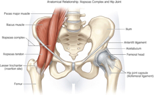
The link between an iliopsoas abscess and an ipsilateral hip prosthesis is uncommon but important. Anatomical studies show communication between the iliopsoas bursa and the hip joint in about 14–40% of adults, and this pathway may widen after arthroplasty through chronic synovitis, polyethylene wear, osteolysis, capsular disruption, or loosening – each easing the spread of infection between joint and compartment [4,5,6,7,8]. An iliopsoas abscess may thus be the first manifestation of a hip PJI or a secondary complication of established infection (Fig. 2 and 3).
Diagnosis is difficult because the early picture is non-specific. Groin pain, fever, gait difficulty, abdominal or lumbar pain, or an isolated inflammatory syndrome commonly point clinicians toward abdominal, urological, or muscular causes before PJI is considered [4,6,7]. Our case followed this pattern: the first CT showed an iliopsoas abscess without clear prosthetic involvement, and only persistent inflammation despite adequate antibiotics – together with repeat imaging showing progressive periprosthetic collections and femoral loosening – established the PJI. Comparable delays recur throughout the literature, and the lesson is consistent: failed drainage or persistent inflammation should send the clinician back to the prosthesis [6, 8].
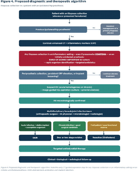
This case argues for a specific, practical rule. In a patient with an initially uninfected hip prosthesis, any iliopsoas collection that appears in an inflammatory setting – even when imaging suggests a simple hematoma – should undergo early CT-guided aspiration. Sampling the collection promptly identifies the organism sooner and allows targeted antibiotics to be started without waiting for the diagnosis to declare itself, shortening the diagnostic delay that so often complicates these cases.
Contrast-enhanced CT is the imaging cornerstone: it maps deep collections, assesses loosening, and guides aspiration or drainage [6]. Imaging alone, however, cannot separate a sterile collection from infection, so microbiological confirmation is mandatory. Guidelines from the Infectious Diseases Society of America, European Bone and Joint Infection Society, and International Consensus Meeting all stress obtaining synovial fluid and multiple intraoperative tissue cultures before definitive treatment whenever feasible [9,10,11,12]. When the prosthesis is removed, sonication of the explant can further improve microbiological yield [13]. Here, aspiration yielded fully susceptible E. coli, which confirmed the diagnosis and directed therapy before surgery.
The organism itself is instructive. Gram-negative PJIs are a minority but cluster in elderly patients and often arise from hematogenous seeding of urinary or gastrointestinal origin [2]. Isolating an enteric organism should therefore trigger a search for an extra-articular source while the prosthetic joint is assessed in parallel – otherwise the primary focus is easily missed.
Treating a hip PJI complicated by an iliopsoas abscess means clearing both the prosthetic and the extra-articular focus. Percutaneous drainage with prolonged antibiotics rarely controls infection while the infected implant stays in place [4,6,7,8]. Persistence or recurrence of the abscess despite apparently adequate drainage is better read as a marker of underlying PJI than as failure of the drainage itself.
Surgical strategy should be individualized to symptom duration, implant stability, organism, bone loss, soft-tissue quality, and host status [9,10]. Debridement, antibiotics, and implant retention suits acute infection with a stable implant and a susceptible organism [14]. Chronic infection with loosening generally requires exchange: one-stage revision gives excellent results in selected patients, while two-stage revision remains the benchmark for many chronic infections because of its high eradication rate [15]. These reconstructions are not suitable for everyone. Once the implant is removed, the optimal route and duration of antimicrobial therapy continue to be refined; recent randomized trials support an early oral switch and shorter total courses in selected patients [16,17].
The patient’s long-term oral anticoagulation deserves particular emphasis. In a patient on oral anticoagulants who develops a psoas hematoma alongside an ipsilateral prosthesis, anticoagulation must be managed proactively – held or reversed around interventions, carefully dose-adjusted, and, where available, monitored with drug-specific assays – to control the hematoma and prevent the kind of expansion that, in this case, required transfusion and repeated surgical evacuation.
The wider literature frames these decisions. Reviews of the diagnostic and therapeutic challenges of PJI remain valuable [18], and debridement with implant retention has been studied specifically in late acute infection [19]. When cross-sectional imaging is equivocal, radionuclide techniques – labeled-leukocyte imaging and fluorodeoxyglucose-positron emission tomography – can add information [20,21]. Contemporary reviews summarize current diagnosis and management [22], and large series have defined the role of implant retention in staphylococcal disease [23]. For infection with loosening, single-stage revision under strict protocols [24] and systematic comparison of one- versus two-stage strategies [25] guide reconstruction, with further reviews addressing diagnosis and management more broadly [26]. The differential for a psoas collection includes pyogenic psoas abscess of varied etiology [27], while the classical description of infection after hip arthroplasty – including the late chronic category relevant to this case – remains a useful framework [28], as do subsequent consensus statements on diagnosis and treatment [29,30].
For our patient, advanced age, cardiovascular comorbidity, chronic anticoagulation, cognitive impairment, limited mobility, and extensive periprosthetic infection with femoral loosening made implant preservation unrealistic. After multidisciplinary discussion within the CRIOAC network, resection arthroplasty without a spacer was judged the safest option [9]. The decision reflects current thinking: treatment should follow physiological reserve and expected functional benefit rather than chronological age alone.
Despite adequate source control, the patient had repeated post-operative complications – a large hematoma needing transfusion and evacuation, recurrent wound breakdown, functional decline, and eventual palliative care. This course is a reminder that death in frail patients with PJI often reflects post-operative complications, immobility, malnutrition, and loss of physiological reserve as much as the infection itself [1,2]. Eradicating infection does not guarantee survival or functional recovery in this population.
Published cases and series of iliopsoas/psoas abscess associated with periprosthetic hip infection are summarized in Table 2. Our case aligns with the published experience on three points. Iliopsoas abscess associated with hip arthroplasty is exceptionally rare and reported mainly as isolated cases or small series [4,5,6,7,8]. Diagnosis is often delayed because the abscess is first treated as a primary retroperitoneal infection. Moreover, durable control is seldom achieved without addressing the infected prosthesis. The case adds the specific challenge of chronic Gram-negative PJI in a very elderly patient with minimal reserve.
| Author (year) | Pathogen | Initial presentation | Prosthesis status | Definitive treatment | Outcome |
|---|---|---|---|---|---|
| Buttaro et al. (2002) [4] | Escherichia coli | Psoas abscess communicating with hip joint | Infected THA (11 year) | Component removal + debridement; ciprofloxacin | Infection eradicated |
| Dauchy et al. (2009) [5] | Various (case-control study) | Psoas abscess in hip PJI | Hip PJI | Observational study (12% had a psoas abscess) | Association underdiagnosed |
| Gunaratne et al. (2016) [7] | Staphylococcus aureus | Groin pain; psoas abscess with sinus to hip | Bilateral hip PJI | Bilateral two-stage revision | Favorable |
| Lawrenz et al. (2017) [6] | Mixed (5-case series) | Iliacus/iliopsoas abscess | Hip PJI | Debridement ± Girdlestone/revision/retention | Poor; refractory, multiple drainages |
| Stone and King (2019) [8] | Not specified | Iliopsoas abscess extending to a metal-on-metal hip | Infected MoM THA | Combined retroperitoneal + posterior debridement | Reported success |
| Present case (2025) | Escherichia coli | Persistent inflammatory syndrome + iliopsoas abscess | Femoral loosening | Explantation without spacer (resection) | Infection controlled; death from post-operative complications and frailty |
THA: Total hip arthroplasty, PJI: Periprosthetic joint infection, MoM: Metal-on-metal. Every entry was checked against the primary source. Narrative reviews and guideline definitions are cited in the text [2, 9, 10, 11, 12] rather than listed as cases
What distinguishes this report is the documented progression from an apparently isolated iliopsoas abscess to microbiologically confirmed chronic E. coli PJI with radiographic loosening. Serial imaging, image-guided aspiration, multidisciplinary decision-making, and definitive explantation trace the full diagnostic pathway – and show that successful infection control can still be followed by a poor outcome when frailty and post-operative complications dominate.
In practice, an iliopsoas abscess in a patient with an ipsilateral hip prosthesis should be treated as PJI until proven otherwise. Persistent inflammation, recurrent collections, failed drainage, or radiographic loosening should prompt immediate image-guided aspiration – of the collection and of the hip – and multidisciplinary evaluation. Early diagnosis and timely surgery shorten delay, spare patients repeated ineffective procedures, and give the best chance of a good result (Fig. 4).
Conclusion
An iliopsoas abscess ipsilateral to a hip prosthesis is uncommon but should always raise suspicion of PJI. Persistent inflammation, recurrent collections, failed image-guided drainage, or radiographic loosening warrant prompt evaluation of the prosthetic joint by image-guided aspiration and culture. In particular, any iliopsoas collection arising in an inflammatory setting on an initially uninfected prosthesis – even when it resembles a hematoma – should undergo early CT-guided aspiration to identify the organism and begin targeted antibiotics.
The case underlines the value of early collaboration among orthopedic surgeons, infectious-disease physicians, microbiologists, and radiologists in reaching the diagnosis and choosing the surgical strategy. Eradicating infection is the primary goal, but treatment must be tailored to implant status, organism, physiological reserve, comorbidities, and functional expectations. In a patient on anticoagulation, proactive control of the hematoma is part of that plan. In a frail patient, controlling infection may not prevent a poor outcome – an honest part of the counseling that should precede surgery.
Clinical Message
In a patient with an ipsilateral hip prosthesis, an iliopsoas abscess is rarely an isolated problem. A persistent inflammatory syndrome or failure of drainage should prompt investigation for PJI. Any iliopsoas collection in this setting – even when it looks like a simple hematoma – warrants early CT-guided aspiration to identify the organism and start targeted antibiotics. Early multidisciplinary management guides diagnosis, directs surgery, and improves outcomes.
Conflict of Interest:
Nil
Source of Support:
Nil
Consent:
The authors confirm that informed consent was obtained from the patient for publication of this article
References
- Zimmerli W, Trampuz A, Ochsner PE. Prosthetic-joint infections. N Engl J Med 2004;351:1645-54. [Google Scholar] | [PubMed]
- Tande AJ, Patel R. Prosthetic joint infection. Clin Microbiol Rev 2014;27:302-45. [Google Scholar] | [PubMed]
- Kurtz SM, Lau E, Watson H, Schmier JK, Parvizi J. Economic burden of periprosthetic joint infection in the United States. J Arthroplasty 2012;27 Suppl 8:61-5.e1. [Google Scholar] | [PubMed]
- Buttaro M, González Della Valle A, Piccaluga F. Psoas abscess associated with infected total hip arthroplasty. J Arthroplasty 2002;17:230-4. [Google Scholar] | [PubMed]
- Dauchy FA, Dupon M, Dutronc H, De Barbeyrac B, Lawson-Ayayi S, Dubuisson V. Association between psoas abscess and prosthetic hip infection: A case-control study. Acta Orthop 2009;80:198-200. [Google Scholar] | [PubMed]
- Lawrenz JM, Mesko NW, Higuera CA, Molloy RM, Simpfendorfer C, Babic M. Treatment challenges of prosthetic hip infection with associated iliacus muscle abscess: Report of 5 cases and literature review. J Bone Jt Infect 2017;2:127-35. [Google Scholar] | [PubMed]
- Gunaratne GD, Khan RJ, Tan C, Golledge C. Bilateral prosthetic hip joint infections associated with a Psoas abscess. A Case Report. J Orthop Case Rep 2016;6:3-6. [Google Scholar] | [PubMed]
- Stone AH, King PJ. Simultaneous combined retroperitoneal and posterior hip approach for the treatment of iliopsoas abscess with extension to a metal-on-metal prosthetic hip joint. Arthroplast Today 2019;5:269-75. [Google Scholar] | [PubMed]
- Osmon DR, Berbari EF, Berendt AR, Lew D, Zimmerli W, Steckelberg JM. Diagnosis and management of prosthetic joint infection: Clinical practice guidelines by the infectious diseases society of America. Clin Infect Dis 2013;56:e1-25. [Google Scholar] | [PubMed]
- McNally M, Sousa R, Wouthuyzen-Bakker M, Chen AF, Soriano A, Vogely HC. The EBJIS definition of periprosthetic joint infection. Bone Joint J 2021;103-B:18-25. [Google Scholar] | [PubMed]
- Parvizi J, Tan TL, Goswami K, Higuera C, Della Valle C, Chen AF. The 2018 definition of periprosthetic hip and knee infection: An evidence-based and validated criteria. J Arthroplasty 2018;33:1309-14.e2. [Google Scholar] | [PubMed]
- Parvizi J, Gehrke T. International Consensus Group on Periprosthetic Joint Infection. Definition of periprosthetic joint infection. J Arthroplasty 2014;29:1331. [Google Scholar] | [PubMed]
- Trampuz A, Piper KE, Jacobson MJ, Hanssen AD, Unni KK, Osmon DR. Sonication of removed hip and knee prostheses for diagnosis of infection. N Engl J Med 2007;357:654-63. [Google Scholar] | [PubMed]
- Grammatopoulos G, Kendrick B, McNally M, Athanasou NA, Atkins B, McLardy-Smith P. Outcome following debridement, antibiotics, and implant retention in hip periprosthetic joint infection-an 18-year experience. J Arthroplasty 2017;32:2248-55. [Google Scholar] | [PubMed]
- Ibrahim MS, Twaij H, Haddad FS. Two-stage revision for the culture-negative infected total hip arthroplasty: A comparative study. Bone Joint J 2018;100-B:3-8. [Google Scholar] | [PubMed]
- Li HK, Rombach I, Zambellas R, Walker AS, McNally MA, Atkins BL. Oral versus intravenous antibiotics for bone and joint infection. N Engl J Med 2019;380:425-36. [Google Scholar] | [PubMed]
- Bernard L, Arvieux C, Brunschweiler B, Touchais S, Ansart S, Bru JP. Antibiotic therapy for 6 or 12 weeks for prosthetic joint infection. N Engl J Med 2021;384:1991-2001. [Google Scholar] | [PubMed]
- Sendi P, Zimmerli W. Challenges in periprosthetic knee-joint infection. Int J Artif Organs 2011;34:947-56. [Google Scholar] | [PubMed]
- Wouthuyzen-Bakker M, Sebillotte M, Lomas J, Taylor A, Palomares EB, Murillo O. Clinical outcome and risk factors for failure in late acute prosthetic joint infections treated with debridement and implant retention. J Infect 2019;78:40-7. [Google Scholar] | [PubMed]
- Love C, Marwin SE, Tomas MB, Krauss ES, Tronco GG, Bhargava KK. Diagnosing infection in the failed joint replacement: A comparison of coincidence detection fluorodeoxyglucose PET and indium-111-labeled leukocyte/marrow imaging. Radiology 2004;232:333-41. [Google Scholar] | [PubMed]
- Glaudemans AW, Galli F, Pacilio M, Signore A. Leukocyte and bacteria imaging in prosthetic joint infection. Eur Cell Mater 2013;25:61-77. [Google Scholar] | [PubMed]
- Peel TN, Buising KL, Choong PF. Prosthetic joint infection: Challenges of diagnosis and treatment. ANZ J Surg 2011;81:32-9. [Google Scholar] | [PubMed]
- Lora-Tamayo J, Murillo O, Iribarren JA, Soriano A, Sanchez-Somolinos M, Baraia-Etxaburu JM. A large multicenter study of methicillin-susceptible and methicillin-resistant Staphylococcus aureus prosthetic joint infections managed with implant retention. Clin Infect Dis 2013;56:182-94. [Google Scholar] | [PubMed]
- Haddad FS, Sukeik M, Alazzawi S. Is single-stage revision according to a strict protocol effective in treatment of chronic knee arthroplasty infections? Clin Orthop Relat Res 2015;473:8-14. [Google Scholar] | [PubMed]
- Kunutsor SK, Whitehouse MR, Blom AW, Beswick AD. INFORM Team. Re-infection outcomes following one- and two-stage surgical revision of infected hip prosthesis: A systematic review and meta-analysis. PLoS One 2015;10:e0139166. [Google Scholar] | [PubMed]
- Cataldo MA, Petrosillo N, Cipriani M, Cauda R, Tacconelli E. Prosthetic joint infection: Recent developments in diagnosis and management. J Infect 2010;61:443-8. [Google Scholar] | [PubMed]
- Ricci MA, Rose FB, Meyer KK. Pyogenic psoas abscess: Worldwide variations in etiology. World J Surg 1986;10:834-43. [Google Scholar] | [PubMed]
- Tsukayama DT, Estrada R, Gustilo RB. Infection after total hip arthroplasty. A study of the treatment of one hundred and six infections. J Bone Joint Surg Am 1996;78:512-23. [Google Scholar] | [PubMed]
- Zmistowski B, Della Valle C, Bauer TW, Malizos KN, Alavi A, Bedair H. Diagnosis of periprosthetic joint infection. J Orthop Res 2014;32 Suppl 1:S98-107. [Google Scholar] | [PubMed]
- Gehrke T, Parvizi J. Proceedings of the International Consensus Meeting on Periprosthetic Joint Infection. Philadelphia, PA: Data Trace Publishing Company; 2013. [Google Scholar] | [PubMed]
© 2026 Journal of Orthopaedic Case Reports - Published by Indian Orthopaedic Research Group
About the Authors
-

Ahmed Ben Rhaïem
-

Abdul Majeed Kikar
-

Mario Ruben Sanguina
Related Articles in Journal of Orthopaedic Case Reports
 September 1, 2026 Angular Improvement with Bracing Over 7 Years in Anterolateral Tibial Bowing without Neurofibromatosis Type 1: A Case Report
 September 1, 2026 Clinical Outcomes of Staged Bilateral Middle Trapezius Transfer for Irreparable Supraspinatus Tendon Tears: A Case Report
 September 1, 2026 Multifactorial Non-union After First Metatarsophalangeal Arthrodesis Revised With Structural Iliac Crest Autograft: A Case Report and Comprehensive Literature Review
 September 1, 2026 Melorheostosis of the Right Lower Limb Presenting as Progressive Painful Swelling in a Young Adult: A Case Report