


Introduction
Pathological femoral neck fractures in young patients pose a challenge due to the need to preserve the native hip joint and ensure long-term function. While typically associated with high-energy trauma, they may also occur in the presence of underlying metabolic or pathological bone conditions [1]. Preservation of the femoral head through internal fixation is generally preferred; however, its success is limited in cases with poor bone quality, delayed presentation, and it always poses an increased risk of fixation failure [2].
Autoimmune polyglandular syndrome (APS) is a rare endocrine disorder characterized by multiple glandular insufficiencies affecting calcium metabolism and bone turnover, thereby predisposing patients to osteoporosis and increased fracture risk. It also introduces important perioperative considerations, including the need for endocrine optimization [3].
Total hip arthroplasty (THA) in young patients remains a subject of ongoing debate. While THA offers reliable pain relief and early functional recovery, concerns persist regarding implant longevity, risk of revision surgery, and activity-related wear over time. Consequently, arthroplasty is often reserved for cases where head-preserving procedures are unlikely to succeed, such as in neglected fractures, severe comminution, or markedly compromised bone stock [4].
Despite the individual challenges posed by pathological femoral neck fractures and systemic metabolic disorders, there is a paucity of literature addressing their combined impact on surgical decision-making and technical execution, particularly in young patients with APS. Existing studies largely focus on either fracture management in young individuals or arthroplasty outcomes in metabolic bone disease, with limited guidance on optimizing surgical technique in such complex clinical scenarios.
This case describes the surgical considerations, intraoperative challenges, and technical nuances of performing THA in a young patient with a pathological neglected femoral neck fracture in the setting of APS.
Clinical Context
A 34-year-old male presented with complaints of left hip pain and inability to bear weight following a trivial trauma, having slipped and fallen from bed at home. The patient reported a delay of approximately 4 months since the initial injury. Clinical examination revealed limb shortening of approximately 1 inch.
A comprehensive metabolic and endocrine evaluation was performed, including assessment of glycemic status, serum calcium, phosphate, parathyroid hormone, Vitamin D levels, serum magnesium, alkaline phosphatase, and renal function. Biochemical analysis demonstrated hypocalcemia and reduced parathyroid hormone levels, consistent with underlying hypoparathyroidism. Glycemic indices and Serum phosphate were elevated, whereas vitamin D levels, serum magnesium, alkaline phosphatase, and renal function parameters were within normal limits.
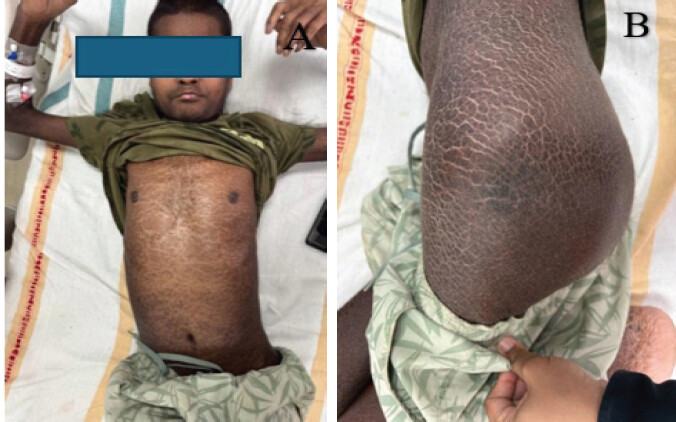
The diagnosis of APS Type 1 (APS-1) was established by the treating endocrinology team based on the characteristic endocrine manifestations, including hypoparathyroidism, chronic hypocalcemia, and Type 1 diabetes mellitus, together with ectodermal features such as ichthyosis vulgaris (Fig. 1). Genetic evaluation demonstrated an AIRE gene mutation, confirming the diagnosis of APS-1. A dual-energy X-ray absorptiometry (DEXA) scan revealed a T-score of −3, consistent with severe osteoporosis. Preoperatively, the patient had severe functional limitation of the affected hip, with a Harris Hip score (HHS) of 25/100.
No clinical, biochemical, or radiological evidence suggestive of malignancy, renal osteodystrophy, osteomalacia, or other secondary causes of pathological fracture was identified. The fracture was therefore attributed to severe osteoporosis associated with APS and chronic hypocalcemia. A multidisciplinary approach was undertaken for pre-operative optimization, given the patient’s underlying endocrine disorder. The patient was evaluated in consultation with the endocrinology team. Glycemic control was optimized, and calcium and phosphate levels were corrected preoperatively. Perioperative steroid coverage was planned to mitigate the risk of adrenal insufficiency despite normal baseline adrenocorticotropic hormone levels.
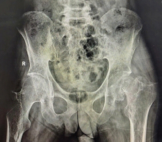
Radiographic assessment included standard anteroposterior radiographs of the pelvis and left hip, which demonstrated a displaced transcervical femoral neck fracture with features of non-union and early femoral head resorption (Fig. 2). Pre-operative templating was performed to estimate appropriate implant size, restore limb length, and achieve optimal offset. Given the neglected nature of the fracture, poor bone quality, and evidence of early femoral head resorption, head-preserving procedures such as internal fixation were considered to have a high risk of failure. Bipolar hemiarthroplasty was also considered suboptimal due to the patient’s young age, higher functional demands, and the risk of progressive acetabular wear. In view of these factors, THA was considered the most reliable option.
The initial plan was made to use uncemented primary cup and stem. However, given the poor bone quality and narrow femoral canal morphology, a contingency plan was made for possible intraoperative conversion from an uncemented to a cemented femoral stem. A cemented stem (MERIL congenital dysplasia of the hip [CDH] stem) and a cemented cup were kept available considering the anticipated difficulty in achieving adequate press-fit fixation. On the acetabular side, a cementless multihole cup (MERIL) with provision for supplementary screw fixation was kept available in case an uncemented cup is used while allowing for long-term biological fixation. In view of the patient’s young age, higher functional demands, and increased risk of post-operative instability, a dual mobility articulation system (MERIL dual mobility system) was selected to reduce the risk of dislocation and improve functional outcomes.
Overall, the surgical plan emphasized achieving stable fixation, restoring biomechanics, and minimizing complications in the setting of compromised bone quality and systemic metabolic disease.
Technique
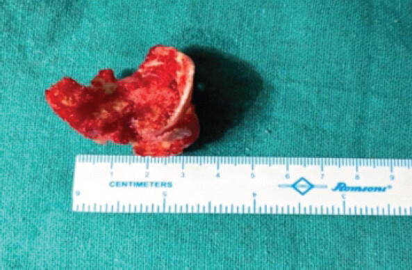
The procedure was performed under combined spinal-epidural anesthesia with the patient positioned in the lateral decubitus position. A standard posterior approach to the hip joint was utilized. The gluteus maximus was split in line with its fibers and the short external rotators, along with the posterior capsule, although torn, were identified and tagged. Intraoperatively, the femoral head was found to be markedly osteoporotic and structurally fragile. During attempts at extraction, the head fragmented due to its poor bone quality and shattered within the acetabulum. Consequently, it was carefully removed in a piecemeal fashion from the acetabular cavity, ensuring complete clearance before proceeding with acetabular preparation (Fig. 3).
Given the pathological nature of the fracture and anticipated poor bone quality, special care was taken during hip dislocation. Excessive torsional forces through the distal limb were avoided. Instead, controlled manipulation of the proximal femur as a single unit was performed to minimize stress concentration and reduce the risk of iatrogenic fracture.
Intraoperative assessment confirmed markedly osteoporotic bone, likely secondary to the underlying APS and associated metabolic derangements. The femoral head was excised. The acetabular preparation was undertaken with sequential reaming, taking care to preserve subchondral bone and avoid over-reaming in an already osteoporotic hip.
Considering the compromised bone stock, a multihole acetabular cup (50 mm) was selected to permit supplementary screw fixation. Five cancellous screws (6.5 mm) were inserted to enhance primary stability and optimize fixation in the osteoporotic acetabular bed. The decision to use an uncemented acetabular cup was made because cemented acetabular components rely entirely on the integrity of the cement-bone interface. This will be suboptimal in osteoporotic bone and can predispose to early loosening under cyclical loading. Furthermore, as the patient was young, a cementless multihole cup with screw augmentation was considered a more durable and biologically favorable option.
Attention was then directed to the femoral side. The femoral canal had two issues. The canal was narrow and also had thin cortices. Initial attempts at achieving press-fit stability with an uncemented stem were unsuccessful due to inadequate axial and rotational stability. In view of this, the decision was made intraoperatively to proceed with a cemented stem.
A cemented femoral stem (size 1) was utilized, given the narrow canal morphology consistent with dysplastic anatomy. Canal preparation was performed carefully, followed by cementation using standard third-generation cementing techniques, including canal lavage, drying, use of a cement restrictor, and retrograde cement insertion to achieve optimal interdigitation and stable fixation.
Given the patient’s young age, higher functional demands, and increased risk of post-operative instability – particularly in the setting of compromised soft tissues and altered biomechanics – a dual mobility articulation system (28 mm/−3.5 head) was employed. This construct was chosen to enhance jump distance, improve range of motion, and reduce the risk of dislocation.
Trial reduction was performed with careful assessment of limb length, offset restoration, and joint stability. Reduction maneuvers were executed gently, again avoiding forceful manipulation of the limb, with controlled guidance of the femur to prevent intraoperative fracture. Following implantation of the final components, hip stability was assessed through a full range of motion. The posterior capsule and short external rotators were meticulously repaired to augment stability. Wound closure was performed in the standard fashion.
Outcome
The patient tolerated the procedure well and was hemodynamically stable in the immediate post-operative period. There were no intraoperative complications. Postoperatively, the patient developed hyperglycemia, which was managed with insulin infusion under close monitoring, following consultation with the endocrinology team.
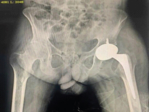
Post-operative radiographs demonstrated a well-positioned acetabular and femoral component with satisfactory alignment. Both components were stable and well-seated, with appropriate restoration of limb length and offset (Fig. 4).
Clinically, the patient reported significant pain relief in the early post-operative period. Given the compromised bone quality, immediate full weight-bearing was deferred. The patient was started on teriparatide therapy to support bone metabolism. Toe-touch weight-bearing was initiated at 2 weeks, followed by progression to full weight-bearing at 4 weeks.
Rehabilitation was uneventful. At 1 month postoperatively, the patient demonstrated progressive improvement in mobility and pain. At 3 months, the patient had satisfactory functional recovery, with a HHS of 78/100 from the initial 25/100. At 6 months postoperatively, the patient demonstrated further functional improvement, with a HHS of 90/100. The patient reported no subjective instability and demonstrated a satisfactory gait without clinically evident abductor insufficiency. Radiographs show no evidence of loosening, osteolysis, heterotopic ossification, or periprosthetic fracture. Limb lengths were clinically comparable. However, the duration of follow-up remains insufficient to assess long-term complications, including implant wear, aseptic loosening, and implant survivorship.
Discussion
Femoral neck fractures in young patients present a significant clinical challenge due to the need for durable functional restoration while preserving the native hip joint [5]. The complexity increases substantially in neglected fractures, where delayed presentation is associated with higher rates of non-union and femoral head compromise [6]. The presence of pathological bone, particularly in the setting of metabolic disorders such as APS, further complicates management by impairing bone quality and healing potential [7]. These combined factors make treatment decisions more demanding and often necessitate deviation from standard head-preserving strategies.
The present case is distinct due to the combination of a neglected fracture, compromised bone quality, and underlying systemic disease. The delayed presentation significantly reduces the potential for fracture union and increases the risk of femoral head resorption. In addition, the presence of severe osteoporosis further compromises the structural integrity of bone, limiting the success of conventional fixation strategies. The coexistence of APS adds another layer of complexity, as metabolic disturbances adversely affect bone turnover and healing, while also necessitating careful perioperative endocrine management. Together, these factors make this a non-routine scenario requiring an individualized surgical approach.
Head-preserving procedures such as internal fixation were considered suboptimal in the present case due to the significantly increased risk of failure. Neglected femoral neck fractures are known to have poor biological healing potential, with delayed presentation associated with higher rates of non-union and femoral head collapse [8]. This risk is further exacerbated in the presence of severe osteoporosis, where reduced bone mineral density compromises screw purchase and fixation stability [9]. In addition, pathological bone conditions adversely affect the vascularity of the femoral head, increasing the likelihood of avascular necrosis [10]. Previous studies have demonstrated that outcomes of internal fixation deteriorate significantly in delayed presentations and in the presence of compromised bone quality, with higher rates of fixation failure and need for revision surgery [11]. In view of these factors, internal fixation in this scenario was associated with an unacceptably high risk of non-union, avascular necrosis, and mechanical failure. Alternative head-preserving options, including valgus osteotomy and vascularized fibular grafting, were also considered; however, their applicability was limited by the neglected fracture, severe osteoporosis, and compromised femoral head.
Bipolar hemiarthroplasty was also considered less suitable in this case. In young and active patients, hemiarthroplasty is associated with a higher risk of progressive acetabular cartilage wear due to continued articulation between the prosthetic head and native acetabulum, leading to pain and functional deterioration over time [12]. Furthermore, long-term outcomes of hemiarthroplasty in younger individuals are generally inferior to THA, with increased likelihood of revision surgery due to acetabular erosion and persistent symptoms [13]. Given the patient’s age, functional demands, and expected longevity, bipolar hemiarthroplasty was deemed a less durable solution.
In the present case, functional recovery was assessed using the HHS. The score improved from 25/100 preoperatively to 78/100 at 3 months and 90/100 at 6 months, demonstrating substantial early functional improvement following THA. THA was preferred in this case as it provides more reliable pain relief and superior functional outcomes compared to other treatment options in complex scenarios. THA has been shown to yield consistent results in cases with poor bone quality and neglected femoral neck fractures, where biological healing potential is compromised. In young patients, although concerns regarding implant longevity exist, several studies have demonstrated favorable functional outcomes and acceptable survivorship when appropriately indicated [14]. Although alternative head-preserving and arthroplasty strategies were considered and discussed, this single-case report was not designed to provide a direct comparative analysis of these treatment modalities.
Implant selection in this case was guided by the underlying poor bone quality and altered femoral anatomy. On the femoral side, a cemented stem was preferred as severe osteoporosis precluded adequate press-fit fixation, and cemented stems have been shown to provide more reliable fixation and reduced risk of early loosening in osteoporotic femurs [15]. The use of a CDH-type stem further facilitated optimal fit within the narrow femoral canal, which is particularly advantageous in stenotic canals and helps reduce the risk of intraoperative fracture while maintaining proper alignment [16]. The use of a cemented femoral stem in a 34-year-old patient warrants careful consideration. Although cemented fixation provided immediate stability in the setting of severe osteoporosis and a narrow femoral canal, concerns remain regarding long-term implant survivorship and the potential complexity of future revision surgery.
On the acetabular side, a cementless multihole cup was selected despite osteoporotic bone, given the patient’s young age and the advantages of long-term biological fixation [17]. Cementless acetabular components have demonstrated superior long-term survivorship in younger patients compared to cemented cups, while the addition of supplementary screw fixation in multihole designs improves primary stability in compromised bone stock [18].
In addition, a dual mobility articulation system was utilized to reduce the risk of post-operative instability. Dual mobility constructs have been widely reported to significantly lower dislocation rates, particularly in high-risk cases such as poor soft tissue envelope, complex primary arthroplasty, and young active individuals, while maintaining good functional outcomes [19].
Given the severe osteoporosis (DEXA T-score −3) and underlying metabolic bone disease, a cautious rehabilitation protocol was adopted. Toe-touch weight-bearing was initiated at 2 weeks, followed by progression to full weight-bearing at 4 weeks based on clinical and radiographic assessment. Although no validated protocol exists for patients with APS-associated osteoporosis undergoing THA for neglected femoral neck fractures, protected early weight-bearing has been described in osteoporotic hip reconstruction to minimize excessive mechanical stress during the initial post-operative period. Teriparatide was prescribed as an adjunctive therapy because of its anabolic effects on bone and its reported ability to preserve periprosthetic bone mineral density and improve implant stability in osteoporotic patients undergoing hip arthroplasty.
Osteoporotic bone is inherently fragile, with reduced cortical thickness and trabecular support, making it highly susceptible to intraoperative fractures during dislocation, preparation, and implantation. Therefore, gentle handling techniques, including controlled dislocation and avoidance of excessive force transmission through the limb, are essential to minimize the risk of iatrogenic fracture and ensure safe component placement [20].
Several studies have demonstrated satisfactory functional outcomes and survivorship of THA in younger individuals when appropriate indications are met. Nevertheless, there is a paucity of literature addressing THA in the setting of systemic endocrine disorders such as APS, especially when combined with neglected pathological femoral neck fractures. Most available studies focus either on fracture management in young patients or arthroplasty outcomes in isolation, with limited guidance on managing such complex, multifactorial cases [21]. This highlights the need for individualised surgical strategies in these rare clinical scenarios.
THA can be a justified and effective treatment option in young patients with neglected pathological femoral neck fractures when the likelihood of successful head-preserving procedures is low [22]. Implant selection should be individualized based on bone quality, anatomical considerations, and intraoperative findings to ensure optimal fixation and long-term outcomes [23]. Furthermore, multidisciplinary management, particularly in patients with underlying endocrine disorders such as APS, is essential to optimize perioperative care and improve overall outcomes [24].
This study has certain limitations. This report describes the management of a single patient and therefore the findings should be interpreted with caution. The unique combination of APS, severe osteoporosis, and a neglected femoral neck fracture makes direct extrapolation to other patients difficult. The present case is intended to highlight surgical decision-making and technical considerations in a rare clinical scenario rather than establish treatment recommendations applicable to all young patients with pathological femoral neck fractures.
Furthermore, the follow-up duration of 6 months is insufficient to evaluate long-term implant performance. Important outcomes such as aseptic loosening, polyethylene wear, osteolysis, periprosthetic fracture, recurrent instability, deep infection, implant survivorship, and the need for revision surgery cannot be assessed within this timeframe. Continued long-term surveillance is required to determine the durability of the reconstruction.
Functional outcome was assessed using the HHS, which improved from 25/100 preoperatively to 78/100 at 3 months and 90/100 at 6 months. However, these assessments were obtained in the context of a single case and relatively short follow-up, limiting the ability to draw conclusions regarding long-term functional outcomes.
Furthermore, being a technical note, the conclusions are based on clinical judgment and intraoperative experience rather than comparative analysis with alternative treatment modalities. Larger studies with longer follow-up are required to validate these findings.
Conclusion
Neglected pathological femoral neck fractures in young patients with APS represent a complex clinical scenario due to compromised bone quality and limited healing potential. This case demonstrates the feasibility of an individualized THA strategy for the management of a neglected pathological femoral neck fracture in a young patient with APS and severe osteoporosis. Satisfactory early clinical and functional outcomes were observed; however, longer follow-up and larger studies are required to establish the durability, safety, and generalizability of this approach.
Clinical Message
Neglected pathological femoral neck fractures in young patients with Autoimmune Polyglandular Syndrome require individualized management rather than routine head-preserving surgery. When severe osteoporosis, delayed presentation, and compromised healing make internal fixation unreliable, THA with appropriate implant selection, meticulous surgical technique, and multidisciplinary endocrine optimization can provide stable fixation, restore function, and achieve satisfactory early clinical outcomes.
Conflict of Interest:
Nil
Source of Support:
Nil
Consent:
The authors confirm that informed consent was obtained from the patient for publication of this article
How to Cite this Article
Chowdhury PN, Medhi M, Kaithoriya P, Malhotra R. Total Hip Arthroplasty for Neglected Pathological Femoral Neck Fracture in a Young Patient with Autoimmune Polyglandular Syndrome. Journal of Orthopaedic Case Reports 2026 October;16(10): 353-360.
References
- Ly TV, Swiontkowski MF. Management of femoral neck fractures in young adults. Indian J Orthop 2008;42:3-12. [Google Scholar] | [PubMed]
- Upadhyay A, Jain P, Mishra P, Maini L, Gautum VK, Dhaon BK. Delayed internal fixation of fractures of the neck of the femur in young adults. A prospective, randomised study comparing closed and open reduction. J Bone Joint Surg Br 2004;86:1035-40. [Google Scholar] | [PubMed]
- Bakkour A, Zakkor MD, Taha Khairy L, Horo R, Mohammed Sharif Ahmed E, Alhussein H. Autoimmune polyglandular syndrome type 2: A case report. Ann Med Surg (Lond) 2022;78:103742. [Google Scholar] | [PubMed]
- Pakos EE, Paschos NK, Xenakis TA. Long term outcomes of total hip arthroplasty in young patients under 30. Arch Bone Jt Surg 2014;2:157-62. [Google Scholar] | [PubMed]
- Gautam VK, Anand S, Dhaon BK. Management of displaced femoral neck fractures in young adults (a group at risk). Injury 1998;29:215-8. [Google Scholar] | [PubMed]
- Elmi A, Tabrizi A, Rouhani A, Mirzatolouei F. Long-term follow-up results of delayed fixation of femoral neck fractures in adults. Trauma Mon 2013;18:8-11. [Google Scholar] | [PubMed]
- Sperling MA, Angelousi A, Yau M. Autoimmune Polyglandular SyndromesFeingold KR, Adler RA, Ahmed SF, Anawalt B, Blackman MR, Chrousos G. Endotext. South Dartmouth, MA: MDText.com, Inc.; 2000. Available from: https://www.ncbi.nlm.nih.gov/books/nbk279152. [Last accessed on 2026 Apr 19].. [Google Scholar] | [PubMed]
- Jaya Raj J, Kow RY, Ganthel Annamalai K, Kunasingh DE, Panicker GK, Lim BC. Outcomes of femoral neck fractures in young patients and the factors associated with complications: A multicenter study from Malaysia. Cureus 2021;13:e18110. [Google Scholar] | [PubMed]
- Mukhopadhaya J, Bhadani JS. Fixation failure in osteoporotic bone: A review of complications and outcomes. Indian J Orthop 2025;59:389-404. [Google Scholar] | [PubMed]
- Konarski W, Poboży T, Śliwczyński A, Kotela I, Krakowiak J, Hordowicz M. Avascular necrosis of femoral head-overview and current State of the art. Int J Environ Res Public Health 2022;19:7348. [Google Scholar] | [PubMed]
- Dhar SA, Ali MF, Dar TA, Sultan A, Butt MF, Kawoosa AA. Delayed fixation of the transcervical fracture of the neck of the femur in the pediatric population: Results and complications. J Child Orthop 2009;3:473-7. [Google Scholar] | [PubMed]
- alotico GG, Matsunaga FT, Filho JS, Moraes VY, Garcia MS, Faloppa F. Total hip arthroplasty versus hemiarthroplasty for displaced femoral neck fracture: An overview of systematic reviews total hip arthroplasty versus hemiarthroplasty for displaced femoral neck fracture: An overview of systematic reviews. J Orthop Surg Res 2025;20:718. [Google Scholar] | [PubMed]
- Abe T, Kaku N, Tabata T, Tagomori H, Tsumura H. Long-term clinical results of bipolar hemiarthroplasty for osteoarthritis and rheumatoid arthritis of the hip: A retrospective study. J Orthop 2020;17:120-3. [Google Scholar] | [PubMed]
- Mei XY, Gong YJ, Safir O, Gross A, Kuzyk P. Long-term outcomes of total hip arthroplasty in patients younger than 55 years: A systematic review of the contemporary literature. Can J Surg 2019;62:249-58. [Google Scholar] | [PubMed]
- Traina F, Tassinari E, Pilla F, De Fine M, Biondi F, Cristofolini L. Outcome of hybrid stem fixation in osteoporotic female patients. A minimum five-year follow-up study. Int Orthop 2009;33:1489-94. [Google Scholar] | [PubMed]
- Salentiny Y, Zwicky L, Ochsner PE, Clauss M. Arch Orthop Trauma Long-term survival of the cemented Müller CDH stem: A minimum follow-up of 10 years Surg 2018;138:1471-7. [Google Scholar] | [PubMed]
- Callaghan J, Teusink M, Donigan J, Warth L, Liu S, Sullivan J. Is cementless acetabular fixation an improvement in total hip arthroplasty performed in younger patients? J Arthroplasty 2007;22:310. [Google Scholar] | [PubMed]
- Fei C, Wang PF, Wei W. Relationship between use of screws and acetabular cup stability in total hip arthroplasty: a meta-analysis. J Int Med Res. 2020;48(2):300060520903649. [Google Scholar] | [PubMed]
- Suarez-Ahedo C, Quintero-Quintero MA, Martinez-Armenta C, Martínez-Gómez LE, Martínez-Nava GA, Pineda C. Modern dual mobility implants in total hip arthroplasty: Stability, survival, and ion safety evaluation: The comprehensive review. JB JS Open Access 2025;10:e2500141. [Google Scholar] | [PubMed]
- Young PS, Patil S, Meek RM. Intraoperative femoral fractures: Prevention is better than cure. Bone Joint Res 2018;7:103-4. [Google Scholar] | [PubMed]
- Dittmar M, Kahaly GJ. Polyglandular autoimmune syndromes: Immunogenetics and long-term follow-up. J Clin Endocrinol Metab 2003;88:2983-92. [Google Scholar] | [PubMed]
- Jain AK, Mukunth R, Srivastava A. Treatment of neglected femoral neck fracture. Indian J Orthop 2015;49:17-27. [Google Scholar] | [PubMed]
- López-López JA, Humphriss RL, Beswick AD, Thom HH, Hunt LP, Burston A. Choice of implant combinations in total hip replacement: Systematic review and network meta-analysis. BMJ 2017;359:j4651. [Google Scholar] | [PubMed]
- Himes CP, Ganesh R, Wight EC, Simha V, Liebow M. Perioperative evaluation and management of endocrine disorders. Mayo Clin Proc 2020;95:2760-74. [Google Scholar] | [PubMed]
© 2026 Journal of Orthopaedic Case Reports - Published by Indian Orthopaedic Research Group
About the Authors
-

Plaban Nath Chowdhury
-

Mridushman Medhi
-

Prateek Kaithoriya
-

Rajesh Malhotra
Related Articles in Journal of Orthopaedic Case Reports
 October 1, 2026 Total Hip Arthroplasty in the Adolescent Population: A Case Series
 October 1, 2026 Posterior Acetabular Reconstruction Can Sometimes Lead To Retroverted Cup Placement In Complex Total Hip Arthroplasty
 October 1, 2026 Humeral Fractures in Metabolically Compromised Bone: Outcomes of Structural Strut Onlay Allograft with Open Reduction and Internal Fixation – A Case Series of Three Patients
 October 1, 2026 Dual Mobility Total Hip Arthroplasty for Recurrent Dislocation After Femoral Neck Fracture in an Elderly Patient: A Case Report