


Introduction
Acetabular fractures are complex injuries that can substantially complicate subsequent total hip arthroplasty (THA). One critical challenge is the management of posterior acetabular bone loss, which predisposes the acetabular component to retroversion. Retroversion, defined as posterior tilt of the acetabular component relative to the sagittal plane, adversely affects implant stability, hip biomechanics, and overall patient outcomes [1].
Posterior bone loss strongly influences acetabular cup orientation. When posterior bone support is compromised, the acetabular component tends to retrovert, increasing contact stresses and edge loading, accelerating polyethylene wear, and predisposing the joint to instability [2,3,4,5]. Retroverted acetabular components are also linked with a heightened risk of anterior impingement, contributing to dislocation and a reduced range of motion [6,7].
Addressing posterior acetabular bone loss requires meticulous pre-operative planning, usually including computed tomography (CT) to assess bone stock and to plan the orientation of the acetabular component. Intraoperative options include structural bone grafting, augmented acetabular components, and custom implants to restore orientation and secure stable fixation [8]. Despite these measures, the long-term outcome of THA in patients with severe posterior bone loss remains a concern, with a higher reported risk of complications than in hips with adequate acetabular bone stock [9].
In this series, we describe three patients in whom uncontained posterosuperior bone loss after an acetabular fracture resulted in retroversion of the acetabular cup at THA despite femoral head structural autograft reconstruction of the posterior wall.
Case Report
Case 1
A 47-year-old male presented 8 months after a road traffic accident that had resulted in a posterior wall fracture of the acetabulum with an associated femoral head fracture and posterior hip dislocation. He had been managed conservatively elsewhere and had no neurological deficit.
Pre-operative radiographs and CT (Fig. 1a, Fig. 1b, c, d) showed a healed posterior wall fracture with significant posterosuperior bone loss (Paprosky grade 2B, American Academy of Orthopaedic Surgeons [AAOS] segmental posterior and superior, Gross type 3). The acetabulum had collapsed posteriorly, and there was dense periarticular fibrosis. THA was planned in view of post-traumatic arthritis and a restricted range of motion.
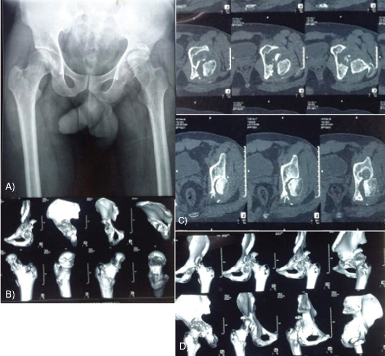
Intraoperatively, the periarticular fibrosis made exposure and positioning of the acetabular cup difficult. C-arm guidance and anatomical landmarks, in particular Ranawat’s triangle, were used to determine cup position. The posterosuperior defect was reconstructed with a femoral head autograft fixed with two partially threaded cannulated cancellous (CC) screws, after which a porous-coated hemispherical acetabular shell with a 10° elevated-rim liner was implanted. An uncemented femoral stem was used, and the hip was reduced.
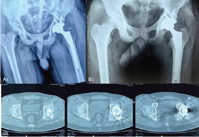
Post-operative radiographs confirmed stable fixation (Fig. 2a). At 12 months, the patient reported good functional recovery with mild discomfort during certain activities, and CT demonstrated mild retroversion of the acetabular cup (Fig. 2b and c).
Case 2
A 35-year-old male presented 3 months after a road traffic accident in which he had sustained a posterior wall and posterior column fracture of the acetabulum with a femoral head fracture and dislocation. He complained of persistent pain and restricted hip mobility.
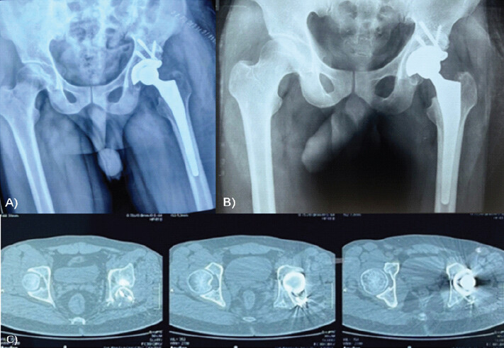
He had been managed conservatively elsewhere, but because of progressive pain and difficulty in ambulation, THA was planned. Pre-operative imaging (Fig. 3a, b, c) showed significant bone loss of the posterosuperior wall (Paprosky grade 2B, AAOS segmental posterior and superior, Gross type 3) with residual deformity from the column fracture.
The posterior wall was reconstructed with femoral head autograft and partially threaded CC screws. A porous-coated hemispherical acetabular shell with a 10° elevated-rim liner was implanted, and the procedure was completed with an uncemented femoral stem and reduction of the hip.
Post-operative radiographs showed adequate fixation of both components (Fig. 4a). At follow-up, the patient reported improvement in pain and hip mobility, although a slight gait abnormality was noted. CT confirmed a retroverted cup (Fig. 4b and c). He remains under close observation for signs of instability or wear.
Case 3
A 59-year-old male sustained a posterior wall and posterior column fracture with dislocation in a road traffic accident 2 months before evaluation. The fracture had been managed conservatively elsewhere, but he continued to have significant pain and functional limitation.
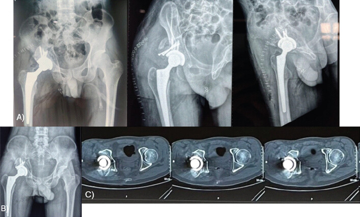
Pre-operative imaging (Fig. 5a and b) demonstrated severe posterior bone loss (Paprosky grade 2B, AAOS segmental posterior and superior, Gross type 3) with displacement of the posterior column. At surgery, the absence of a stable posterior wall necessitated reconstruction with a femoral head autograft fixed with partially threaded CC screws and a reconstruction plate (Fig. 5c).
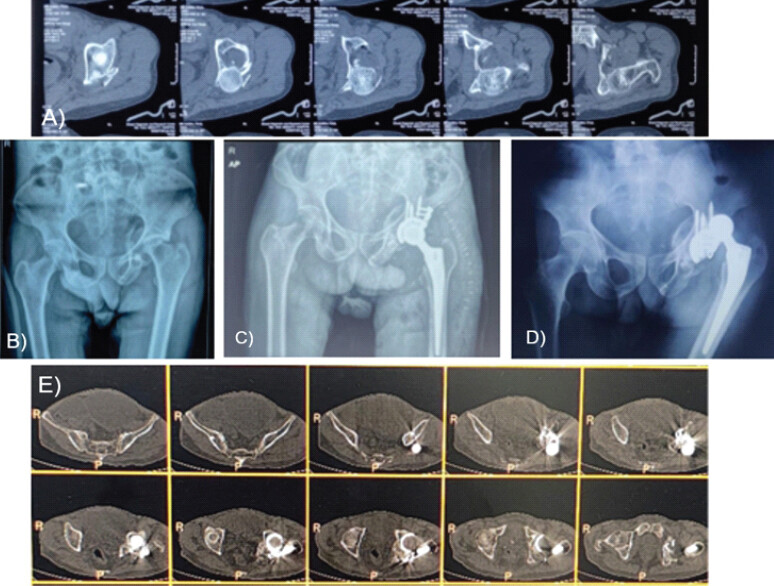
Recovery was uneventful, and at follow-up he reported significant improvement in pain and range of motion. Two months after THA, however, he presented with posterior dislocation of the prosthetic hip. CT showed retroversion of the acetabular cup (Fig. 5d). Closed reduction was performed, and the patient was kept under regular review.
The key characteristics of the three cases are summarized in Table 1.
| Case | Age (years)/Sex | Fracture pattern | Bone defect (Paprosky/AAOS/Gross) | Reconstruction | Cup retroversion | Complication | Outcome |
|---|---|---|---|---|---|---|---|
| 1 | 47/M | Posterior wall+femoral head fracture+dislocation (Pipkin 4) | 2B/Segmental posterior and superior/Type 3 | FH autograft+two CC screws | 5° | Mild discomfort | Good function at 12 months |
| 2 | 35/M | Posterior wall+column+femoral head fracture+dislocation (Pipkin 4) | 2B/Segmental posterior and superior/Type 3 | FH autograft+two CC screws | 19° | Slight gait abnormality | Improved pain and mobility; under observation |
| 3 | 59/M | Posterior wall+column+dislocation | 2B/Segmental posterior and superior/Type 3 | FH autograft+CC screws+reconstruction plate | 7° | Posterior dislocation at 2 months (closed reduction) | Improved ROM and pain; under regular review |
AAOS: American Academy of Orthopaedic Surgeons, CC: Cannulated cancellous, FH: Femoral head, M: Male, ROM: Range of motion, THA: Total hip arthroplasty
Discussion
A search of MEDLINE through PubMed (from inception to August 2026) using the keywords “acetabular fracture”, “total hip arthroplasty”, “posterior bone loss”, “acetabular cup version”, “retroversion”, and “structural autograft”, together with a review of the bibliographies of the retrieved articles, did not identify any report specifically addressing persistent cup retroversion after femoral head autograft reconstruction of an uncontained posterior wall defect.
In our series, two patients had femoral head fractures associated with acetabular wall and column fractures (Pipkin type 4), and one had a posterior wall and column fracture alone. As all three presented late, THA was the more viable option, and the posterior (Kocher–Langenbeck) approach was used throughout. A recent meta-analysis showed that primary THA after an acetabular fracture is associated with notably lower rates of post-operative complications and better functional outcomes than conversion THA [10].
All three hips had uncontained posterosuperior defects (Paprosky grade 2B, AAOS segmental posterior and superior, Gross type 3). Gross and Goodman noted that uncontained defects involving <50% of the acetabulum (type III) can be managed with a structural graft; alternatives include a high hip center, an oblong or asymmetric cup, and metal augments. The advantage of a structural graft is that it restores the anatomical center of rotation and preserves bone stock for future surgery. Because such grafts support <50% of the acetabular component, uncemented cups can be used without the additional support of a ring or cage. These minor column or shelf grafts have shown excellent long-term results, with a 94% survival rate at 10 years [11].
We used femoral head autograft fixed with two partially threaded CC screws in all three hips. Superior and posterosuperior segmental defects usually retain a residual bony shelf that supports the graft, and lag-screw fixation alone is generally sufficient. In the third patient, the autograft and the remaining posterior wall were supplemented with a reconstruction plate for additional support. After fixation, the inner surface of the autograft and the acetabular bed were contoured with reamers. A porous-coated hemispherical acetabular cup supplemented with screws, an uncemented femoral stem, and a 10° elevated-rim liner were used in every case.
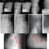
Loss of posterior support has several implications for THA, including difficulty in maintaining correct cup orientation and a tendency for the acetabular component to assume a retroverted position. Retroversion most probably occurs when the surgeon attempts to obtain a press fit from the superior and posterior walls in an inadequately reconstructed posterior wall. Retroversion was confirmed by CT in all three hips, which showed mild-to-moderate retroversion of the acetabular cup (Case 1, 5°; Case 2, 19°; and Case 3, 7° from neutral) (Fig. 6).
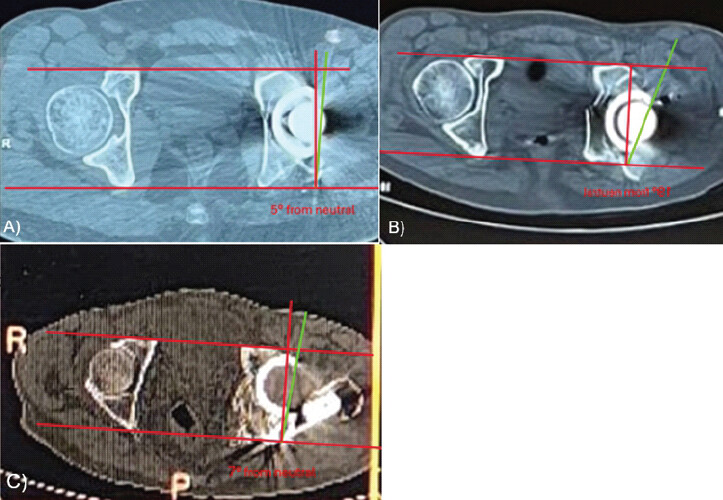
A retroverted cup can result in several mechanical problems. Anterior impingement may occur between the femoral neck and the anterior rim of the acetabular component during flexion, and the altered mechanics of the joint increase the risk of dislocation, particularly during activities involving hip flexion and internal rotation [12,13]. One of our patients developed a posterior prosthetic dislocation 2 months after THA. Lai et al. similarly reported three dislocations among 31 hips after THA for acetabular fracture [14]. A retroverted cup also alters the distribution of forces across the joint, potentially accelerating wear of the acetabular liner and contributing to early loosening of the implant [15].
Although the short-term outcomes in this series were relatively favorable, the long-term consequences of cup retroversion remain a concern. Retroversion has been associated with increased wear, liner edge loading, and impingement, and these patients may therefore face a higher risk of dislocation and of revision surgery for wear-related problems or loosening. Prosthesis durability and the occurrence of late complications in this cohort will be addressed in a future study.
Conclusion
Posterior bone loss in acetabular fractures presents a significant challenge during THA, particularly in achieving correct orientation of the acetabular component. Retroversion of the cup is a frequent outcome in these hips and is associated with increased mechanical risk, including impingement, instability, and altered load distribution. This series underlines the importance of thorough pre-operative planning and of intraoperative techniques that minimize the degree of retroversion. Future work should focus on surgical techniques and implants that better address posterior bone loss in acetabular fractures.
Clinical Message
In acetabular fractures with substantial posterior bone loss, surgeons performing total hip arthroplasty should anticipate a high likelihood of cup retroversion even after structural autograft reconstruction. Intraoperative confirmation of cup version using fluoroscopy and anatomical landmarks such as Ranawat’s triangle is essential. Where posterior support is inadequate, additional measures such as enhanced posterior buttress reconstruction, navigation assistance, or stability-enhancing implants, including dual-mobility bearings, should be considered to reduce the risk of impingement and dislocation. Close post-operative monitoring with cross-sectional imaging is advisable in these complex reconstructions.
Conflict of Interest:
Nil
Source of Support:
Nil
Consent:
The authors confirm that informed consent was obtained from the patient for publication of this article
How to Cite this Article
Agrawal AC, Chauhan S, Singh J, Jayan JT, Bhattacharyya S, Temker DM. Posterior Acetabular Reconstruction Can Sometimes Lead To Retroverted Cup Placement In Complex Total Hip Arthroplasty. Journal of Orthopaedic Case Reports 2026 October;16(10):129-136.
References
- Barrack RL, Lavernia C, Ries M, Thornberry R, Tozakoglou E. Virtual reality computer animation of the effect of component position and design on stability after total hip arthroplasty. Orthop Clin North Am 2001;32:569-77, vii. [Google Scholar] | [PubMed]
- Lewinnek GE, Lewis JL, Tarr R, Compere CL, Zimmerman JR. Dislocations after total hip-replacement arthroplasties. J Bone Joint Surg Am 1978;60:217-20. [Google Scholar] | [PubMed]
- Kennedy JG, Rogers WB, Soffe KE, Sullivan RJ, Griffen DG, Sheehan LJ. Effect of acetabular component orientation on recurrent dislocation, pelvic osteolysis, polyethylene wear, and component migration. J Arthroplasty 1998;13:530-4. [Google Scholar] | [PubMed]
- Blondel B, Parratte S, Tropiano P, Pauly V, Aubaniac JM, Argenson JN. Pelvic tilt measurement before and after total hip arthroplasty. Orthop Traumatol Surg Res 2009;95:568-72. [Google Scholar] | [PubMed]
- Dorr LD, Malik A, Dastane M, Wan Z. Combined anteversion technique for total hip arthroplasty. Clin Orthop Relat Res 2009;467:119-27. [Google Scholar] | [PubMed]
- Callanan MC, Jarrett B, Bragdon CR, Zurakowski D, Rubash HE, Freiberg AA. The John Charnley award: Risk factors for cup malpositioning: Quality improvement through a joint registry at a tertiary hospital. Clin Orthop Relat Res 2011;469:319-29. [Google Scholar] | [PubMed]
- Esposito CI, Gladnick BP, Lee YY, Lyman S, Wright TM, Mayman DJ. Cup position alone does not predict risk of dislocation after hip arthroplasty. J Arthroplasty 2015;30:109-13. [Google Scholar] | [PubMed]
- Murphy SB. Management of acetabular bone stock deficiency. J Arthroplasty 2005;20 4 Suppl 2:85-90. [Google Scholar] | [PubMed]
- Alberton GM, High WA, Morrey BF. Dislocation after revision total hip arthroplasty: An analysis of risk factors and treatment options. J Bone Joint Surg Am 2002;84:1788-92. [Google Scholar] | [PubMed]
- Shaker F, Esmaeili S, Nakhjiri MT, Azarboo A, Shafiei SH. The outcome of conversion total hip arthroplasty following acetabular fractures: A systematic review and meta-analysis of comparative studies. J Orthop Surg Res 2024;19:83. [Google Scholar] | [PubMed]
- Gross AE, Goodman S. The current role of structural grafts and cages in revision arthroplasty of the hip. Clin Orthop Relat Res 2004;429:193-200. [Google Scholar] | [PubMed]
- Makridis KG, Obakponovwe O, Bobak P, Giannoudis PV. Total hip arthroplasty after acetabular fracture: Incidence of complications, reoperation rates and functional outcomes: Evidence today. J Arthroplasty 2014;29:1983-90. [Google Scholar] | [PubMed]
- Raj M, Jaiman A, Chopra RK. Evaluation of version of acetabular component following total hip replacement on conventional radiograph and computed tomography. J Orthop Trauma Rehabil 2020;27:179-85. [Google Scholar] | [PubMed]
- Lai O, Yang J, Shen B, Zhou Z, Kang P, Pei F. Midterm results of uncemented acetabular reconstruction for posttraumatic arthritis secondary to acetabular fracture. J Arthroplasty 2011;26:1008-13. [Google Scholar] | [PubMed]
- Sai Sathikumar A, Jacob G, Thomas AB, Varghese J, Menon V. Acetabular cup positioning in primary routine total hip arthroplasty-a review of current concepts and technologies. Arthroplasty 2023;5:59. [Google Scholar] | [PubMed]
© 2026 Journal of Orthopaedic Case Reports - Published by Indian Orthopaedic Research Group
About the Authors
-

Alok Chandra Agrawal
-

Shivam Chauhan
-

Jagprit Singh
-

Jayakrishnu Thundathil Jayan
-

Sunit Bhattacharyya
-

Darshan Madan Temker
Related Articles in Journal of Orthopaedic Case Reports
 December 1, 2025 Total Hip Arthroplasty for Neglected Hip Dislocation – Surgical Technique and Results
 September 1, 2025 Total Hip Arthroplasty with Cage Application for Complex Posterior Hip Dislocation and Acetabular Fracture in a Patient with Prior Patellectomy: A Case Report
 October 1, 2026 A Study of Spinopelvic Relationship and its Impact on Functional Outcome of Total Hip Arthroplasty
 October 1, 2026 Late Diagnosis of Femoral Shaft Perforation During Total Hip Arthroplasty: Case Report