


Introduction
Insertional Achilles tendinopathy (IAT) is a common cause of posterior heel pain in active individuals and accounts for approximately one-fifth to one-third of Achilles tendon disorders [1,2]. It is characterized by degenerative changes at the calcaneal insertion and is often associated with intratendinous calcification, posterosuperior calcaneal prominence (Haglund deformity), and retrocalcaneal bursitis [2]. Tightness of the gastrocnemius–soleus complex, assessed clinically using the Silfverskiöld test, is increasingly recognized as a contributor to the pathogenesis of insertional disease and to the associated bony and intratendinous changes [3].
First-line management is non-operative and includes activity modification, heavy slow-resistance loading and eccentric strengthening, extracorporeal shockwave therapy and, in selected athletes, injectable treatments such as platelet-rich plasma (PRP). Surgery is generally reserved for patients with persistent symptoms despite at least 6 months of well-conducted conservative treatment [2]. Isolated endoscopic calcaneoplasty can reliably address bony impingement but does not adequately treat intratendinous calcification or significant tendon degeneration [4,5]. In such cases, tendon detachment and reinsertion may be indicated. When more than half of the insertion is detached, double-row suture-bridge fixation is recommended because it restores a broad insertional footprint and provides greater biomechanical strength than single-row repair [6], with favorable clinical and radiological outcomes reported [7,8].
Open reinsertion, however, is associated with wound-healing complications and slower recovery, both of which are particularly relevant in elite athletes. The endoscopic calcaneal SpeedBridge™, also known as “Snake technique,” performs calcaneoplasty, tendon debridement, and knotless double-row reinsertion through portals [9]. This minimally invasive approach has been associated with a lower complication rate [10] and faster early recovery than open surgery [11,12]. Concomitant gastrocnemius tightness can be addressed by proximal lengthening [13].
Despite increasing experience with minimally invasive surgery for IAT, return to sprinting at the highest level is rarely documented. In addition, whether an associated cavovarus hindfoot with a high calcaneal pitch requires correction remains controversial. We describe a single-stage minimally invasive surgery combining endoscopic SpeedBridge™ reinsertion with proximal medial gastrocnemius lengthening in an elite sprinter, with particular emphasis on return to the previous level of sport and on the deliberate decision not to correct the associated bony cavus.
Case Report
History and clinical examination
A 24-year-old elite international-level female track sprinter and non-smoker presented with chronic pain at the right Achilles tendon insertion, which had evolved over approximately 2 years and progressively limited both training and competition. A well-conducted conservative program, including physiotherapy with eccentric strengthening without dorsiflexion beyond neutral and heavy slow-resistance loading, and three PRP injections had failed to provide sustained relief. On clinical examination, she had a structural pes cavus and a positive Silfverskiöld test: Ankle dorsiflexion measured 25° with the knee flexed but decreased to 0° with the knee extended, consistent with isolated gastrocnemius tightness as the main driver of the insertional overload. Tenderness was localized to the calcaneal insertion of the Achilles tendon, without proximal main-body involvement.
Imaging
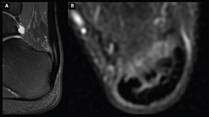
Weight-bearing lateral radiography of the foot demonstrated a calcaneal pitch of 40°, markedly above the usual range of approximately 20–25°, confirming the high-arched hindfoot (Fig. 1). Magnetic resonance imaging (MRI) showed insertional tendinopathy with intratendinous calcification, without major retrocalcaneal bursitis or bone-marrow edema, findings that may be associated with a poorer prognosis when present (Fig. 2).
Figure 1: Pre-operative weight-bearing lateral radiograph of the right foot showing a calcaneal pitch of 40° (high-arched hindfoot; normal approximately 20–25°).
Surgical technique
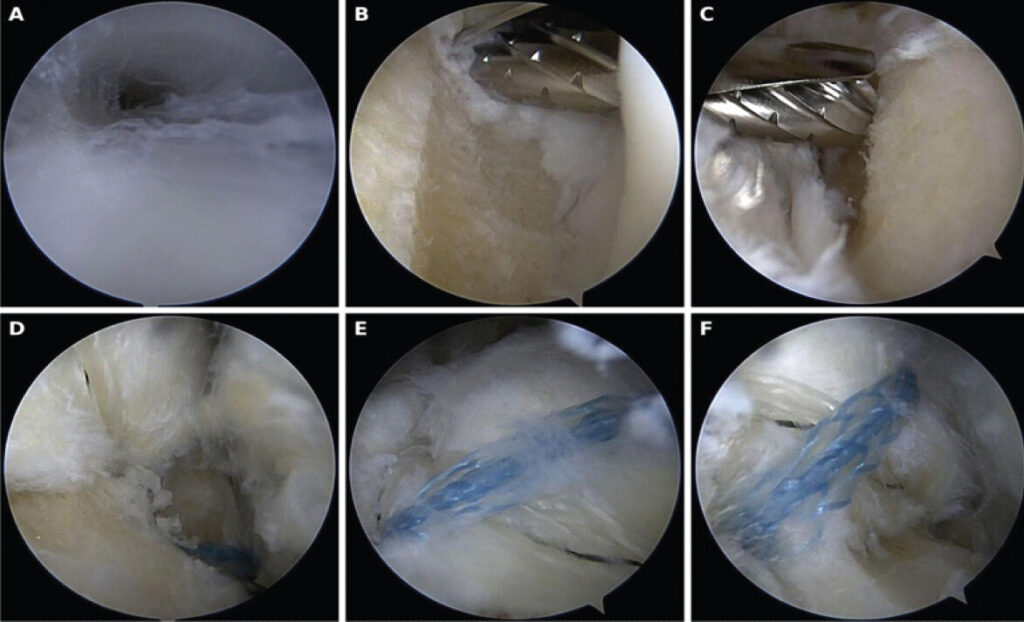
Under spinal anesthesia, with the patient prone and a thigh tourniquet applied, a single-stage two-part procedure was performed. First, proximal medial gastrocnemius lengthening was carried out through a transverse popliteal approach, with longitudinal section of the crural fascia and proximal aponeurotomy of the medial gastrocnemius (Barouk technique), followed by layered closure. Second, endoscopic calcaneal SpeedBridge™ reinsertion was performed [9]. Posterolateral and posteromedial portals, as described for posterior ankle arthroscopy by Van Dijk, allowed tibiotalar and subtalar release and creation of a working space with a 3.5-mm shaver. The peroneal tendons and flexor hallucis longus were identified as lateral and medial anatomical limits, respectively. The conflicting posterosuperior calcaneal corner was resected (calcaneoplasty), and the intratendinous calcifications were debrided (Fig. 3). Through an accessory distal midline portal, the distal central portion of the Achilles tendon was detached from its calcaneal insertion. A knotless double-row repair was then performed using 4.75-mm SwiveLock anchors (Arthrex, Naples, FL) and FiberTape (Arthrex, Naples, FL), with the tape passed transtendinously. The skin portals were closed with simple sutures.
Post-operative course and rehabilitation
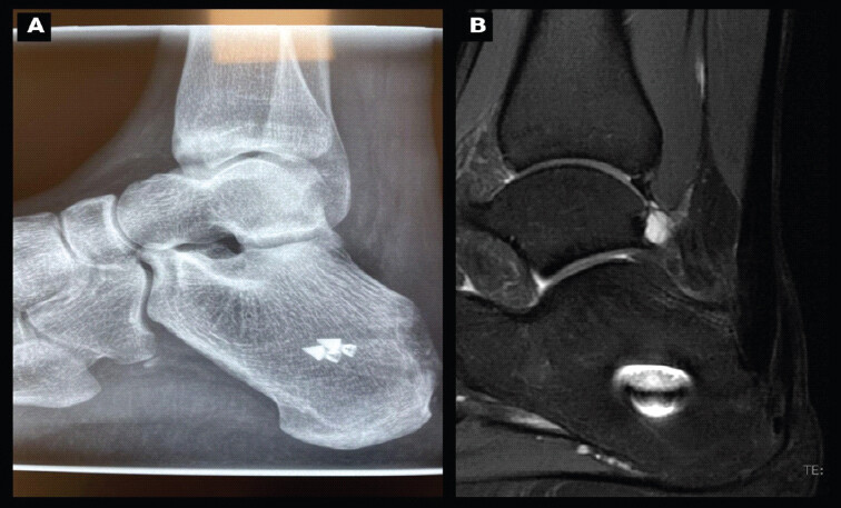
Immediate full weight-bearing was permitted in a walking boot, which was worn day and night for 1 month, in accordance with functional rehabilitation protocols reported after double-row reinsertion [14]. Thromboprophylaxis was prescribed for 21 days, and physiotherapy was initiated at 2 weeks, with emphasis on passive followed by active ankle dorsiflexion to optimize gastrocnemius lengthening. The early postoperative course was uneventful, with no wound complications and excellent scar healing. At 1 month, the patient was walking with a single crutch and a normal heel-to-toe pattern. Radiographs confirmed satisfactory anchor positioning (Fig. 4a).
Return to sport and outcomes
Rehabilitation progressed without complication. Running was reintroduced in water and on an anti-gravity treadmill from approximately 4 months postoperative, allowing video analysis and correction of running mechanics. The patient resumed jogging at 6 months, when the Victorian Institute of Sport Assessment–Achilles (VISA-A) score was 81, and follow-up MRI was normal (Fig. 4b). High-intensity running on grass was achieved by 9 months, with only transient discomfort at the distal anchor site when running on the track. Sprint-specific training, including starting-block starts, was resumed by 13 months, at which point the patient was pain-free. She returned to official competition at 16 months of follow-up, with a national level of performance. Twenty months after surgery, the patient was competing at the same chronometrical level as before intervention.
The clinical course and rehabilitation are summarized in Table 1. No surgical or thromboembolic complication occurred during follow-up.
Clinical course and rehabilitation milestones after combined surgery.
| Time point | Assessment | Findings / Milestone |
|---|---|---|
| Day 0 | Surgery | Proximal medial gastrocnemius lengthening + endoscopic calcaneal SpeedBridge™ (calcaneoplasty, debridement of calcifications, double-row reinsertion). Immediate full weight-bearing in walking boot. |
| 2 weeks | Rehabilitation | Physiotherapy started; passive then active ankle dorsiflexion. |
| 1 month | Clinical + radiograph | Uneventful healing; walking with one crutch, normal heel-to-toe gait; satisfactory anchor position. |
| 4 months | Clinical | No limp; running reintroduced in water / anti-gravity treadmill with gait-video analysis. |
| 6 months | MRI + PROM | Normal control MRI; jogging resumed; VISA-A 81. |
| 9 months | Clinical | High-intensity running on grass; transient discomfort at distal anchorage on the track. |
| 13 months | Clinical | Pain-free; sprint-specific training including starting- block starts resumed. |
| 16 months | Performance | Returned to official competition (national level of performance) |
| 20 months | Performance | Returned to pre-injury level (same chronometrical level as before surgery) |
Discussion
The main message of this case is twofold. First, a minimally invasive single-stage procedure combining endoscopic double-row Achilles tendon reinsertion with proximal medial gastrocnemius lengthening allowed an elite sprinter to return to official competition at 16 months. Second, a good functional result was achieved despite a markedly increased calcaneal pitch that was deliberately left uncorrected. The surgical strategy instead focused on the diseased tendon and the tight posterior muscle chain.
Surgical management for recalcitrant IAT remains debated, and no single technique has been shown to be superior across the literature [7, 8]. Isolated endoscopic calcaneoplasty can reliably address bony impingement but does not adequately treat intratendinous calcification or substantial tendon degeneration [4, 5]. In such cases, partial tendon detachment, debridement, and reinsertion may be required. When more than half of the insertion is detached, double-row fixation is generally preferred because it restores a broader insertional footprint and provides greater biomechanical strength than single-row repair [6]. The endoscopic SpeedBridge™ integrates these principles, allowing calcaneoplasty, tendon debridement, and knotless double-row reinsertion through portals [9].
Fradet and Lopes reported a lower post-operative complication rate with the endoscopic technique than with the open approach [10]. Similarly, a prospective multicenter study from the Francophone Arthroscopy Society found significantly better early VISA-A and functional scores with the endoscopic approach at 3 months, with comparable favorable outcomes thereafter [11]. A recent systematic review and meta-analysis also found comparable functional outcomes between open and endoscopic approaches, but with a lower complication rate and faster recovery after endoscopic surgery, enabling return to sport within 12–18 months in highly active individuals, compared with 20–30 months after open surgery [12]. The uneventful healing and progressive functional recovery observed in the present case are consistent with these findings and are particularly relevant in an elite athlete, for whom a posterior wound complication could be career-threatening.
The role of gastrocnemius tightness deserves emphasis. A positive Silfverskiöld test, as documented in this patient, indicates isolated gastrocnemius contracture and may contribute to increased tensile load at the Achilles tendon insertion. Isolated gastrocnemius recession has shown favorable patient-reported outcomes in IAT, although recovery of strength may be variable [15]. Combining a proximal medial gastrocnemius release with tendon debridement and double-row reinsertion has also been reported to provide good results in calcified IAT [13] and may improve ankle function and athletic performance [16]. In a power-dependent sprinter, proximal medial gastrocnemius lengthening was chosen to reduce posterior-chain overload while preserving as much push-off strength as possible. The favorable clinical outcome and absence of perceived weakness are encouraging, although objective plantarflexion strength was not formally measured.
Perhaps the most instructive element of this case is the management of the cavus deformity. A calcaneal pitch of 40° represents a marked structural deformity, and dorsal closing-wedge calcaneal osteotomy is sometimes advocated to reduce mechanical stress at the Achilles insertion. In this case, however, the cavus was not corrected, yet the patient returned to pre-injury level of sport. This suggests that, in selected young high-level athletes without MRI findings associated with a poor prognosis, addressing the tendon insertion and gastrocnemius tightness may be sufficient, while avoiding the additional morbidity and longer recovery associated with a bony procedure. However, whether an uncorrected high calcaneal pitch increases the long-term risk of recurrence remains unknown and requires longer follow-up.
This report has the limitations inherent to a single case. Preoperative patient-reported outcomes were not formally recorded, and the VISA-A score [17] was available only from 6 months postoperatively, limiting direct before-and-after comparison. Plantarflexion strength and sprint performance metrics were not objectively measured. The follow-up duration is therefore insufficient to confirm durability and recurrent risk. Nonetheless, this case adds to the limited literature on minimally invasive surgery for IAT in elite athletes and highlights a tendon- and muscle-chain-focused strategy that may be appropriate in selected cases.
Conclusion
In this elite female sprinter with chronic recalcitrant IAT, a single-stage combination of endoscopic calcaneal SpeedBridge™ reinsertion and proximal medial gastrocnemius lengthening resulted in uncomplicated healing, return to official competition at 16 months, and return to pre-injury performance level at 20 months of follow-up. This outcome was achieved without correction of the markedly increased calcaneal pitch, supporting a tendon- and muscle-chain–focused strategy with minimal soft-tissue morbidity in selected elite athletes. This case may be of particular interest to foot-and-ankle and sports surgeons and adds to the growing evidence that minimally invasive Achilles reinsertion can support high-level athletic rehabilitation.
Clinical Message
Recalcitrant IAT in an elite athlete with a positive Silfverskiöld test may be managed with a single-stage minimally invasive procedure combining endoscopic double-row SpeedBridge™ reinsertion and proximal medial gastrocnemius lengthening. In selected cases, a coexisting high calcaneal pitch may not require bony correction to achieve a good functional outcome and return to pre-injury level of sport.
Conflict of Interest:
Nil
Source of Support:
Nil
Consent
The authors confirm that informed consent was obtained from the patient for publication of this article
How to Cite this Article
Lopes R, Bruneau A, Gaillaud M, Santos-Moreira A. Combined Endoscopic SpeedBridge™ Reinsertion and Proximal Medial Gastrocnemius Lengthening for Chronic Insertional Achilles Tendinopathy in an Elite Sprinter: A Case Report. Journal of Orthopaedic Case Reports 2026 October;16(10): 227-232
References
- Maffulli N, Sharma P, Luscombe KL. Achilles tendinopathy: Aetiology and management. J R Soc Med 2004;97:472-6. [Google Scholar] | [PubMed]
- Irwin TA. Current concepts review: Insertional Achilles tendinopathy. Foot Ankle Int 2010;31:933-9. [Google Scholar] | [PubMed]
- Silfverskiöld N. Reduction of the uncrossed two-joint muscles of the leg to one-joint muscles in spastic conditions. Acta Chir Scand 1924;56:315-30. [Google Scholar] | [PubMed]
- Jerosch J, Schunck J, Sokkar SH. Endoscopic calcaneoplasty (ECP) as a surgical treatment of Haglund's syndrome. Knee Surg Sports Traumatol Arthrosc 2007;15:927-34. [Google Scholar] | [PubMed]
- Vega J, Baduell A, Malagelada F, Allmendinger J, Dalmau-Pastor M. Endoscopic achilles tendon augmentation with suture anchors after calcaneal exostectomy in haglund syndrome. Foot Ankle Int 2018;39:551-9. [Google Scholar] | [PubMed]
- Beitzel K, Mazzocca AD, Obopilwe E, Boyle JW, McWilliam J, Rincon L. Biomechanical properties of double- and single-row suture anchor repair for surgical treatment of insertional Achilles tendinopathy. Am J Sports Med 2013;41:1642-8. [Google Scholar] | [PubMed]
- Greiner F, Trnka HJ, Chraim M, Neunteufel E, Bock P. Clinical and radiological outcomes of operative therapy in insertional Achilles tendinopathy with debridement and double-row refixation. Foot Ankle Int 2021;42:1115-20. [Google Scholar] | [PubMed]
- Maciel R, Castilho R, Baumfeld D, Baumfeld T. A comparative study of single- vs. Double-row technique in surgical treatment of insertional achilles tendinopathy. J Foot Ankle 2021;15:8-13. [Google Scholar] | [PubMed]
- Lopes R, Padiolleau G, Fradet J, Vieira TD. Endoscopic speedbridge procedure for the treatment for insertional achilles tendinopathy: The snake technique. Arthrosc Tech 2021;10:e2127-34. [Google Scholar] | [PubMed]
- Fradet J, Lopes R. Endoscopic calcaneal speedbridge technique: Decreased postoperative complication rate in insertional achilles tendinopathy. Orthop Traumatol Surg Res 2024;110:103916. [Google Scholar] | [PubMed]
- Lopes R, Ancelin D, Boniface O, Ghorbani A, Amouyel T, Andrieu M. Comparison of endoscopic and open achilles speedbridge techniques in the treatment of insertional achilles tendinopathy: A prospective multicenter study of 89 patients by the francophone arthroscopy society. Orthop Traumatol Surg Res 2025;111:104220. [Google Scholar] | [PubMed]
- Chen PY, Tzeng IS, Yang KC, Wang CC. Endoscopic versus open surgery for insertional achilles tendinopathy: A systematic review and meta-analysis of comparative outcomes. J Exp Orthop 2025;12:e70374. [Google Scholar] | [PubMed]
- Saraiva D, Knupp M, Freitas D, Rodrigues AS, Pato T, Tulha J. Outcomes of combined proximal medial gastrocnemius release and achilles tendon debridement and reinsertion for calcified insertional achilles tendinopathy. Foot Ankle Orthop 2025;10. 24730114251348194[tps2.1][RL2.2].. [Google Scholar] | [PubMed]
- Baumfeld DS, De Oliveira GM, Pereira OV, Oliveira TB, Baumfeld TS, Nery C. What is the best: Functional rehabilitation or traditional immobilization after double-row suture anchor repair for insertional achilles tendinopathy?. J Foot Ankle Surg 2025;64:494-7. [Google Scholar] | [PubMed]
- Purnell J, Johnson AH, Cody EA. Outcomes of isolated open gastrocnemius recession for the treatment of chronic insertional achilles tendinopathy: A retrospective cohort study. Foot Ankle Int 2023;44:1105-11. [Google Scholar] | [PubMed]
- Stumpner T, Recheis S, Gruber MS, Kindermann H, Mattausch D, Bock P. Achilles tendon debridement, calcaneoplasty and double-row tendon footprint reconstruction improve ankle function and athletic performance in patients with insertional achilles tendinopathy. Foot Ankle Surg 2024;30:673-80. [Google Scholar] | [PubMed]
- Robinson JM, Cook JL, Purdam C, Visentini PJ, Ross J, Maffulli N. The VISA-A questionnaire: A valid and reliable index of the clinical severity of achilles tendinopathy. Br J Sports Med 2001;35:335-41. [Google Scholar] | [PubMed]
© 2026 Journal of Orthopaedic Case Reports - Published by Indian Orthopaedic Research Group
About the Authors
-

Ronny Lopes
-

Antoine Bruneau
-

Michel Gaillaud
-

André Santos-Moreira
Related Articles in Journal of Orthopaedic Case Reports
 October 1, 2026 Blood Flow Restriction-Assisted Rehabilitation for Persistent Osgood-Schlatter Disease in an Adolescent Footballer: A Case Report with Isokinetic Evaluation
 September 1, 2026 Opportunistic Biological Augmentation during Acute Achilles Tendon Repair: A Novel Surgical Concept With Three-Year Follow-Up—A Case Report
 February 1, 2026 Early Return to Competitive Sport Following a Surgically Managed Mandibular Fracture: A Multimodal Ultrasound-Guided Rehabilitation Case Report
 April 10, 2016 Acromioclavicular Joint Dislocation with Ipsilateral Mid Third Clavicle, Mid Shaft Humerus and Coracoid Process Fracture – A Case Report