


Introduction
Tendon xanthomas are benign, non-neoplastic lesions of the tendons, synovium, and subcutaneous tissues. They are an important clinical manifestation of defective lipid metabolism, most commonly familial hypercholesterolemia (FH) [1] and familial coronary artery disease [2]. Elevated low-density lipoprotein (LDL) is oxidized and engulfed by macrophages, resulting in the formation of foam cells [3].
Xanthomatous deposition occurs most frequently in tendons close to the skin, notably the Achilles tendon, the patellar tendon, and the extensor tendons of the hand [4]. Lesions are most commonly seen in the third decade of life, with a slight female predominance (4:3) [2], and are bilateral in about 90% of cases [5].
Most lesions are initially asymptomatic but may become symptomatic as they enlarge. Common presenting complaints include weakness of plantar flexion, difficulty walking, and cosmetic concerns, which may prompt surgery [6]. The tendon may become swollen and its function impaired, causing achillodynia, cosmetic disfigurement, and very rarely, spontaneous rupture [7].
Magnetic resonance imaging (MRI) can distinguish collagen fibers (low signal intensity) from infiltrating foam cells and inflammatory cells (high signal intensity) [8]. Although imaging demonstrates the typical features of Achilles tendon xanthoma, histology remains the definitive diagnostic modality, typically showing clusters of multinucleated giant cells, foamy macrophages (histiocytes), and hemosiderin deposits [9].
Initial treatment is medical, with hypolipidemic agents (statins), which may induce regression. A reduction in xanthoma size has also been reported in pregnancy, coinciding with normalization of LDL levels, an effect attributed to the associated hormonal (estrogen-related) changes [10]. In advanced cases, surgical resection is required when the patient presents with a mass, pain, or gait disturbance. The recurrence rate after partial resection is reported to be 12–15% [11], and total resection of the tendon from the musculotendinous junction to the calcaneum is recommended to reduce recurrence [12]. We report a patient with bilateral Achilles tendon xanthoma treated with complete excision and gastrosoleus turn-down reconstruction of the more symptomatic, right-sided lesion.
Case Report
A 33-year-old woman presented to the outpatient department with a 3-year history of swelling over the posterior aspect of both ankles, just above the heel, with pain over the swelling for the same duration; both were worse on the right. The swelling was insidious in onset and progressive, with a dull, aching, non-radiating pain of moderate-to-severe intensity that was aggravated by walking and relieved by rest.
She also reported frequent, non-watery, non-foul-smelling, sticky stools (3–4/day) and intermittent yellowish discoloration of the skin and sclera (four episodes since the age of 15 years). There was no history of xanthelasma, chronic abdominal pain, weight loss, tremors, palpitations, heat intolerance, yellow discoloration of urine, or pruritus. There was no history of sudden cardiac death, peripheral vascular disease, or other comorbidity in the patient or her family. She had undergone laparoscopic cholecystectomy in 2020 for symptomatic gallstone disease.
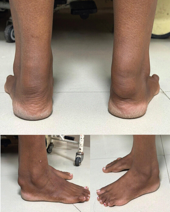
On examination, she walked unassisted with a symmetrical gait. There was gross swelling over the insertion of both Achilles tendons, measuring 8 × 6 cm on the right and 6 × 4 cm on the left, with irregular surfaces and margins, firm-to-hard consistency, and adherence to the underlying tendon. The overlying skin was pinchable, and the swellings were non-reducible and non-translucent (Fig. 1). Tenderness was present over the right swelling only, with no local rise in temperature. Ankle and subtalar range of motion were normal, with no muscle wasting or neurovascular deficit. The Matles test, O’Brien test, and Thompson squeeze test were negative. Activities of daily living were markedly affected by difficulty in wearing shoes and pain on walking.
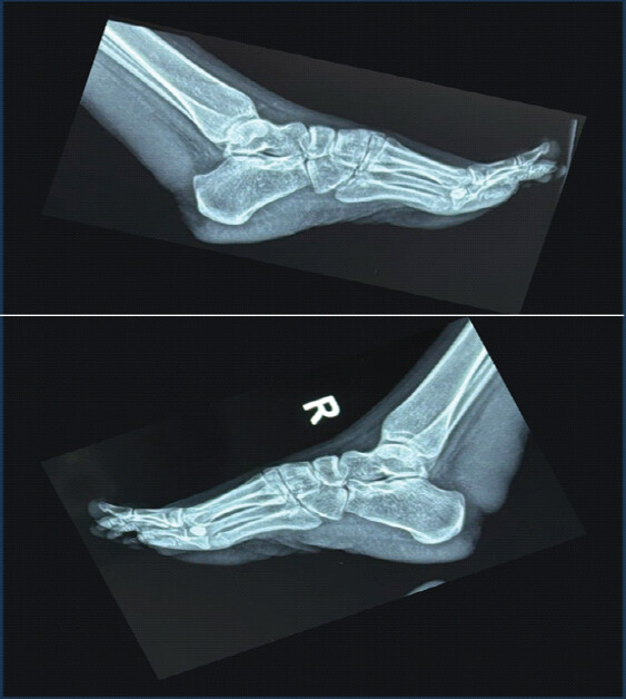
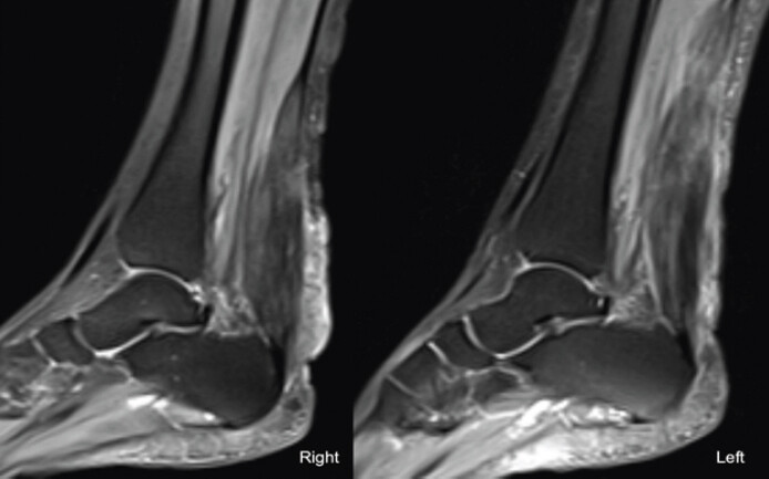
Complete blood count, erythrocyte sedimentation rate, C-reactive protein, and thyroid profile were within normal limits. Liver function tests, including serum bilirubin, serum glutamate pyruvate transaminase, serum glutamic-oxaloacetic transaminase, and alkaline phosphatase, were also within normal limits. The lipid profile showed a total cholesterol of 228 mg/dL (reference range, 123–200 mg/dL); high-density lipoprotein cholesterol, LDL cholesterol, and triglycerides were within their respective reference ranges. Radiographs of both ankles showed no bony abnormality but revealed thickened, non-calcified soft-tissue shadows in the region of both Achilles tendons (Fig. 2). MRI showed diffuse, symmetrical, fusiform thickening of both Achilles tendons with heterogeneous, increased intratendinous signal intensity, and loss of the normal anterior concavity (anteroposterior diameter, 2.1 cm on the right and 2.3 cm on the left), without a tear – findings consistent with bilateral Achilles tendon xanthoma (Fig. 3).
Given the greater symptom severity on the right, surgical excision was planned for the right side. The left-sided lesion was managed conservatively, as it was not associated with significant pain.
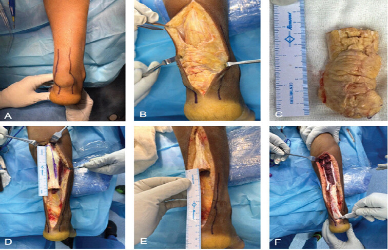
Surgery was performed under spinal anesthesia with the patient prone and a tourniquet applied. A midline incision was made over the swelling. The procedure comprised two steps: Excision of the xanthoma, followed by reconstruction of the defect. The xanthoma was freed from adhesions and excised en bloc from the Achilles tendon insertion, leaving a 10-cm tendon defect. To bridge the defect, a central, full-thickness, inverted U-shaped flap, 13.5 cm in length, was fashioned from the tendinous portion of the gastrosoleus complex, preserving 2 cm of the distal end of the proximal stump, which was sutured in place to prevent inadvertent separation of the flap. The turn-down flap was advanced distally and secured to the calcaneal insertion with a suture anchor under appropriate tension (Fig. 4). After hemostasis, the wound was closed in layers, and a dorsal slab was applied with the ankle in 20° of plantarflexion.
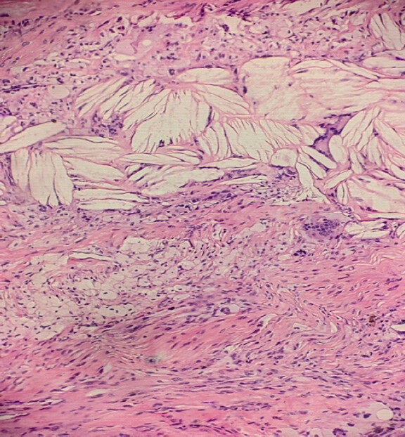
Histopathological examination showed dense collagenous tendon tissue infiltrated by sheets of foamy histiocytes with abundant vacuolated cytoplasm. Numerous cholesterol clefts were surrounded by foreign body-type multinucleated giant cells, with focal chronic inflammatory infiltrates – features consistent with tendon xanthoma (Fig. 5).
Post-operative care
Recovery was uneventful. The patient was mobilized non-weight-bearing in a below-knee slab with the ankle in 20° of plantarflexion. Sutures were removed on post-operative day 14, and the wound was healthy. A below-knee fiberglass cast was then applied with the ankle in neutral, and full weight-bearing was permitted. The cast was removed at 6 weeks, after which active and passive ankle range-of-motion exercises were started; strengthening exercises began at 3 months. Because her lipid levels were only modestly elevated, she was managed with a low-fat diet and lifestyle modification rather than pharmacological lipid-lowering therapy, under endocrinology guidance.
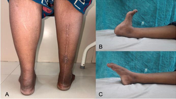
At 1-year follow-up, there were no skin complications. Pain improved from a pre-operative Visual Analog Scale score of 7–2 at final follow-up, and the American Orthopaedic Foot and Ankle Society score improved from 52 to 90. Ankle motion ranged from 20° of dorsiflexion to 30° of plantarflexion, single-leg heel raise was possible, and there was no clinical recurrence. She walked independently and performed activities of daily living without difficulty (Fig. 6).
Discussion
Xanthomas are benign, non-neoplastic lesions formed by the accumulation of collagen, lipid-laden macrophages, giant cells, and inflammatory cells in response to tissue deposition of LDL. They occur in the skin, particularly the eyelids, and are less commonly seen in the tendons and synovium [13]. In patients with FH, tendon xanthomas are associated with an approximately threefold higher risk of cardiovascular disease [14] and contribute to the clinical diagnosis of FH [15].
The clinical manifestations of Achilles tendon xanthoma depend chiefly on lesion size. Small lesions may go unnoticed, whereas larger lesions cause visible disfigurement and may produce localized pain, irritation, and restricted movement. The differential diagnosis of a bulky, enlarged Achilles tendon includes xanthoma, tendinopathy, chronic degeneration, tendinitis, peritendinitis, bursitis, trauma, rheumatoid nodules, tophaceous gout, infection, and neoplasm [16].
Our patient’s total cholesterol was only modestly elevated, and LDL cholesterol was within the reference range. Her history of chronic loose stools, recurrent jaundice since adolescence, and cholelithiasis raises the possibility of lipid-handling disorders like FH. Additional possibilities can be cerebrotendinous xanthomatosis or sitosterolemia, both of which can produce tendon xanthomas with a near-normal standard lipid profile [17]. Achilles tendon xanthomas have also been reported in patients with an entirely normal lipid profile [18]. Genetic testing for FH was not pursued in this patient, and this represents a limitation of the present report; it should be considered in similar future cases.
Plain radiography is often the first imaging modality, and lateral views allow assessment of the anteroposterior diameter of the tendon [19]. MRI is the most informative modality: it distinguishes low-signal collagen fibers from the higher-signal infiltrate of foam and inflammatory cells [8] and defines the extent of intratendinous involvement, which is essential for surgical planning. Histopathology, however, remains the definitive diagnostic test [9].
Initial management comprises lipid-lowering measures, chiefly statins together with dietary modification, in patients with significant hyperlipidemia [3]. Surgery is challenging and is generally reserved for patients with pain, gait disturbance, restricted ankle motion, or severe disfigurement. Because partial resection is associated with recurrence [11,12], complete resection combined with tendon reconstruction is regarded as the treatment of choice for large lesions.
Two surgical strategies have been described: Complete excision with reconstruction of the defect using tendon grafts [20] and subtotal intralesional resection [21]. Complete resection with reconstruction yields lower recurrence rates [22], at the cost of greater postoperative morbidity, whereas subtotal resection allows faster recovery and earlier return to work but carries a higher recurrence rate [23].
Various reconstructive options have been used after complete excision, including a short FHL graft fixed to the calcaneum with a bioabsorbable screw [24], tensor fascia lata autograft harvested from the opposite thigh [25], and FHL transfer, either alone or combined with a Bosworth turn-down flap [23]. The gastrosoleus turn-down flap (Bosworth technique) [26] uses a strip of gastrosoleus aponeurosis, hinged and turned distally, to bridge the defect, and is well established for reconstructing large defects after chronic Achilles tendon rupture, including in combination with V-Y advancement for extensive gaps [27]. In the setting of xanthoma, however, its use appears to have been reported only in combination with FHL transfer [23]; to our knowledge, no prior report describes an isolated gastrosoleus turn-down reconstruction, without an adjunct tendon transfer, after excision of an Achilles tendon xanthoma. In our patient, the technique bridged a 10-cm defect using local autologous tissue, avoided a second donor site, and was followed by restoration of ankle motion and independent walking at 1 year.
This report is limited by its single-case design, unilateral surgical treatment, and 1-year follow-up, together with the absence of a confirmatory genetic test for FH discussed above. Because recurrence has been reported after resection [11,12], long-term surveillance is warranted.
Conclusion
Achilles tendon xanthomas are uncommon. Patients presenting with heel swelling, particularly if bilateral, should be evaluated with a lipid profile. Because a tendon xanthoma may signal an underlying lipid-handling disorder, imaging should be used for accurate diagnosis, together with lipid evaluation and cardiovascular risk assessment. Medical and surgical management should be tailored to symptom severity. In symptomatic large lesions, complete excision with gastrosoleus turn-down reconstruction is a viable option.
Clinical Message
Achilles tendon xanthoma should be considered in patients with bilateral, progressive tendon swellings. Early recognition should prompt lipid evaluation and cardiovascular risk assessment. In symptomatic, extensive lesions, complete excision with gastrosoleus turn-down reconstruction offers a viable means of restoring tendon continuity and function.
Conflict of Interest:
Nil
Source of Support:
Nil
Consent
The authors confirm that informed consent was obtained from the patient for publication of this article
How to Cite this Article
Gaurav A, Meena R, Kanwar S, Bondarde P, Gupta S, Meena P. Complete Excision and Gastrosoleus Turn-Down Reconstruction for Achilles Tendon Xanthoma: A Case Report. Journal of Orthopaedic Case Reports 2026 October;16(10): 302-307.
References
- Van Den Bosch HC, Vos LD. Images in clinical medicine. Achilles'-tendon xanthoma in familial hypercholesterolemia. N Engl J Med 1998;338:1591. [Google Scholar] | [PubMed]
- Fahey JJ, Stark HH, Donovan WF, Drennan DB. Xanthoma of the achilles tendon: Seven cases with familial hyperbetalipoproteinemia. J Bone Joint Surg Am 1973;55:1197-211. [Google Scholar] | [PubMed]
- Tsouli SG, Kiortsis DN, Argyropoulou MI, Mikhailidis DP, Elisaf MS. Pathogenesis, detection and treatment of achilles tendon xanthomas. Eur J Clin Invest 2005;35:236-44. [Google Scholar] | [PubMed]
- Khot R, Nagpure K. Familial hypercholesterolemia with different types of xanthomas. J Assoc Physicians India 2018;66:86. [Google Scholar] | [PubMed]
- Rekha A, Rai DK. Tendon xanthomas. Foot (Edinb) 2010;20:85-6. [Google Scholar] | [PubMed]
- Lin GT. Surgical excision of the tendon xanthoma in familial hypercholesterolemia--a case report. Kaohsiung J Med Sci 1999;15:441-6. [Google Scholar] | [PubMed]
- Haacke H, Parwaresch MR. Spontaneous rupture of the achilles tendon - a sign of hyperlipoproteinaemia (HLP) type II. Klin Wochenschr 1979;57:397-400. [Google Scholar] | [PubMed]
- Dussault RG, Kaplan PA, Roederer G. MR imaging of achilles tendon in patients with familial hyperlipidemia: Comparison with plain films, physical examination, and patients with traumatic tendon lesions. AJR Am J Roentgenol 1995;164:403-7. [Google Scholar] | [PubMed]
- Kruth HS. Lipid deposition in human tendon xanthoma. Am J Pathol 1985;121:311-5. [Google Scholar] | [PubMed]
- Mabuchi H, Sakai Y, Watanabe A, Haba T, Koizumi J, Takeda R. Normalization of low-density lipoprotein levels and disappearance of xanthomas during pregnancy in a woman with heterozygous familial hypercholesterolemia. Metabolism 1985;34:309-15. [Google Scholar] | [PubMed]
- Friedman MS. Xanthoma of the achilles tendon. J Bone Joint Surg Am 1947;29:760-6. [Google Scholar] | [PubMed]
- Tomita T, Ochi T, Fushimi H, Matsuzawa Y, Ono K. Reconstruction of the Achilles tendon for xanthoma: Findings at operative re-exploration. A case report. J Bone Joint Surg Am 1994;76:444-7. [Google Scholar] | [PubMed]
- Weiss SW, Goldblum JR. Benign fibrohistiocytic tumorsIn: Enzinger and Weiss's Soft Tissue Tumors. 4th ed. St Louis, MO: Mosby; 2001. 471-4. [Google Scholar] | [PubMed]
- Oosterveer DM, Versmissen J, Yazdanpanah M, Hamza TH, Sijbrands EJ. Differences in characteristics and risk of cardiovascular disease in familial hypercholesterolemia patients with and without tendon xanthomas: A systematic review and meta-analysis. Atherosclerosis 2009;207:311-7. [Google Scholar] | [PubMed]
- Nordestgaard BG, Chapman MJ, Humphries SE, Ginsberg HN, Masana L, Descamps OS. Familial hypercholesterolaemia is underdiagnosed and undertreated in the general population: Guidance for clinicians to prevent coronary heart disease: Consensus statement of the European atherosclerosis society. Eur Heart J 2013;34:3478-90. [Google Scholar] | [PubMed]
- Rodriguez CP, Goyal M, Wasdahl DA. Best cases from the AFIP: Atypical imaging features of bilateral Achilles tendon xanthomatosis. Radiographics 2008;28:2064-8. [Google Scholar] | [PubMed]
- Sastri VR, Ravindranath VS, Metikala S, Kumar M. Bilateral xanthomas of tendoachilles in a patient of cerebrotendinous xanthomatosis - a rare case report and review of literature. J Orthop Case Rep 2014;4:40-3. [Google Scholar] | [PubMed]
- Lakkireddy M, Eppakayala S, Satapathy D, Maley DK, Ifthekar S. Massive tendo achilles xanthoma treated with en bloc excision and reconstruction with flexor hallucis longus tendon transfer: A case report. J Orthop Case Rep 2025;15:199-203. [Google Scholar] | [PubMed]
- Blankenhorn DH, Meyers HI. Radiographic determination of achilles tendon xanthoma size. Metabolism 1969;18:882-6. [Google Scholar] | [PubMed]
- Huang L, Miao XD, Yang DS, Tao HM. Bilateral achilles tendon enlargement. Orthopedics 2011;34:e960-4. [Google Scholar] | [PubMed]
- Eil W, Faber FW. Resection of bilateral giant achilles tendon xanthomas with a tendon sparing technique-a case report. Clin Res Foot Ankle 2014;2014:1-3. [Google Scholar] | [PubMed]
- Carranza-Bencano A, Fernádez-Centeno M, Leal-Cerro A, Duque-Jimeno V, Gomez-Arroyo JA, Zurita-Gutierrez M. Xanthomas of the achilles tendon: Report of a bilateral case and review of the literature. Foot Ankle Int 1999;20:314-6. [Google Scholar] | [PubMed]
- Moroney PJ, Besse JL. Resection of bilateral massive Achilles tendon xanthomata with reconstruction using a flexor hallucis longus tendon transfer and bosworth turndown flap: A case report and literature review. Foot Ankle Surg 2012;18:e25-8. [Google Scholar] | [PubMed]
- Sahu NK, Nanda SN, Tripathy M, Samant S, Tripathy SK, Gachhayat AK. Xanthoma of tendoachilles: Management and follow-up. Case Rep Orthop Res 2021;4:166-72. [Google Scholar] | [PubMed]
- Senthil L, Jambu N, Chittaranjan S. Treatment of large recurrent bilateral xanthomatosis of achilles tendon-a case report. J Orthop Case Rep 2015;5:11-4. [Google Scholar] | [PubMed]
- Bosworth DM. Repair of defects in the tendo achillis. J Bone Joint Surg Am 1956;38-A:111-4. [Google Scholar] | [PubMed]
- Ponnapula P, Aaranson RR. Reconstruction of achilles tendon rupture with combined V-Y plasty and gastrocnemius-soleus fascia turndown graft. J Foot Ankle Surg 2010;49:310-5. [Google Scholar] | [PubMed]
© 2026 Journal of Orthopaedic Case Reports - Published by Indian Orthopaedic Research Group
About the Authors
-

Ankit Gaurav
-

Rajesh Meena
-

Sachin Kanwar
-

Parshwanath Bondarde
-

Sanchit Gupta
-

Pradeep Meena
Related Articles in Journal of Orthopaedic Case Reports
 September 1, 2025 Massive Tendo Achilles Xanthoma Treated with en bloc Excision and Reconstruction with Flexor Hallucis Longus Tendon Transfer: A Case Report
 June 1, 2026 Neglected but Negotiated – A Case Report of Multiple Zone VII Extensor Tendon Injuries Reconstructed Using Fascia Lata Autograft
 May 1, 2026 Semitendinosus Autograft Reconstruction for Neglected Patellar Tendon Rupture: Technical Simplicity with Excellent Outcome
 March 1, 2026 Considerations Regarding the Family Members in the Management of Patients with Familiar Hypercholesterolemia