


Introduction
Achilles tendon rupture is among the most common tendon injuries encountered in orthopedic practice. However, the vast majority of reported cases involve closed spontaneous or degenerative ruptures in middle-aged recreational athletes, typically males in the fourth to fifth decade of life [1]. Open traumatic Achilles tendon ruptures secondary to sharp penetrating objects are much less commonly reported, carrying the added risks of wound contamination, tendon exposure, and concurrent injury to adjacent neurovascular structures.
In the pediatric population, Achilles tendon ruptures of any etiology are relatively uncommon. The pediatric Achilles tendon is characteristically more elastic and resilient than its adult counterpart, with the calcaneal apophysis – rather than the tendon itself – being the weakest link in the musculotendinous unit during childhood [2]. Consequently, avulsion injuries at the calcaneal apophysis are more frequently reported in children [3]. Open traumatic ruptures in children have very limited published literature [4].
To identify relevant literature, we searched PubMed/MEDLINE and Google Scholar using combinations of terms including “Achilles tendon rupture,” “pediatric,” “adolescent,” “tendon repair,” and “imaging.” Relevant case reports, reviews, epidemiological studies, and biomechanical studies were reviewed, and reference lists of relevant articles were also screened for additional studies.
The Achilles tendon is anatomically flanked by neurovascular bundles medially, which are vulnerable in sharp lacerating injuries of the posterior ankle region. Failure to recognize and manage concurrent neurovascular injury in conjunction with tendon repair may result in chronic sensory deficits, ischemic complications, and suboptimal functional recovery.
A focused review of the published literature shows that very few cases of open Achilles tendon rupture in the pediatric age group have been reported. Reporting the management and outcomes of such cases may help clinicians managing similar injuries [5, 6, 7].
The modified Kessler technique, originally developed for flexor tendon repair in the hand, has been widely adopted for Achilles tendon reconstruction because it provides good repair strength with less gap formation compared to simpler suture configurations [8]. We present this case to describe our experience with primary modified Kessler repair in an open pediatric Achilles tendon rupture (Table 1).
Published cases of open Achilles tendon rupture in the pediatric age group
| Author (year) | Age/sex | Mechanism | NV deficit | Outcome | Repair technique |
|---|---|---|---|---|---|
| Supanich MA (2023) | 10/M | Traumatic laceration | Not reported | Good | Krackow |
| Kouassi-Driai (2026) | 14/M | Bilateral spontaneous | None | Excellent | Not mentioned |
| Vasileff WK (2014) | 10/M | Sports injury (partial) | None | Good | Not mentioned |
| Present case (2026) | 9/M | Sharp metal object | None (vessel intact) | Excellent —AOFAS 95 | Modified Kessler |
M: Male, NV: Neurovascular
Case Report
Patient presentation
A 9-year-old male child was brought to the Emergency Department by his parents with acute pain, swelling, and inability to walk following an accidental laceration to the right posterior ankle by a sharp metallic object approximately 2 h before presentation. There was no history of prior tendon pathology, systemic illness, metabolic disorder, or medication use (including fluoroquinolones or corticosteroids). The child’s vitals were stable.
Local examination
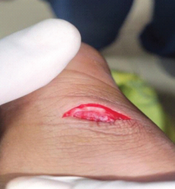
Examination of the right ankle and posterior leg revealed a 3 × 1 × 1 cm contaminated laceration wound over the posterior aspect of the ankle, located approximately 2–3 cm proximal to the calcaneal insertion of the Achilles tendon. Wound margins were sharply incised. Visible tendon ends were identifiable through the wound with active capillary ooze (Fig. 1).
A palpable gap was appreciated at the site of Achilles tendon transection on gentle palpation adjacent to the wound. Thompson’s test was positive. The child was weakly able to plantar-flex the right ankle actively.
Pre-operative planning and consent
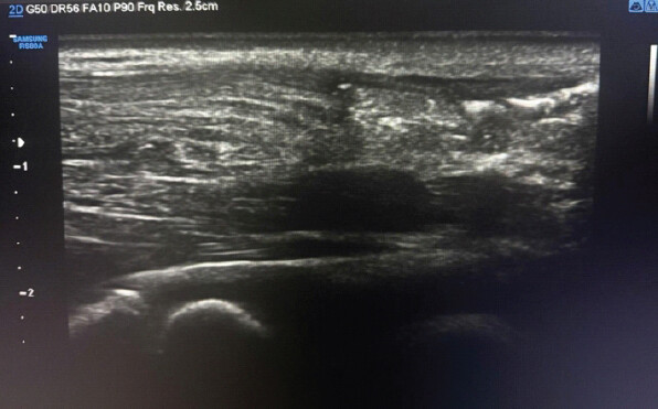
Pre-operative ultrasonography demonstrated complete discontinuity of the Achilles tendon approximately 4.5 cm proximal to its calcaneal insertion, confirming the diagnosis.
Parental informed written consent was obtained, including consent for clinical photography and video documentation for academic publication. Surgical risks, anesthesia risk, and post-surgical complication were discussed. The child was kept nil by mouth as per pediatric anesthesia guidelines. Intravenous cefazolin 25 mg/kg (age-adjusted) was administered as antibiotic prophylaxis 30 min pre-incision. Tetanus prophylaxis was administered at presentation.
Operative procedure
The child was taken to the operating room and positioned prone on the operating table under general anesthesia (endotracheal intubation). The right lower limb was scrubbed, painted, and draped in standard sterile fashion.
The traumatic wound was extended using a posteromedial approach to provide adequate exposure with care taken to avoid injury to nearby neurovascular structures. The wound was copiously irrigated with normal saline and betadine solution under pressure. Minimal debridement of non-viable tissue was performed.
Intraoperative findings: Complete transverse laceration of the Achilles tendon approximately 4.5 cm from its calcaneal insertion. Both tendon ends were retrievable. No active arterial injury was identified (Figs. 2 and 3).
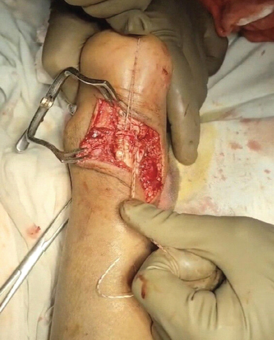
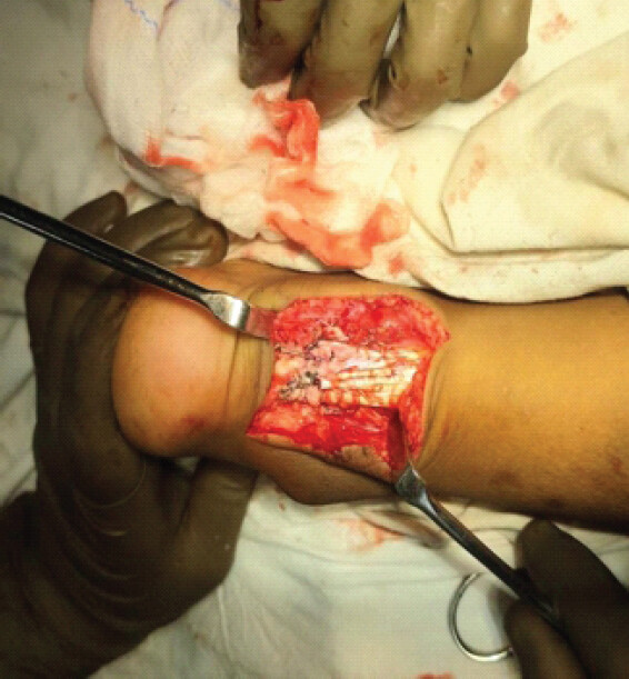
Achilles tendon repair: Primary end-to-end repair was performed using the modified Kessler technique. A No. 2-0 FiberWire® suture was used to place a locking Kessler core suture through each tendon stump. Because of the smaller tendon size, smaller suture bites were taken and proportionally placed to maximize purchase without splitting the tendon. The repair was tensioned with the ankle in approximately 15–20° of plantar flexion. A continuous epitendinous circumferential suture using 1-0 Vicryl was placed to reinforce the repair and smooth the coaptation site. The repair was stable without visible gapping.
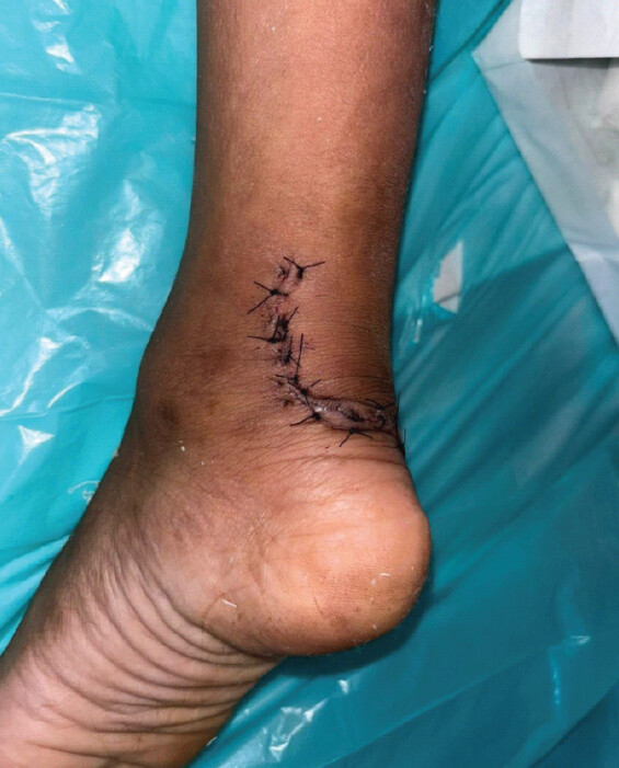
Hemostasis was confirmed. Wound closure was performed in layers: Subcutaneous tissue with 2-0 Vicryl, and skin with interrupted 3-0 Ethilon mattress sutures. A well-padded below-knee plaster slab was applied with the ankle maintained in 20° plantar flexion.
Post-operative management
Postoperatively, the child was monitored in the pediatric ward. A standardized post-operative protocol was followed (Table 2 and Fig. 4).
A standardized post-operative protocol was followed
| Phase/timeframe | Management |
|---|---|
| Day 1–5 (in-patient) | IV Cefazolin 25 mg/kg TDS; IV paracetamol for analgesia; limb elevation; NV observations 4-hourly; wound inspection day 3. |
| Day 5–14 | Discharge on oral cephalexin 25 mg/kg/day BD; analgesia as needed; slab maintained in 20° PF; suture removal day 14; wound check. |
| Week 3 | Slab at 10° plantar flexion; no weight-bearing; physiotherapy: Isometric quadriceps and hip exercises. |
| Week 4–6 | Slab at neutral (0°); toe-touch weight-bearing with crutches commenced. |
| Week 6–10 | Slab removed; transition to removable boot; physiotherapy: Passive and active ROM, plantar flexion strengthening, hydrotherapy. |
| Month 4–6 | Full unrestricted physical activity at 6 months |
PF: Plantar flexion, ROM: Range of motion, NV: Neurovascular, IV: Intravenous
Discussion
This case is unusual because of the patient’s age, open injury pattern. First, it represents a rare occurrence of complete open Achilles tendon rupture in a 9-year-old child. Second, it demonstrates that with prompt surgical management, excellent functional outcomes (AOFAS score 95) including full return to childhood physical activity are achievable.
Achilles tendon rupture is less common in children. Unlike adults, where degeneration and eccentric loading predispose the tendon to failure, the pediatric Achilles tendon retains high elasticity and intrinsic tensile strength. The weakest point in the pediatric posterior chain is the calcaneal apophysis – leading to Sever’s disease or apophyseal avulsions rather than midsubstance tendon failure [2]. In open injuries, however, sharp lacerating injuries can still result in complete tendon transection.
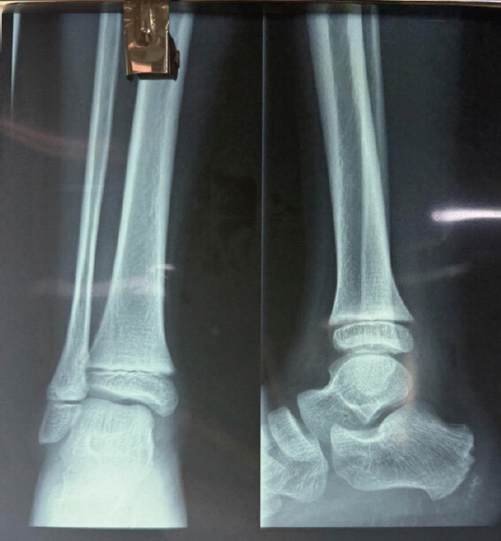
The diagnosis of complete Achilles tendon rupture was established clinically by a positive Thompson test and confirmed by preoperative ultrasonography. Ultrasonography is the first-line imaging modality of choice for Achilles tendon injuries – it is rapid, cost-effective, dynamic, and avoids ionizing radiation, which is particularly pertinent in the pediatric population [9]. Magnetic resonance imaging, though more detailed, was not required in this acute setting given the clear clinical and sonographic diagnosis. Plain radiography was performed to exclude bony injury and avulsion fractures at the calcaneal apophysis, which are more common in children [3] (Figs. 5 and 6).
The neurovascular bundle lies in close proximity to the Achilles tendon and is particularly vulnerable in posterior ankle lacerations.
The posterior tibial vessels, which course medially to the Achilles tendon, showed adventitial contusion without frank arterial injury in our case. Vascular integrity was confirmed intraoperatively. Had there been a complete vascular transection, immediate vascular surgical intervention would have been required to prevent limb ischemia. In surgeons managing such injuries, vascular assessment should be performed when vascular injury is suspected.
Regarding tendon repair, the modified Kessler technique has been studied in biomechanical models. Hockenbury and Johns (1990) demonstrated superior tensile load-to-failure and minimal gap formation with Kessler-based repairs compared to Bunnell or simple suture configurations in cadaveric Achilles tendons [8]. Compared with the Krackow technique, the modified Kessler repair provides adequate tensile strength with smaller suture bulk, making it particularly suitable for the pediatric population [10]. In our case, suture caliber and bite size were adjusted proportionally to account for the smaller tendon diameter, to avoid damage to the smaller tendon.
The post-operative rehabilitation protocol used in this case followed a progressive plantar-flexion reduction and early functional mobilization model. Careful wound management was important in this case because of contamination risk. Pediatric skin is more pliable and vascular than adult skin, facilitating wound healing; however, contaminated wounds still carry infection risk. Our protocol of copious irrigation, conservative debridement, layered primary closure, and targeted antibiotic prophylaxis resulted in uneventful wound healing with no infective complications. Post-operative recovery was uneventful, with normal walking and return to activities by 6 months after surgery with an AOFAS ankle-hindfoot score of 95, indicating excellent functional recovery. A limitation of this report is the relatively short follow-up duration; however, early functional recovery was excellent.
In summary, this case demonstrates that early repair of open Achilles tendon injury in children can result in good functional recovery.
Conclusion
Open traumatic Achilles tendon rupture, though uncommon in children, demands prompt recognition and surgical management. Primary Achilles tendon repair using the modified Kessler technique, combined with intra-operative neurovascular assessment, wound irrigation, and structured progressive rehabilitation, results in excellent functional outcomes. Compared with the Krackow technique, the modified Kessler repair provides adequate tensile strength with smaller suture bulk, making it particularly suitable for the pediatric population.
In this 9-year-old patient, full plantar flexion strength, complete return to physical activity were achieved by 6 month follow-up. This case adds to the limited pediatric literature on open Achilles tendon injuries and highlights the importance of clinician awareness of this rare but serious injury pattern.
Clinical Message
Early primary repair with the modified Kessler technique gives a strong, low-bulk construct well suited to the small pediatric tendon, and with structured rehabilitation a full return to activity is a realistic goal.
Conflict of Interest:
Nil
Source of Support:
Nil
Consent:
The authors confirm that informed consent was obtained from the patient for publication of this article
How to Cite this Article
Farooqui M, Game N. Open Traumatic Achilles tendon Rupture in a Pediatric Patient Managed by Modified Kessler Technique: A Case Report. Journal of Orthopaedic Case Reports 2026 October;16(10): 34-39.
References
- Lantto I, Heikkinen J, Flinkkilä T, Ohtonen P, Leppilahti J. Epidemiology of Achilles tendon ruptures: Increasing incidence over a 33-year period. Scand J Med Sci Sports 2015;25:e133-8. [Google Scholar] | [PubMed]
- Scharfbillig RW, Jones S, Scutter SD. Sever's disease: What does the literature really tell us?. J Am Podiatr Med Assoc 2008;98:212-23. [Google Scholar] | [PubMed]
- Haines M, Pirlo L, Bowles KA, Williams CM. Describing frequencies of lower-limb apophyseal injuries in children and adolescents: A systematic review. Clin J Sport Med 2022;32:433-9. [Google Scholar] | [PubMed]
- Ashebo L, Stevens AC, MacAlpine EM, Wittstein JR, Bradley KE, Lawrence JT. Achilles tendon injuries in the pediatric population. J Pediatr Orthop 2023;43:e513-8. [Google Scholar] | [PubMed]
- Supanich MA, Bodine M, Milshteyn M. Case report: Traumatic distal Achilles tendon rupture in a pediatric patient. J Orthop Case Rep 2023;13:137-41. [Google Scholar] | [PubMed]
- Kouassi-Dria AK, Obro RB, Ouattara JJ, Gonebo KA, Aké YL, Moh N. Bilateral Achilles tendon injury in a 14-year-old adolescent: Case report. Int J Surg Case Rep 2026;138:133-5. [Google Scholar] | [PubMed]
- Vasileff WK, Moutzouros V. Unrecognized pediatric partial Achilles tendon injury followed by traumatic completion: A case report and literature review. J Foot Ankle Surg 2014;53:485-8. [Google Scholar] | [PubMed]
- Hockenbury RT, Johns JC. A biomechanical in vitro comparison of open versus percutaneous repair of tendon Achilles. Foot Ankle 1990;11:67-72. [Google Scholar] | [PubMed]
- Bleakney RR, White LM. Imaging of the Achilles tendon. Foot Ankle Clin 2005;10:239-54. [Google Scholar] | [PubMed]
- Diniz P, Pacheco J, Fernandes RM, Pereira H, Castelo Ferreira F, Kerkhoffs GM. Modified triple Kessler with least risk of elongation among Achilles tendon repair techniques: A systematic review and network meta-analysis of human cadaveric studies. Knee Surg Sports Traumatol Arthrosc 2023;31:1644-57. [Google Scholar] | [PubMed]
© 2026 Journal of Orthopaedic Case Reports - Published by Indian Orthopaedic Research Group
About the Authors
-

Mubasshir Farooqui
-

Nikhil Game
Related Articles in Journal of Orthopaedic Case Reports
 September 1, 2026 Comparing Bridge-Enhanced Anterior Cruciate Ligament Repair to Standard Autograft for Anterior Cruciate Ligament Reconstruction: Biomechanical and Clinical Outcomes
 February 1, 2026 Proximal Hamate Autograft in Non-union Proximal Scaphoid Fractures, A Novel Technique – A Case Report
 September 1, 2026 Osteoblastoma of the Hamate: A Case Report
 May 1, 2025 Understanding the Levels of Evidence in Medical Research