


Introduction
We present the case of a patient with a rare synovial sarcoma (SS) of the tibial nerve. So far, only 4 cases of patients with SS originating from the tibial nerve have been described in the literature [1, 2, 3, 4, 5].
Case Report
On date July 09, 2024, a 17-year-old male presented with a 1-day history of acute-onset, sharp, shooting pain in the left calf, radiating along the plantar aspect of the left foot. On physical examination, the patient exhibited an antalgic gait with the left foot maintained in an equinus posture.
Palpation revealed a markedly tender mass measuring approximately 3 cm × 1 cm located in the posteromedial compartment of the left leg, situated deep to the investing fascia along the anatomical course of the tibial nerve. Passive dorsiflexion of the left ankle elicited severe pain. Neurological assessment demonstrated hypoesthesia within the tibial nerve sensory distribution; no focal motor deficits were identified in the left lower extremity. Baseline functional evaluation using the Musculoskeletal Tumor Society (MSTS) scoring system yielded a score of 5/30 (16.7%) on Day 1 (Table 1).
MSTS score breakdown
| Category | Finding | Initial Date July 09, 2024 | Follow-up at 2 years and 4 months |
|---|---|---|---|
| Pain | No pain | 0 | 5 |
| Function | Recreational restriction | 0 | 3 |
| Emotional acceptance | Intermediate (satisfied) | 2 | 3 |
| Supports | No support | 1 | 5 |
| Walking | Intermediate | 1 | 3 |
| Gait | Intermediate | 1 | 3 |
| Total raw score | May-30 | 22/30 | |
| Percentage | 16.6 | 73.3 |
MSTS: Musculoskeletal tumor society
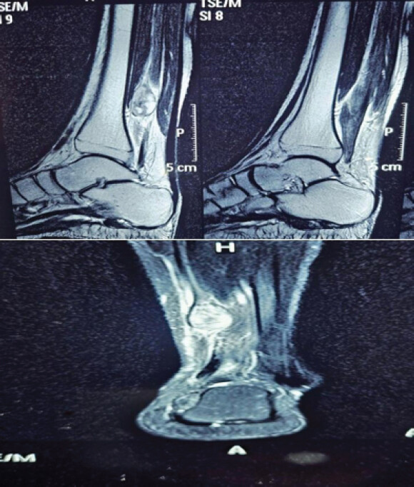
Plain radiographs of the left lower extremity revealed no significant osseous abnormalities. Subsequent ultrasonography demonstrated a well-defined, hypoechoic, solid mass within the tibial nerve measuring 24 × 19 × 14 mm, raising suspicion for a peripheral nerve sheath tumor. Magnetic resonance imaging (MRI) of the left leg further characterized a well-demarcated, T2-weighted hyperintense lesion measuring 14 × 15 × 25 mm within the posteromedial compartment. The mass was anatomically continuous with the tibial nerve, highly suggestive of a benign neurogenic tumor, most likely a schwannoma (Fig. 1).
On August 07, 2024, the patient was scheduled for a total excisional biopsy of the mass. The procedure was initiated through a short longitudinal skin incision placed over the lesion along the course of the tibial nerve. However, intraoperative exploration revealed an unencapsulated, poorly demarcated mass. Consequently, the planned excision was aborted; instead, a representative incisional biopsy was obtained for histopathological evaluation, and definitive surgical management was deferred.
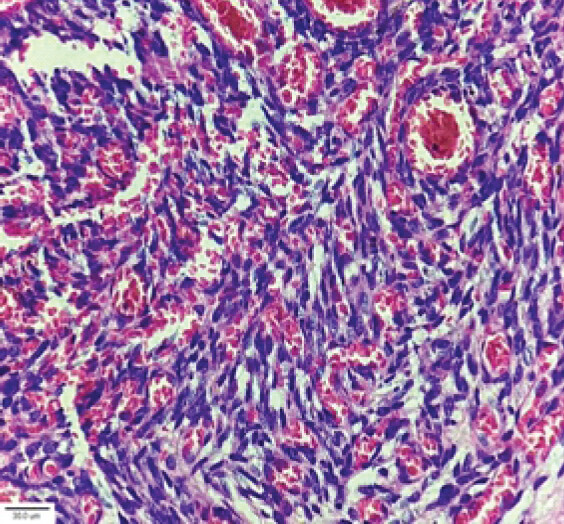
“On August 10, 2024, the histopathological examination revealed a cellular lesion composed of spindle-shaped cells arranged in classic Verocay bodies, consistent with a benign schwannoma (Fig. 2). There was no evidence of histological malignancy.”
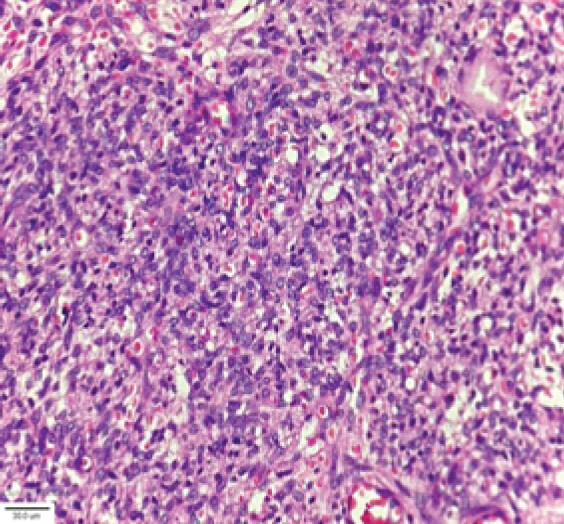
“To seek an expert second opinion, the patient’s histopathological tissue block was referred to an external pathologist. The subsequent review on August 22, 2024, described a markedly cellular spindle cell neoplasm with malignant and sarcomatous features (Fig. 3), favoring a malignant diagnosis over a benign process.”
Subsequent immunohistochemical analysis was performed for definitive classification.”
Combined case narrative (full sequence so far)
“On August 07, 2024, the patient was scheduled for a total excisional biopsy of the mass. The procedure commenced with a short longitudinal incision over the lesion along the course of the tibial nerve. However, intraoperative exploration revealed an unencapsulated, poorly demarcated mass, prompting the abandonment of total excision. Instead, a representative incisional biopsy was obtained, and definitive surgical management was deferred.
Initial histopathological evaluation on August 10, 2024, suggested a benign schwannoma characterized by spindle cells forming Verocay bodies (Fig. 2). To obtain an expert second opinion, the tissue block was referred to an external pathologist. The revised report on August 22, 2024, described a markedly cellular spindle cell neoplasm with sarcomatous features (Fig. 3), favoring malignancy over a benign process. A comprehensive immunohistochemical study was subsequently performed to further characterize the lesion.”
Immunohistochemical and molecular findings
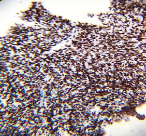
Immunohistochemical analysis supported the diagnosis of SS (Fig. 4), demonstrating strong positivity for epithelial membrane antigen (EMA) and TLE1 (Fig. 5), alongside focal/patchy immunoreactivity for CD99 and pan-keratin. Molecular testing was positive for the SS18::SSX fusion protein product, confirming the pathognomonic balanced translocation t(X;18) (p11.2;q11.2) [6].
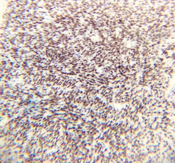
Alternative spindle cell malignancies were excluded based on negative staining for CD34 (solitary fibrous tumor), S100 and SOX10 (malignant peripheral nerve sheath tumor and melanocytic neoplasms), and desmin (rhabdomyosarcoma/leiomyosarcoma/desmoplastic spindle cell neoplasms) [6].
The Ki-67 proliferation index evaluates cellular proliferation by quantifying the percentage of actively dividing tumor cells [7]. In the present case, the patient exhibited a Ki-67 index of approximately 20%, markedly exceeding the established prognostic threshold of 9–10% [8]. Ki-67 expression above this cutoff correlates significantly with an increased risk of local recurrence, disease progression, and decreased metastasis-free survival [9].
On September 05, 2024, a whole-body fluorodeoxyglucose (FDG) positron emission tomography and contrast-enhanced computed tomography (CT) scan was performed for disease staging. The scan revealed a well-defined, enhancing intramuscular soft-tissue mass in the posterior compartment of the left lower leg measuring approximately 1.5 × 1.2 cm, demonstrating heterogeneous FDG avidity with a maximum standardized uptake value (SUVmax 4.05).
Based on the Fédération Nationale des Centres de Lutte Contre le Cancer (FNCLCC) histologic grading system for soft-tissue sarcomas and the findings in these pathology reports of the patient, this tumor classified as FNCLCC Grade 2 (Intermediate).
All SSs receive a mandatory differentiation of Score 3 (Tumor Differentiation).
Biopsy notes: “Only occasional mitosis present” (Ki-67 proliferation index ~20%). Occasional mitoses correlate to <10 mitoses/10 HPF (Score 1 = 0–9 mitoses/10 HPF). Score 1 (Mitotic Count).
Gross and microscopic examinations show cystic cavity/foci of hemorrhage and foreign body giant cell reaction, but report no frank tumor necrosis (Score 0 = 0% necrosis). Score 0 (Tumor Necrosis).
• Total cumulative score: 3 + 1 + 0 = 4
• FNCLCC grade: Grade 2 (Total score of 4 or 5 corresponds to Grade 2; Grade 1 is 2–3, Grade 3 is 6–8).
Based on the TNM staging system, the lesion was classified as Stage IIA (G2 T1 N0 M0) [10]. Histopathologic examination and Ki-67 immunohistochemical evaluation confirmed an intermediate-to-high grade tumor (G2). MRI revealed an intercompartmental neoplasm along the course of the tibial nerve measuring <5 cm (T1), with no regional lymph node involvement (N0) and no evidence of distant metastasis on PET imaging (M0) [11].
The surgical plan aimed for limb preservation through a wide resection with clear margins, including excision of the previous surgical scar. Through a longitudinal incision incorporating the scar island (Fig. 6a), a wide-margin excision was performed. The resected specimen included a segment of the tibial nerve along with a cuff of normal surrounding tissue, achieving surgical margins of 1.8 cm superiorly, 1.0 cm inferiorly, 1.0 cm medially, 0.8 cm laterally, and 0.5 cm at the deep base. Histopathological examination confirmed R0 tumor-free margins in all directions.
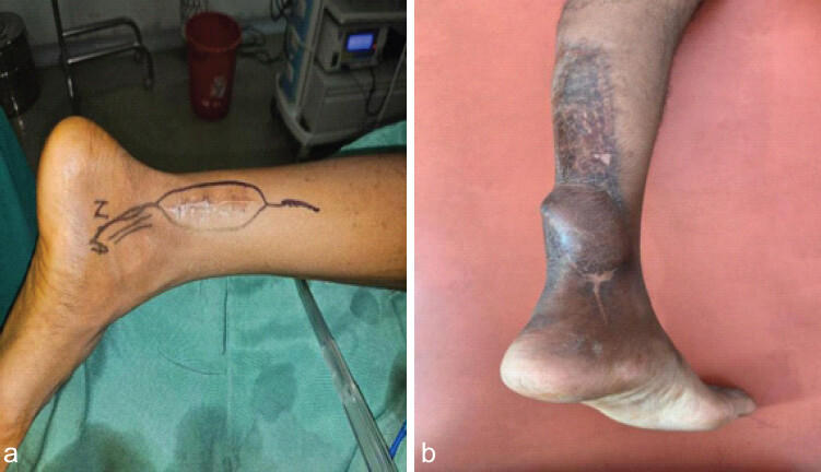
For reconstruction, a sural nerve cable graft harvested from the contralateral leg was used for end-to-end fascicular coaptation to bridge the proximal and distal ends of the resected tibial nerve. Soft-tissue coverage of the primary defect was achieved using a distally based neurovascular sural pedicled neurocutaneous local flap. A split-thickness skin graft was then applied to cover the donor site and any remaining open areas. All surgical wounds healed uneventfully within 20 days.
Postoperatively, adjuvant radiation therapy was indicated due to multiple high-risk clinicopathologic features:
• Histopathologic nature: SS is an aggressive, high-grade malignancy with a high propensity for local recurrence and distant metastasis.
• Tumor depth: The lesion was situated deep to the deep fascia, which independently increases the risk of recurrence.
• Surgical margins: Although an R0 resection was achieved, the basal margin was close at 0.5 cm (<1.0 cm), warranting adjuvant irradiation to optimize local control.
Consequently, the patient received adjuvant radiotherapy with a total dose of 64 Gy delivered in 32 daily fractions.
In accordance with the EORTC 62931 trial (“Adjuvant chemotherapy with doxorubicin, ifosfamide, and lenograstim for resected soft-tissue sarcoma: a multicenter randomised controlled trial”), the absolute overall survival benefit of adjuvant anthracycline and ifosfamide in localized soft-tissue sarcoma is not sufficient to outweigh the substantial risks of treatment-related morbidity [12].
At the 28-month post-operative follow-up, the patient remained pain-free but demonstrated a pes cavus deformity with clawing of the toes and a mildly altered gait (Fig. 6b). Physical examination revealed mild, functionally non-limiting sensory blunting, an absent ipsilateral Achilles tendon reflex, and intrinsic foot muscle weakness (MRC grade 4/5 for toe abduction and adduction). The MSTS functional score [10] was 22/30 (73.3%), corresponding to a “Good” clinical outcome (Table 1).
Follow-up imaging – including soft-tissue sonography, MRI, and chest X-ray – revealed no abnormalities.
Discussion
Sss are malignant mesenchymal tumors, arising from connective tissue, schwannomas, neurofibromas, or malignant tumors arise from Schwann cells. A perineurium forms the second envelop, made up of epithelioid cells, from which perineurinomas can arise. The last sheath is the epineurium, formed by fibrous tissue and mesenchymal cells, and from where sarcomas can originate [13]. SSs account for 5–10% of all soft-tissue sarcomas [14]. However, the name SS is misleading, because the tumor does not originate from synovial cells, but rather from primitive mesenchymal cells [11]. The name most likely originated from the localization around the large joints on the limbs, more often on the lower ones, in the area of the knee joints [15]. There are generally two types of SS. Monophasic sarcoma, i.e., made up of only one type of cells, namely, spindle or glandular cells, and biphasic sarcoma, made up of both types of cells. Soft-tissue sarcomas are usually fast-growing, painless tumors, but SSs often grow more slowly, are painful, can cause joint contractures, swelling, and the average time from the onset of symptoms to the established diagnosis is 2 years [16]. Pain was the dominant symptom in our patient as well. SSs often appear in adolescents and young adults, and affects both sexes equally [17,18]. The basis for diagnosis is a magnetic resonance examination and then a biopsy, which should be performed before surgery so that a sufficiently radical procedure can then be performed. Cytogenetically, SS is characterized by the chromosomal translocation t(X;18) (p11.2; q11.2), which leads to the fusion of two genes – SS18 (SYT) on chromosome 18 and SSX on chromosome X, and to the formation of SYT-SSX fusion transcripts (SS18-: SSX1, SS18-: SSX2, and rarely SS18: -SSX4). Even in our patient, fluorescence in situ hybridization (FISH) analysis confirmed a break in the SS18 gene, testifying to this chromosomal translocation. The immunohistochemical profile of SSs characteristically shows signs of epithelial differentiation.
The detection of pancytokeratins, positive in up to 90% of cases [19], and the expression of the EMA are typical. Both of these markers can be commonly identified in the vast majority of epithelial Tumors [20]. EMA positivity itself is non-specific in soft-tissue pathology. The same is true of pancytokeratins [21]. Due to this limited specificity (approximate 64–85% [pooled average: ~73–81%]), TLE1 is recommended as part of an immunohistochemical panel (e.g., with EMA, CD34, BCL2, and keratins) and cannot be considered completely diagnostic on its own [22], while immunohistochemical detection of other proteins (Bcl2, CD56 and CD99) is currently only of limited importance due to low specificity. The focal positivity of the S100 protein can be identified in approximately 1/3 of SSs [23]. S100 protein is a typical marker of neuroectodermal tumors, and a similar focal positivity is often observed in malignant Tumors from peripheral nerve sheaths, which also share a similar spindle cell morphology with SSs. The most reliable means to distinguish between the two units is the proof of a break in the SS18 gene using FISH, reverse transcription polymerase chain reaction, or the massively parallel sequencing method. The main treatment of SS is surgical treatment, which should take place only after a biopsy with histological examination of the tumor so that it is sufficiently radical and does not have to undergo an additional reoperation, as happened in the case of our patient. Primary less radical excision without prior biopsy verification leads to a higher risk of local recurrence, even if a proper re-excision was performed immediately after biopsy verification of the sarcoma [24,25]. Neoadjuvant radiotherapy is recommended especially for larger tumors (>5 cm) or in cases where the resection margin has been reduced, for example due to an ongoing vessel, nerve or bone [26]. Radiotherapy seems to prolong the overall survival of patients [27,28,29]. SSs appear to be more chemo-sensitive than other soft-tissue sarcomas. Chemotherapy is recommended for younger patients or patients with unresectable or already generalized sarcomas [30,31,32]. Patient’s prognosis is uncertain; reported 5-year survival is around 30–50%, and only 10% of patients survive more than 10 years. Negative prognostic factors include tumor invasion into bone or blood vessels, advanced age, larger tumor size, and less radical excision [33, 34, 35]. Primary insufficiently radical excision increases the risk of local recurrence and thus worse prognosis.
Conclusion
This case report draws attention to a rare type of malignant nerve tumor, which both clinically and radiologically can mimic benign peripheral nerve sheath tumors. SS situated under deep fascia is a significant negative prognostic factor which requires a timely diagnosis using MR imaging and a biopsy with histological examination to start treatment quickly.
Clinical Message
Deep-seated soft-tissue masses must be treated with a high index of suspicion for malignancy, regardless of size, and the heterogeneity of the tumor can pose a diagnostic challenge, even for pathologists specializing in sarcoma.
Conflict of Interest:
Nil
Source of Support:
Nil
Consent:
The authors confirm that informed consent was obtained from the patient for publication of this article
How to Cite this Article
Mehta VR, Mehta R, Parmar R, Mehta SR. Synovial Sarcoma of the Tibial Nerve in the Distal Leg: A Case of Diagnostic Ambiguity and Multimodal Management Protocol for Limb Salvage and Nerve Reconstruction – A Case Report. J Orthop Case Rep 2026 November;16(11): 20-26. doi: 10.13107/jocr.2026.v16.i11.8374.
References
- Spielmann A, Janzen DL, O'Connell JX, Munk PL. Intraneural synovial sarcoma. Skeletal Radiol 1997;26:677-81. [Google Scholar] | [PubMed]
- Scheithauer BW, Amrami KK, Folpe AL, Silva AI, Edgar MA, Woodruff JM . Synovial sarcoma of nerve. Hum Pathol 2011;42:568-77. [Google Scholar] | [PubMed]
- Larque AB, Bredella MA, Nielsen GP, Chebib I. Synovial sarcoma mimicking benign peripheral nerve sheath tumor. Skeletal Radiol 2017;46:1463-8. [Google Scholar] | [PubMed]
- Hashimoto K, Nishimura S, Fujii K, Kakinoki R, Akagi M. Intraneural synovial sarcoma of the tibial nerve. Rare Tumours 2018;10:2036361318776495. [Google Scholar] | [PubMed]
- Hemerková P, Matulová H, Vališ M, Soukup J, Kanta M, Jandura J. Synovial sarcoma of the tibial nerve - case report of a rare tumor in a rare location requiring early diagnosis. BMC Neurol 2023;23:65. [Google Scholar] | [PubMed]
- Fisher C. Synovial sarcoma. Ann Diagn Pathol 1998;2:401-21. [Google Scholar] | [PubMed]
- Li LT, Jiang G, Chen Q, Zheng JN. Ki67 is a promising molecular target in the diagnosis of cancer (review). Mol Med Rep 2015;11:1566-72. [Google Scholar] | [PubMed]
- Wojcik E, Kulik A, Koscielniak E, Rychłowska-Myszka M, Bilska K, Karpatko O . Ki-67 expression in soft tissue sarcomas in children. Nowotwory J Oncol. 2012;62:181-6. [Google Scholar] | [PubMed]
- Skytting BT, Bauer HC, Perfekt R, Nilsson G, Larsson O. Ki-67 is strongly prognostic in synovial sarcoma: Analysis based on 86 patients from the Scandinavian sarcoma group register. Br J Cancer 1999;80:1809-14. [Google Scholar] | [PubMed]
- Enneking WF, Dunham W, Gebhardt MC, Malawar M, Pritchard DJ. A system for the functional evaluation of reconstructive procedures after surgical treatment of tumors of the musculoskeletal system. Clin Orthop Relat Res 1993;286:241-6. [Google Scholar] | [PubMed]
- WHO Classification of Tumours Editorial Board Soft Tissue and Bone Tumours. WHO Classification of Tumours Series. 5th ed 3 Lyon International Agency for Research on Cancer;2020:290-3. [Google Scholar] | [PubMed]
- Woll PJ, Reichardt P, Le Cesne A, Bonvalot S, Azzarelli A, Hoekstra HJ, Leahy M, Van Coevorden F, Verweij J, Hogendoorn PC, Ouali M, Marreaud S, Bramwell VH, Hohenberger P. EORTC Soft Tissue and Bone Sarcoma Group and the NCIC Clinical Trials Group Sarcoma Disease Site Committee Adjuvant chemotherapy with doxorubicin, ifosfamide, and lenograstim for resected soft-tissue sarcoma: a randomised controlled trial. Lancet Oncol 2012;13:1045-54 [Last accessed on 2026 Oct 05]. [Google Scholar] | [PubMed]
- Belakhoua SM, Rodriguez FJ. Diagnostic pathology of tumors of peripheral nerve. Neurosurgery 2021;88:443-56. [Google Scholar] | [PubMed]
- Toro JR, Travis LB, Wu HJ, Zhu K, Fletcher CD, Devesa SS. Incidence patterns of soft tissue sarcomas, regardless of primary site, in the surveillance, epidemiology and end results program, 1978-2001: An analysis of 26,758 cases. Int J Cancer 2006;119:2922-30. [Google Scholar] | [PubMed]
- Sultan I, Rodriguez-Galindo C, Saab R, Yasir S, Casanova M, Ferrari A . Comparing children and adults with synovial sarcoma in the surveillance, Epidemiology, and end results program, 1983 to 2005: An analysis of 1268 patients. Cancer 2009;115:3537-47. [Google Scholar] | [PubMed]
- Chotel F, Unnithan A, Chandrasekar CR, Parot R, Jeys L, Grimer RJ. Variability in the presentation of synovial sarcoma in children. J Bone Jt Surg Br 2008;90-B:1090-6. [Google Scholar] | [PubMed]
- De Silva MV, Barrett A, Reid R. Premonitory pain preceding swelling: A distinctive clinical presentation of synovial sarcoma which may prompt early detection. Sarcoma 2003;7:131-5. [Google Scholar] | [PubMed]
- Aytekin MN, Öztürk R, Amer K, Yapar A. Epidemiology, incidence, and survival of synovial sarcoma subtypes: SEER database analysis. J Orthop Surg (Hong Kong) 2020;28:2309499020936009. [Google Scholar] | [PubMed]
- Miettinen M, Limon J, Niezabitowski A, Lasota J. Patterns of keratin polypeptides in 110 biphasic, monophasic, and poorly differentiated synovial sarcomas. Virchows Arch 2000;437:275-83. [Google Scholar] | [PubMed]
- Michal M. Basic immunohistochemical panel for the diagnosis of soft tissue tumors. Ceska Patol 2021;57:12-8. [Google Scholar] | [PubMed]
- Miettinen M. Immunohistochemistry of soft tissue tumours - review with emphasis on 10 markers. Histopathology 2014;64:101-18. [Google Scholar] | [PubMed]
- Foo WC, Cruise MW, Wick MR, Hornick JL. Immunohistochemical staining for TLE1 distinguishes synovial sarcoma from histologic mimics. Am J Clin Pathol 2011;135:839-44. [Google Scholar] | [PubMed]
- Pelmus M, Guillou L, Hostein I, Sierankowski G, Lussan C, Coindre JM. Monophasic fibrous and poorly differentiated synovial sarcoma: Immunohistochemical reassessment of 60 t(X;18)(SYT-SSX)-positive cases. Am J Surg Pathol 2002;26:1434-40. [Google Scholar] | [PubMed]
- Chandrasekar R, Wafa H, Grimer RJ, Carter SR, Tillman RM, Abudu A. The effect of an unplanned excision of a soft-tissue sarcoma on prognosis. J Bone Jt Surg Br 2008;90:203-8. [Google Scholar] | [PubMed]
- Potter BK, Adams SC, Pitcher JD Jr. Local recurrence of disease after unplanned excisions of high-grade soft tissue sarcomas. Clin Orthop Relat Res 2008;466:3093-100. [Google Scholar] | [PubMed]
- Von Mehren M, Randall RL, Benjamin RS, Boles S, Bui MM, Conrad EU 3rd . Soft tissue sarcoma, version 2.2016, NCCN clinical practice guidelines in oncology. J Natl Compr Canc Netw 2016;14:758-86. [Google Scholar] | [PubMed]
- Gingrich AA, Marrufo AS, Liu Y, Li CS, Darrow MA, Monjazeb AM . Radiotherapy is associated with improved survival in patients with synovial sarcoma undergoing surgery: A national cancer database analysis. J Surg Res 2020;255:378-87. [Google Scholar] | [PubMed]
- Naing KW, Monjazeb AM, Li CS, Lee LY, Yang A, Borys D . Perioperative radiotherapy is associated with improved survival among patients with synovial sarcoma: A SEER analysis. J Surg Oncol 2015;111:158-64. [Google Scholar] | [PubMed]
- Song S, Park J, Kim HJ, Kim IH, Han I, Kim HS . Effects of adjuvant radiotherapy in patients with synovial sarcoma. Am J Clin Oncol 2017;40:306-11. [Google Scholar] | [PubMed]
- Canter RJ, Qin LX, Maki RG, Brennan MF, Ladanyi M, Singer S. A synovial sarcoma-specific preoperative nomogram supports a survival benefit to ifosfamide-based chemotherapy and improves risk stratification for patients. Clin Cancer Res 2008;14:8191-7. [Google Scholar] | [PubMed]
- Vlenterie M, Litière S, Rizzo E, Marréaud S, Judson I, Gelderblom H . Outcome of chemotherapy in advanced synovial sarcoma patients: Review of 15 clinical trials from the european organisation for research and treatment of cancer soft tissue and bone sarcoma group; setting a new landmark for studies in this entity. Eur J Cancer 2016;58:62-72. [Google Scholar] | [PubMed]
- Spurrell EL, Fisher C, Thomas JM, Judson IR. Prognostic factors in advanced synovial sarcoma: An analysis of 104 patients treated at the Royal Marsden hospital. Ann Oncol 2005;16:437-44. [Google Scholar] | [PubMed]
- De Silva MV, McMahon AD, Reid R. Prognostic factors associated with local recurrence, metastases, and Tumor-related death in patients with synovial sarcoma. Am J Clin Oncol 2004;27:113-21. [Google Scholar] | [PubMed]
- Trassard M, Le Doussal V, Hacène K, Terrier P, Ranchère D, Guillou L . Prognostic factors in localized primary synovial sarcoma: A multicenter study of 128 adult patients. J Clin Oncol 2001;19:525-34. [Google Scholar] | [PubMed]
- Kawai A, Woodruff J, Healey JH, Brennan MF, Antonescu CR, Ladanyi M. SYT-SSX gene fusion as a determinant of morphology and prognosis in synovial sarcoma. N Engl J Med 1998;338:153-60. [Google Scholar] | [PubMed]
- . Available from: https://www.ncbi.nlm.nih.gov/books/nbk65923.1/table/CDR0000062934__749/?report=objectonly [Last accessed on 2026 Oct 05]. [Google Scholar] | [PubMed]
- Beaino ME, Jupiter DC, Assi T, Rassy E, Lazar AJ, Araujo DM . Diagnostic value of TLE1 in synovial sarcoma: A systematic review and meta-analysis. Sarcoma 2020;2020:7192347. [Google Scholar] | [PubMed]
© 2026 Journal of Orthopaedic Case Reports - Published by Indian Orthopaedic Research Group
About the Authors
-

Varun R Mehta
-

Rajkumar Mehta
-

Rahul Parmar
-

Sonal R Mehta
Related Articles in Journal of Orthopaedic Case Reports
 October 1, 2026 Arthroscopic Piecemeal Excision of a Large Intra-articular Infrapatellar Osteochondroma of the Knee: A Case Report
 August 1, 2026 Intramuscular Glomus Tumor of Foot Presenting as Neuroma of Sural Nerve: A Case Report
 August 1, 2026 Understanding Diagnostic Challenges in Ewing Sarcoma – A Report of Two Cases
 June 1, 2026 Comparison of Radial Head Prosthesis versus Excision in Comminuted Radial Head Fractures: A Retrospective Comparative Study