


Introduction
Epidural catheterization is a common practice for perioperative analgesia in orthopedic, obstetric, and general surgical practice. Although considered a safe procedure, infrequent complications of epidural catheter kinking, knotting, migration, and fracture have been reported. Epidural catheter breakage is an uncommon incident with an estimated incidence of approximately 0.002% [1]. Despite its low frequency, retention of a catheter fragment within the paraspinal tissues or spinal canal may result in a challenging clinical dilemma.
Retained epidural catheter fragments have traditionally been regarded as biologically inert foreign bodies, and conservative management has been proposed for asymptomatic adult patients [2,3]. Several reports have documented uneventful outcomes following observation alone [4]. However, delayed complications, including radiculopathy, nerve root compression, fragment migration, and epidural fibrosis have also been described, necessitating subsequent surgical intervention [5,6]. When a retained fragment lies within or adjacent to the spinal canal, adherence to the dura and surrounding structures due to granulation tissue may pose a challenge to delayed retrieval in selected cases [7]. Published literature describes successful outcomes with both conservative observation and surgical retrieval, highlighting the absence of a universally accepted treatment strategy.
Given the rarity of these complications, consensus regarding optimal management remains limited. Decisions are typically influenced by fragment location, neurological status, radiological findings, patient factors, and feasibility of safe retrieval [3,8]. Kasiamnham further highlighted the ongoing controversy surrounding retained epidural catheter fragments, noting that both conservative and surgical retrieval have been advocated depending on clinical and fragment location [9]. We present a case of a retained epidural catheter fragment in a polytrauma patient managed surgically and review the present literature with emphasis on management decisions.
Case Report
A 31-year-old male polytrauma patient presented to the emergency department with a history of a road traffic accident. Clinical and radiographic evaluation revealed a right-sided closed fracture of the neck of the femur with an ipsilateral midshaft femur fracture, a left closed fracture of the midshaft humerus with radial nerve palsy, and a left closed distal one-third femoral shaft fracture.
Initially, the patient was planned for closed reduction and internal fixation of the right neck of femur and ipsilateral midshaft femur fracture with a proximal femoral nail anti-rotation. For perioperative analgesia, epidural catheter placement was attempted at the L3-L4 level, under aseptic precautions using an 18-gauge Tuohy needle. During the catheter insertion, resistance was encountered, and during repositioning, the catheter fractured, leaving a retained segment. Upon comparison with the original catheter, a segment of 15 cm in length had been retained. The epidural procedure was abandoned, and the surgery was subsequently performed under general anesthesia. Post-operatively, the patient remained neurologically intact, with no sensory deficit, motor weakness, or bowel and bladder dysfunction. Plain radiographs and ultrasonography scans failed to localize the retained catheter fragment. Therefore, a computed tomography (CT) scan was obtained as it provided precise localization of the retained fragment relative to bony landmarks and was more cost-effective in our setting. It demonstrated a linear retained catheter fragment at the L3-L4 level extending from the interspinous region toward the posterior epidural space (Fig. 1).
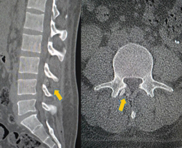
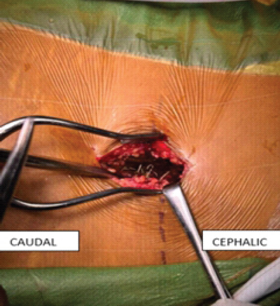
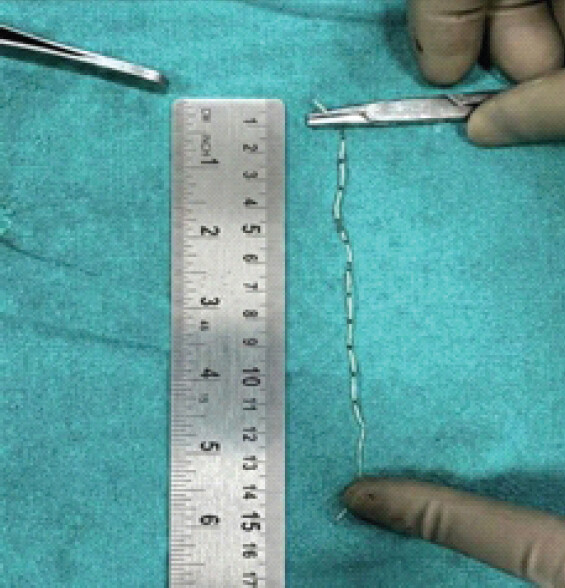
On post-operative day 3, the patient underwent planned fixation of the left distal femoral shaft fracture with a distal femoral nail. Following multidisciplinary discussion and review of the imaging findings, retrieval of the retained epidural fragment was planned during the same operative session. A posterior midline spinal approach was utilized. A 4 cm incision was made over the L3-L4 interspace corresponding to the site of catheter insertion. Following careful dissection through the subcutaneous tissues and paraspinal musculature, the retained catheter fragment was identified under direct visualization (Fig. 2). Intraoperatively, the catheter was found extending between the interspinous region and posterior epidural space. The retained segment was retrieved in its entirety, measuring approximately 15 cm in length (Fig. 3). There was no dural breach, cerebrospinal fluid leak, or neural element injury identified. Hemostasis was achieved and the wound closed in layers.
Three days later, the patient underwent open reduction and internal fixation of the left midshaft humerus with radial nerve exploration. The recovery from the surgery was uneventful and no neurological symptoms attributable to the retained catheter or its retrieval were observed. The patient was discharged in a stable condition and remained asymptomatic at follow-up after 1, 6, and 12 weeks and at 6 months, with no back pain, radicular symptoms, sensory or motor deficits, or bowel and bladder dysfunction.
Discussion
Epidural catheter fracture is a rare complication of neuraxial analgesia, with a limited prevalence [1]. Most reported cases occur during catheter insertion or removal when resistance is encountered. Proposed mechanisms include entrapment within the ligamentum flavum, interspinous ligaments, laminae, or paraspinal tissues, as well as knotting or looping of the catheter within the epidural space [2,8].
The management of retained epidural fragments remains controversial. Kasiamnham [9] reviewed the available evidence and emphasized that the decision to remove a retained epidural catheter fragment should be individualized, with surgical intervention generally reserved for patients with neurological symptoms, infection, or radiological evidence suggesting a high-risk location, while asymptomatic patients may be suitable for observation. Mitra and Fleischmann [2] noted that conservative management may be appropriate in asymptomatic adult patients because epidural catheter materials are generally considered biologically inert. Similarly, Abujayeh et al. [4] reported successful non-operative management without neurological sequelae.
However, delayed complications, including radicular pain, nerve root compression, migration, and dural adhesions, have been described. Tarukado et al. [7] described fragment migration and dense adhesions that complicated delayed retrieval. Gompels et al. [1] and Hippalgaonkar et al. [5] suggested that removal may be considered when imaging demonstrates a retained fragment within or extending into the spinal canal. Subith et al. [10] reported successful retrieval of an epidural catheter following accurate pre-operative localization.
In the present case, the patient remained neurologically intact, and CT localized the retained fragment to L3-L4, extending through the interspinous space into the posterior epidural space. Given its deep location, the potential for delayed adhesion or migration, and the option for retrieval during planned orthopedic fixation, the fragment was removed during the same operation without dural injury or neurological deficit. It highlights the importance of radiological localization, multidisciplinary assessment and individual decision-making based on fragment location, patient factors and feasibility of safe retrieval.
Conclusion
Retained epidural catheter fragments represent a rare complication of neuraxial analgesia for which no universally accepted guidelines exist. The available literature supports both conservative observation and surgical retrieval in selected circumstances. Careful clinical assessment, accurate radiological localization and multidisciplinary evaluation remain essential when determining the most appropriate management strategy.
Clinical Message
Retained epidural catheter fragments present a unique management challenge. Detailed clinical evaluation, accurate radiological localization, and an understanding of the fragment’s anatomical relationship to surrounding structures are essential for planning appropriate management.
Conflict of Interest:
Nil
Source of Support:
Nil
Consent
The authors confirm that informed consent was obtained from the patient for publication of this article
How to Cite this Article
Singla A, Rai A, Barwar N, Sharma PK, Rathi U, Chahal H. Management of Retained Epidural Catheter Fragment in a Polytrauma Patient: A Case Report. J Orthop Case Rep 2026 November;16(11): 50-53. doi: 10.13107/jocr.2026.v16.i11.8384.
References
- Gompels B, Rusby T, Slater N. Fractured epidural catheter with retained fragment in the epidural space-a case study and proposed management algorithm. BJA Open 2022;4:100095. [Google Scholar] | [PubMed]
- Mitra R, Fleischmann K. Management of the sheared epidural catheter: Is surgical extraction really necessary?. J Clin Anesth 2007;19:310-4. [Google Scholar] | [PubMed]
- Fragneto RY. The broken epidural catheter: An anesthesiologist's dilemma. J Clin Anesth 2007;19:243-4. [Google Scholar] | [PubMed]
- Reena, Vikram A. Fracture of epidural catheter: a case report and review of literature. Saudi J Anaesth. 2017;11 1:108-110. doi:10.4103/1658-354X.197359.. [Google Scholar] | [PubMed] | [CrossRef]
- Hippalgaonkar AV, Kudalkar AG, Gaikwad SM, Modak S, Gupta HB, Tendolkar BA. Successful management of a broken epidural catheter !!!. Saudi J Anaesth 2017;11:228-31. [Google Scholar] | [PubMed]
- Ugboma S, Au-Truong X, Kranzler LI, Rifai SH, Joseph NJ, Salem MR. The breaking of an intrathecally-placed epidural catheter during extraction. J Clin Anesth 2002;14:62-4. [Google Scholar] | [PubMed]
- Tarukado K, Oda T, Tono O, Suetsugu H, Doi T. A retained epidural catheter fragment treated by surgery. Asian Spine J 2015;9:461-4. [Google Scholar] | [PubMed]
- Sharma J, Murmu S, Samagh N, Grewal A. Management of an epidural catheter rent: An anesthesiologist's dilemma. Indian J Pain 2024;38:S534. [Google Scholar] | [PubMed]
- Kasiamnham R. The broken epidural catheter: To remove or not to remove?. Br J Hosp Med (Lond) 2012;73:710-1. [Google Scholar] | [PubMed]
- Subith S, Dussa KR, Kokate S, Mukadam M, Zafar S, Choudhari A. When the unexpected happens: The intriguing retrieval of a broken epidural catheter. J Orthop Case Rep 2024;14:136-9. [Google Scholar] | [PubMed]
© 2026 Journal of Orthopaedic Case Reports - Published by Indian Orthopaedic Research Group
About the Authors
-

Ayush Singla
-

Ankit Rai
-

Nilesh Barwar
-

Pankaj Kumar Sharma
-

Uma Rathi
-

Himanshu Chahal
Related Articles in Journal of Orthopaedic Case Reports
 November 1, 2026 Metastatic Lung Cancer to Non-vascular Soft Tissue and Lumbar Spine Mimicking Postoperative Scar at L4–5 Non-union: A Case Report
 November 1, 2026 Second and Third Carpometacarpal Joint Dislocation in a Polytrauma Patient: Successful Closed Reduction and 2-Year Follow-Up
 October 1, 2026 Bilateral Cedell Fractures of the Talus Associated with Ankle Fractures in a Polytrauma Patient: A Rare Injury Managed with Damage Control Orthopedics and Staged Fixation
 October 1, 2026 Degloving in Disguise: Managing a Delayed Spinal Morel-Lavallée Lesion after Thoracic Fracture-Dislocation