

When congenital tibial pre-pseudarthrosis remains unchanged during carefully monitored bracing, and the child cannot tolerate repeated brace modification, operative correction may be considered on an individualized basis; this case documents durable 9-year correction without claiming superiority over continued non-operative care.
Farhad Mustafa Mousa Atrushi, Department of Physiotherapy, College of Health Sciences, Hawler Medical University, Erbil, Kurdistan Region, Iraq. E-mail: farhadm.atrushi@gmail.com
Abstract
Introduction: Congenital tibial pre-pseudarthrosis is the intact dysplastic stage of the congenital pseudarthrosis spectrum. Management before fracture is unsettled: Bracing is commonly used, whereas prophylactic surgery is reserved for selected circumstances.
Case Report: A 2-year-and-2-month-old boy had painless anterolateral bowing and dysplastic narrowing of the left tibia without pathological fracture or established pseudarthrosis. Bracing was continued for approximately 5.5 months. Three brace configurations were tried and modified to improve fit and tolerance, with serial clinical and radiographic review showing no meaningful correction of the deformity. The child repeatedly developed apparent pain, restlessness, and crying during brace use, particularly at night; these symptoms settled after the brace was removed. After counseling, the parents declined further bracing and preferred surgery. An open-wedge tibial osteotomy was performed with drill recanalization of the medullary canals and fixation with a 2.5-mm Kirschner wire. A 1.5-cm segment of the ipsilateral distal fibula was resected, divided into two autologous grafts, and placed in the tibial wedge. At 9 years, the tibial osteotomy and fibular donor site were completely healed and remodeled, alignment was maintained, and no pathological fracture or established pseudarthrosis had occurred. The patient was pain-free, had full knee and ankle motion, and had an asymptomatic 11-mm shortening of the left lower limb.
Conclusion: This case provides long-term documentation of an individualized prophylactic reconstruction after a prolonged, poorly tolerated trial of bracing. The result supports the technical feasibility of open-wedge correction with local fibular autograft and simple Kirschner-wire fixation, but a single case cannot establish that surgery is preferable to bracing.
Keywords: Congenital tibial pre-pseudarthrosis, anterolateral tibial bowing, fibular autograft, osteotomy, pediatric orthopedics.

Congenital pseudarthrosis of the tibia (CPT) represents a spectrum of congenital tibial dysplasia. The earliest radiographic stage may consist of anterolateral bowing and segmental narrowing while the tibia remains intact; with progression, pathological fracture and established pseudarthrosis may occur [1]. This intact stage has been described as anterolateral tibial bowing, pre-fracture disease, or pre-pseudarthrosis [1,2,3,4,5,6].
Treatment before fracture remains controversial. Bracing is widely used to protect the dysplastic tibia, although the literature does not show that an orthosis reliably corrects the deformity or abolishes fracture risk [1,3]. Several prophylactic operations have also been reported, including bypass grafting with prolonged bracing, Ilizarov-based correction, and, more recently, guided growth [2,3,4,5]. The limited and heterogeneous evidence makes patient selection and timing difficult.
We report the 9-year course of a child in whom bracing was pursued for approximately 5.5 months and was repeatedly modified but remained poorly tolerated, while serial clinical and radiographic assessment showed no meaningful improvement in the deformity. The subsequent reconstruction combined an open-wedge tibial osteotomy, drill recanalization, local ipsilateral fibular autograft, and 2.5-mm Kirschner-wire fixation. The report follows the CARE framework [7,8,9].
Clinical history and non-operative management
The patient was a boy with a visible, painless deformity of the left leg. At the time of surgery in July 2017, he was 2 years and 2 months old. There was no known family history of a similar skeletal disorder. No café-au-lait macules or Lisch nodules were documented, and the available clinical record did not identify an associated neurological, vascular, or metabolic disorder.
Non-operative treatment with bracing was continued for approximately 5.5 months. Three separate brace configurations were used during this period. Each brace was adjusted or modified in an attempt to improve fit and tolerance. The child was reviewed clinically and with serial radiographs, but no meaningful improvement in the deformity was identified. According to the parents, wearing the brace repeatedly provoked apparent pain and marked discomfort, with restlessness and frequent crying during the day and especially at night. The child settled after brace removal. Because the same pattern persisted despite three brace trials and repeated modification, the parents declined further bracing.
The family was counseled about continued non-operative care and the uncertainties, risks, and potential benefits of prophylactic surgery in an intact dysplastic tibia. They expressed a preference for operative correction, and surgery proceeded with their informed agreement. Pre-operative limb-length discrepancy, mechanical-axis deviation, rotational profile, and standardized functional scores had not been quantified.
Imaging and diagnosis
Pre-operative anteroposterior radiography demonstrated pronounced anterolateral bowing and dysplastic narrowing of the left tibia without a frank pathological fracture or an established pseudarthrosis (Fig. 1). The ipsilateral fibula was also bowed, while the right lower limb appeared grossly normal on the available projection. The findings were considered consistent with congenital tibial pre-pseudarthrosis. Because the source radiographs were available as non-calibrated photographs rather than Digital Imaging and Communications in Medicine studies, reliable angular and length measurements could not be reconstructed.
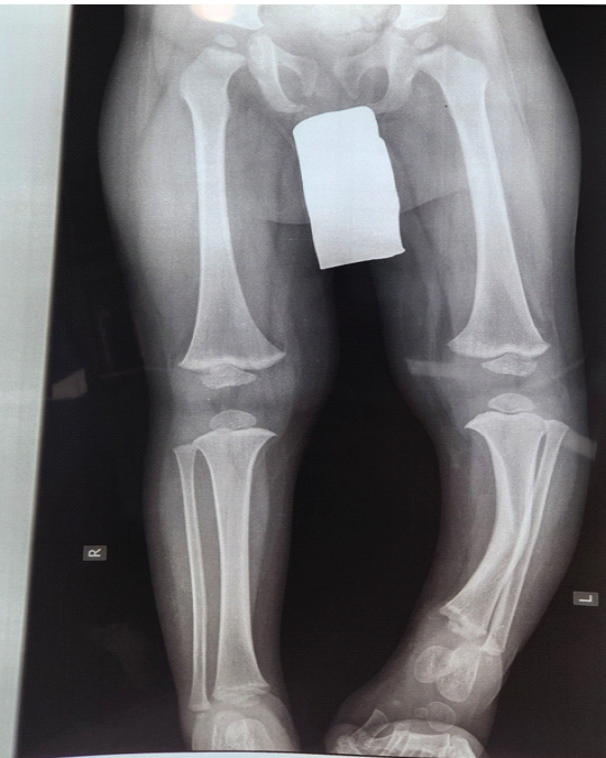
Figure 1: Pre-operative anteroposterior radiograph. Pre-operative anteroposterior radiograph of both lower limbs at age 2 years. The left tibia demonstrates marked anterolateral bowing and dysplastic narrowing without a pathological fracture or established pseudarthrosis on the available projection. The left fibula is also bowed; the right lower limb is grossly unremarkable.
The differential diagnosis included osteofibrous dysplasia, post-traumatic deformity, and metabolic causes of tibial bowing. The patient’s age, unilateral anterolateral deformity, and dysplastic tibial narrowing favored congenital pre-pseudarthrosis. The absence of clinical stigmata of neurofibromatosis type 1 at this young age was not considered sufficient to exclude later recognition of the disorder [10].
Surgical treatment
Surgery was performed on July 10, 2017, when the child was 2 years, 2 months, and 25 days old. Under general anesthesia, with the patient supine and a pneumatic tourniquet in place, the limb was prepared and draped in the usual sterile fashion. A separate incision was made over the distal fibula. The fibular periosteum was split longitudinally and reflected, and a 1.5-cm cylindrical segment was resected. The segment was divided into two pieces and retained as a local autologous graft.
A second incision was centered over the apex of the tibial deformity. The tibial periosteum was opened, and an open-wedge osteotomy was made with an oscillating saw. The proximal and distal medullary canals were opened with a drill. After correction of alignment, the two fibular grafts were placed within the tibial wedge, and the reconstruction was stabilized with a 2.5-mm Kirschner wire. Fluoroscopy confirmed satisfactory alignment. The tibial periosteum was repaired; the reflected fibular periosteum was not repaired. No demineralized bone matrix, platelet-rich plasma, bone marrow aspirate or concentrate, or other biological adjunct was used.
The wounds were closed in layers, and an above-knee fiberglass cast was applied. A window was fashioned in the cast while the child remained anesthetized so that the wounds could later be inspected without using a cast saw close to the awake child. Radiographs on the first post-operative day showed improved tibial alignment, stable Kirschner-wire fixation, and the expected distal fibular donor-site defect (Fig. 2).
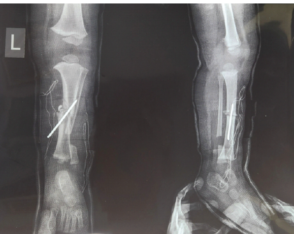
Figure 2: First post-operative-day radiographs. Anteroposterior and lateral radiographs through the above-knee fiberglass cast on the 1st post-operative day. Tibial alignment is corrected and maintained with a 2.5-mm Kirschner wire. The two grafts prepared from the resected 1.5-cm distal fibular segment lie within the tibial open wedge, and the expected fibular donor-site defect is visible.
Post-operative course
Intravenous antibiotics were administered for 2 days, followed by oral antibiotics for 8 days. The wounds were reviewed through the cast window, and the sutures were removed on post-operative day 10.
At 1 month, the Kirschner wire was removed under mild sedation and sterile conditions. The above-knee cast was then shortened to a below-knee cast. Fifteen days later, 45 days after surgery, immobilization was discontinued. No formal physiotherapy program was prescribed because of the child’s age; progressive spontaneous activity was allowed after cast removal.
Follow-up and outcome
Radiographs at 1 month showed progressive consolidation of the tibial osteotomy and early osseous bridging at the fibular donor site (Fig. 3). At the 9-year review, anteroposterior and lateral radiographs showed complete healing and mature remodeling of the tibial osteotomy with maintained alignment (Fig. 4). There had been no pathological fracture and no progression to established pseudarthrosis. The 1.5-cm fibular donor-site defect had also reconstituted, with solid continuity and no visible residual resection line.
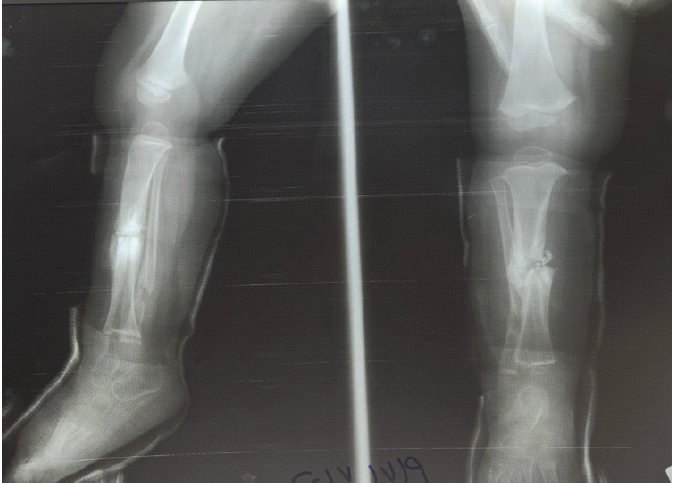
Figure 3: One-month post-operative radiographs. Anteroposterior and lateral radiographs at 1 month, after Kirschner-wire removal. The tibial reconstruction shows progressive consolidation and early cortical continuity. Callus and osseous bridging are also developing across the distal fibular donor site.
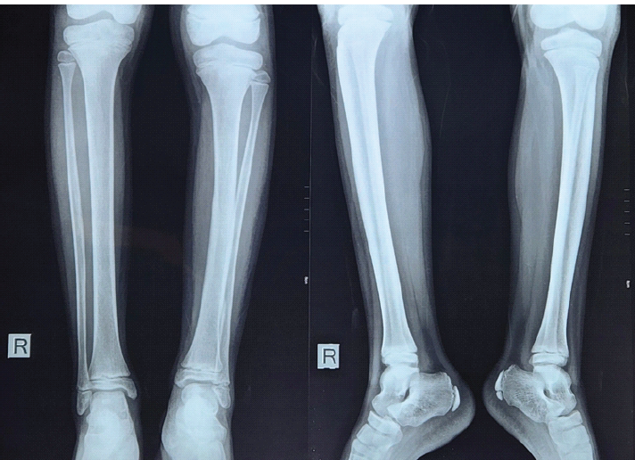
Figure 4: Nine-year follow-up radiographs. Final anteroposterior and lateral radiographs at 9 years. The tibial osteotomy is completely healed and remodeled with maintained alignment and no pathological fracture or progression to established pseudarthrosis. The previous 1.5-cm fibular defect has also healed completely, with solid bridging and restored continuity and contour.
Clinically, the patient was pain-free and had full knee and ankle motion. The only residual finding was an asymptomatic 11-mm shortening of the left lower limb, without an apparent limp. No surgical-site infection, wound dehiscence, recurrent deformity, Achilles tendon contracture, or loss of motion had been documented. Formal gait analysis and a validated patient-reported outcome measure were not available.
The principal events are summarized in Table 1.
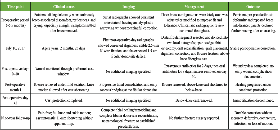
Table 1: Clinical timeline
The most important observation in this case is the sustained course of an intact dysplastic tibia after prophylactic reconstruction. Nine years after surgery, alignment was preserved, the osteotomy had remodeled, the fibular graft had incorporated, and neither pathological fracture nor established pseudarthrosis had developed. Because the tibia had not fractured before treatment, this outcome is better described as durable correction and fracture avoidance than as “union” of a pre-existing pseudarthrosis.
Bracing remains the conventional first approach for many children with an intact dysplastic tibia. In this child, however, the decision to operate did not follow a single unsuccessful fitting. Bracing had been continued for approximately 5.5 months, three configurations had been tried and modified, and the deformity had been followed clinically and radiographically without meaningful improvement. More importantly for day-to-day care, the child repeatedly became distressed while wearing the brace and settled when it was removed. These factors influenced shared decision-making, but they should not be interpreted as universal indications for prophylactic surgery.
Published experience confirms that surgery before fracture is not unprecedented. Ofluoglu et al. reported ten children treated with a posteromedial fibular allograft bypass and long-term bracing; tibial fracture or pseudarthrosis was avoided at final follow-up, although graft-related complications and later corrective osteotomy occurred [2]. El-Mowafi et al. treated six intact Crawford type II tibiae with Ilizarov-based excision and correction and reported correction of bowing, with the expected burden of external fixation and docking-site complications [3]. More recently, Dong et al. described a modified McFarland allograft-bypass strategy in seven Crawford type I/II patients [4], while Zhou et al. reported guided growth in 18 children, with angular improvement and three revisions for staple displacement during relatively short follow-up [5]. These series differ substantially in disease severity, fixation, graft choice, and follow-up; they establish precedent, not comparative superiority.
The technical feature of the present reconstruction was the use of the ipsilateral fibula for two purposes. Resection of the bowed distal fibular segment facilitated correction, and the same segment supplied local autograft for the tibial open wedge. The medullary canals were opened with a drill, and fixation was intentionally simple. At long-term review, the fibular donor site had reconstituted completely. Specific evidence on segmental fibular donor-site regeneration in pre-pseudarthrosis is limited; observations from osteotomy healing in CPT provide biological context but are not directly equivalent [11,12,13,14].
Radiographic classification also matters. El-Rosasy et al. emphasized the distinction between resolving congenital segmental tibial dysplasia and a pre-pseudarthrosis phenotype with adverse radiographic features, such as sclerosis, medullary obliteration, cystic change, or progressive deformity [6]. That distinction is important when interpreting reports of intervention before fracture, because an intact dysplastic tibia should not be described as though an established pseudarthrosis were already present.
This report has several strengths: The pre-fracture phenotype was documented, conservative treatment was attempted over several months with repeated brace modification, the operative details are available, and the clinical and radiographic follow-up extends to 9 years. Its limitations are equally important. It is a single retrospective case; the pre-operative radiographs were not calibrated; serial angular measurements, genetic testing, histopathology, formal gait analysis, and validated outcome scores were unavailable. We also cannot know whether longer bracing would have led to an equally favorable course. The absence of early neurofibromatosis type 1 stigmata does not definitively exclude later recognition of the condition [10].
Selected pre-fracture reports are summarized in Table 2.
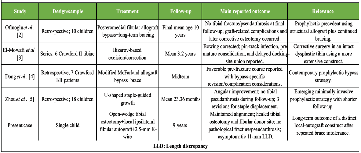
Table 2: Selected pre-fracture literature
In this child, approximately 5.5 months of monitored bracing, including three modified brace trials, neither improved the deformity nor became tolerable. Individualized operative correction was followed by maintained alignment, complete tibial and fibular remodeling, full pain-free knee and ankle motion, and no pathological fracture or established pseudarthrosis at 9 years. The case adds long-term experience with a simple open-wedge reconstruction using local ipsilateral fibular autograft and Kirschner-wire fixation. It does not establish that prophylactic surgery is superior to continued bracing.
An intact dysplastic tibia should be distinguished from established pseudarthrosis. When bracing is repeatedly poorly tolerated, and the deformity remains unchanged, prophylactic surgery may be discussed as an individualized option, with the limits of the available evidence made explicit.
References
- 1. Siebert MJ, Makarewich CA. Anterolateral tibial bowing and congenital pseudoarthrosis of the tibia: Current concept review and future directions. Curr Rev Musculoskelet Med 2022;15:438-46. [Google Scholar] [PubMed]
- 2. Ofluoglu O, Davidson RS, Dormans JP. Prophylactic bypass grafting and long-term bracing in the management of anterolateral bowing of the tibia and neurofibromatosis-1. J Bone Joint Surg Am 2008;90:2126-34. [Google Scholar] [PubMed]
- 3. El-Mowafi H, Abulsaad M, El-Adl W. Prophylactic surgical correction of Crawford’s type II anterolateral bowing of the tibia using Ilizarov’s method. Acta Orthop Belg 2005;71:577-81. [Google Scholar] [PubMed]
- 4. Dong C, Li C, Brückner U, Hellmich H, Krieg AH. Preventing of nonunion in congenital pseudarthrosis of the tibia cases of Crawford type I and II through the use of allograft bypass and a brace: Midterm findings. J Child Orthop 2024;18:187-99. [Google Scholar] [PubMed]
- 5. Zhou X, Liu Y, Chen H, Hu X, Zhu G, Liu K, et al. Clinical study of the ‘U’-shaped staple-guided growth technique in the treatment of congenital anterolateral bowing of the tibia in children. J Pediatr Orthop B 2025;34:608-15. [Google Scholar] [PubMed]
- 6. El-Rosasy MA, Hammad ME, Nada AA. Congenital segmental tibial dysplasia and late onset pseudarthrosis of the tibia. J Orthop 2022;32:25-30. [Google Scholar] [PubMed]
- 7. Kesireddy NR, Kheireldin RK, Lu A, Cooper J, Liu J, Ebraheim NA. Current treatment of congenital pseudarthrosis of the tibia: A systematic review and meta-analysis. J Pediatr Orthop B 2018;27:541-50. [Google Scholar] [PubMed]
- 8. Gagnier JJ, Kienle G, Altman DG, Moher D, Sox H, Riley D, et al. The CARE guidelines: Consensus-based clinical case reporting guideline development. J Med Case Rep 2013;7:223. [Google Scholar] [PubMed]
- 9. Riley DS, Barber MS, Kienle GS, Aronson JK, Von Schoen-Angerer T, Tugwell P, et al. CARE guidelines for case reports: Explanation and elaboration document. J Clin Epidemiol 2017;89:218-35. [Google Scholar] [PubMed]
- 10. Van Royen K, Brems H, Legius E, Lammens J, Laumen A. Prevalence of neurofibromatosis type 1 in congenital pseudarthrosis of the tibia. Eur J Pediatr 2016;175:1193-8. [Google Scholar] [PubMed]
- 11. Shah H, Rousset M, Canavese F. Congenital pseudarthrosis of the tibia: Management and complications. Indian J Orthop 2012;46:616-26. [Google Scholar] [PubMed]
- 12. Paley D. Congenital pseudarthrosis of the tibia: Biological and biomechanical considerations to achieve union and prevent refracture. J Child Orthop 2019;13:120-33. [Google Scholar] [PubMed]
- 13. Johnston CE, Birch JG. A tale of two tibias: A review of treatment options for congenital pseudarthrosis of the tibia. J Child Orthop 2008;2:133-49. [Google Scholar] [PubMed]
- 14. Nahm NJ, Makarewich CA, Rosenwasser KA, Herzenberg JE, McClure PK. Does an osteotomy performed in congenital pseudarthrosis of the tibia heal? J Pediatr Orthop 2022;42:e630-5. [Google Scholar] [PubMed]
Related Articles in Journal of Orthopaedic Case Reports
 July 1, 2026 Step-cut and Butterfly Femoral Osteotomies Following Intraoperative Fracture Propagation during Distraction Osteogenesis: Report of Two Cases
 April 1, 2026 Late-onset Legg-Calvé-Perthes Disease Treated with Varus Derotation Femoral Osteotomy: A Case Report
 November 1, 2026 Slipped Capital Femoral Epiphysis Associated with Congenital Coxa Vara: A Case Report
 November 1, 2026 Identical Twins with Legg-Calve-Perthes Disease: A Report of Two Cases