
Giant lipomas (>5 cm) are rare and can mimic malignant tumours. Deep variants may cause neurovascular compression, resulting in sensory or motor deficits. MRI is the imaging modality of choice for lesion characterisation and preoperative planning, while histopathology confirms the diagnosis. Complete marginal excision provides definitive diagnosis and excellent outcomes.
Dr Antigoni Gkoudina, Academic Department of Trauma and Orthopaedics, University General Hospital of Alexandroupolis, St Niarhos 1, Dragana, 68100, Alexandroupolis, Greece. E-mail: antigkou@gmail.com
Abstract
Introduction: Even though lipomas are the most common benign soft tissue tumours, their occurrence in the hand is relatively uncommon. Giant lipomas, defined as those larger than 5 cm, are exceedingly rare in this location. In the hand, lipomas may occur in subcutaneous tissue, within intramuscular or intermuscular spaces, and may occasionally present with functional impairment or digital paraesthesia due to compression of adjacent neurovascular structures. While most lesions are benign, large or atypical masses require careful evaluation to exclude malignancy. Magnetic resonance imaging is the preferred imaging modality for preoperative assessment, while histopathological examination confirms the diagnosis.
Case Report: We present the case of a 41-year-old Greek Caucasian woman with a progressively enlarging mass in the left hand over a 3-month period, associated with the recent onset of pain and paraesthesia. Imaging findings were consistent with a lipomatous lesion, which was successfully treated with marginal excision. Histopathological analysis confirmed the presence of a benign lipoma.
Conclusion: Giant lipomas of the hand are rare and may mimic malignant tumours; therefore, their evaluation requires careful clinical assessment, appropriate imaging, and histopathological confirmation for accurate diagnosis and optimal management.
Keywords: Giant lipoma, hand lipoma, hand tumours, hand surgery.
In 1856, Sir James Paget was the first to document a lipoma within the trapezius muscle. Later, in 1925, Bufalini described an area of radiolucency within the soft tissues, now referred to as the “Bufalini sign” [1]. Lipomas are the most common benign mesenchymal tumours, affecting approximately 2% of the population. However, their occurrence in the hand is relatively uncommon, accounting for less than 1% of cases. Giant lipomas are defined as those exceeding 5 cm in size and are usually asymptomatic [2]. In some cases, the expansion in deeper anatomical spaces and compression of surrounding structures may cause pain, functional impairment, or neurological symptoms. While the majority of hand tumours are benign in nature, aggressively enlarging and painful masses should raise suspicion for malignancy [3]. Magnetic resonance imaging (MRI) is the imaging modality of choice for evaluating soft-tissue masses, providing detailed information regarding lesion characteristics and anatomical relationships [4]. In cases of large or atypical lesions, histopathologic analysis is essential to confirm diagnosis and rule out the presence of atypia [5].
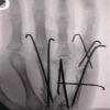
A 41-year-old right-hand-dominant woman sought medical attention after 3 months of a progressively enlarging mass on the volar aspect of her left hand. Over the past 6 days, she had experienced pain and a tingling sensation along the ulnar side of her middle finger and the radial side of her ring finger. She reported no history of trauma to the area, and her medical history was unremarkable. Clinical examination revealed a smooth, relatively mobile, and mildly tender mass on the volar aspect of the 3rd and 4th metacarpal bones (Fig. 1). Examination for light touch sensation revealed numbness in the aforementioned fingers, but otherwise, the patient was neurovascularly intact and displayed a normal range of motion. An initial lateral X-ray of the left hand depicted the “Bufalini sign”, while laboratory testing appeared normal (Fig. 2). Consequently, MRI was performed to better define the lesion’s nature and anatomical boundaries. MRI findings demonstrated a large soft-tissue mass measuring 4.2 × 2.2 × 3.6 cm on the volar aspect of the 2nd, 3rd, and 4th metacarpal bones. The mass exhibited high-signal intensity on both T1- and T2-weighted MRI sequences, with signal suppression on the short tau inversion recovery sequence—findings characteristic of a lipomatous lesion (Fig. 3). Surgical excision was performed through a midline volar incision extending from the proximal border of the transverse carpal ligament to the distal transverse palmar crease. A well-encapsulated multilobular mass 5.5 × 4.5 cm (Fig. 4) with macroscopic characteristics of a lipoma was carefully dissected from the surrounding tissues and excised en bloc. The mass was found to exert pressure on the adjacent common palmar digital nerve and proper digital nerves for the middle and ring finger, explaining the symptoms of the patient (Fig. 5). Histopathological examination demonstrated mature adipose tissue arranged in lobules separated by thin fibrous septa, without evidence of cellular atypia. Immunohistochemical staining showed no overexpression of CDK4 or MDM2, findings consistent with a diagnosis of benign lipoma. Following surgical intervention, the patient experienced immediate relief from pain and tingling sensations, with no signs of motor or vascular compromise, though slight hypaesthesia persisted. At 6 weeks, the patient resumed regular daily activities, achieving full sensory recovery and demonstrating a full, painless range of finger motion. The patient was followed up at 1, 2, 6 weeks, 6 months, and 12 months post-surgery, with no neurological deficits or local recurrence noted.
Lipomas represent the most common benign tumours of mesenchymal origin in humans, as noted by Higgs et al. [6]. The highest incidence is observed between the ages of 40 and 60 and is slightly more prevalent in men than in women [1]. The most common areas of presentation are the upper back, neck, abdomen, and extremities, with proximal locations being more frequent than distal ones [7]. However, their occurrence in the hand region is relatively rare. Giant hand lipomas, defined as those exceeding 5 cm in size, are exceptionally uncommon [2]. When present, they are most frequently located in the thenar and hypothenar regions, while their occurrence in the fingers accounts for only 1% [8,9]. The aetiology of lipoma formation remains unclear, though genetic, metabolic, and traumatic factors have been implicated [10]. Soft-tissue lipomas are classified as superficial or deep according to their anatomical position. Superficial lipomas form in the subcutaneous tissue, whereas deep lipomas arise in regions such as the carpal tunnel, Guyon’s canal, and the deep palmar space [11]. Rarely, parosteal lipomas emerge near bone surfaces, occasionally causing cortical erosion [10]. In cases where no limiting anatomical structures such as bone, fascia, or muscle are present, lipomas are round in shape. Yet, these tumours tend to project into deeper anatomical planes and, as a result, give rise to lipomas of varying size and shape. Deep lipomas tend to be larger and less well-defined, primarily due to the concealing effect of the palmar fascia [11]. Macroscopically, lipomas represent well-margined lobulated masses with a yellow, greasy surface and a thin fibrous capsule, while microscopically, they comprise lobules of mature adipocytes. Soft-tissue lipomas are usually asymptomatic, presenting as a singular, soft, mobile, non-tender mass growing slowly over time. Up to a quarter of superficial hand lipomas can exhibit symptoms including altered sensation, localised discomfort, and muscle weakness [3,5]. Some initially asymptomatic patients may develop clinical signs secondary to lesion enlargement, exerting a mass effect on adjacent neurovascular structures. Deep palmar space lipomas may resemble carpal tunnel syndrome owing to median nerve compression, although lipoma-induced carpal tunnel syndrome is uncommon. Although most hand tumours are benign, lesions demonstrating rapid or progressive enlargement, particularly when associated with pain or neurological symptoms, should be carefully evaluated, as certain malignant lipomatous tumours may exhibit metastatic potential [12,13]. Numerous recent cytogenetic investigations have focused on adipose-derived neoplasms such as lipomas and well-differentiated liposarcomas (WDLPS), showing that these tumours exhibit chromosomal aberrations in regions 12q13-15 and 6p13q [14]. However, malignant transformation of lipomas is considered highly unlikely [11]. Liposarcomas comprise roughly 20% of soft tissue sarcomas. WDLPS represents 40–45% of cases, with the lipoma-like variant being the most prevalent subtype [15]. Atypical lipomatous tumours (ALT) exhibit a predilection for limb involvement, making it a likely diagnosis in cases of malignant giant lipomas of the hand. Both WDLPS and ALT are locally aggressive and share identical histological characteristics but do not metastasise. In contrast, dedifferentiated liposarcoma, myxoid round cell liposarcoma, and pleomorphic liposarcoma exhibit higher metastatic (17–30%) and recurrence rates (34–45%). While superficial soft-tissue masses <5 cm do not require imaging, in cases of large lipomatous lesions of the hand, advanced preoperative imaging is strongly advised in light of the risk of possible malignancy. MRI plays a central role in the evaluation of soft tissue masses, offering high diagnostic accuracy and aiding surgical planning (reported up to 94%) [4,16]. Lipomas show a uniform fat-intense signal across all MRI sequences and may occasionally contain thin septa or show minimal enhancement with gadolinium [17]. These typical imaging features allow for a confident diagnosis without the need for biopsy. However, atypical MRI findings such as heterogeneous signals, non-fat-like intensity, enhancement, or necrosis should raise suspicion for other conditions [18]. Computed tomography is a suitable alternative for patients who cannot undergo MRI due to medical contraindications [17]. Ultrasound imaging usually reveals a well-defined, homogenous, hyperechoic or isoechoic lesion, while plain X-rays may demonstrate a well-margined radiolucent area (“Bufalini sign”) [1,10]. Calcifications, seen in approximately 11% of cases, are more commonly linked to liposarcomas [17]. Histopathological confirmation via excisional tissue biopsy is highly recommended in cases of giant lipomas, serving both diagnostic and therapeutic purposes. Asymptomatic lipomas may be observed, whereas enlarging, symptomatic, or cosmetically concerning lesions warrant surgical removal [6]. The optimal therapeutic modality is marginal excision, requiring meticulous dissection to preserve neurovascular structures. Surgery complications depend on tumour location and may include neurovascular injury, contractures, scar hypersensitivity, infection, and complex regional pain syndrome [2]. Recurrence is rare (<5%), usually due to incomplete excision [19]. Liposarcomas require multidisciplinary care, with treatment focused on wide surgical excision, supported by radiation and possibly chemotherapy, to minimise recurrence [1]. Unlike the majority of reported cases, which are typically located in the thenar or hypothenar regions [2], the present case involved the central palmar space and was associated with early digital nerve compression. Notably, a discrepancy between MRI findings and intraoperative tumour dimensions was observed, underscoring the importance of clinical correlation in surgical planning.
Giant lipomas of the hand are rare entities that may pose diagnostic challenges given their resemblance to malignant tumours. While most lipomas are benign and asymptomatic, those presenting with rapid or progressive enlargement, pain, or neurological symptoms require thorough evaluation. MRI is essential for preoperative assessment, while histopathological examination is vital for diagnostic certainty, particularly in large or atypical lesions. Marginal excision remains the treatment of choice, with excellent outcomes when performed with appropriate surgical technique. A multidisciplinary approach is essential in suspected malignant cases to guide appropriate management and optimise prognosis.
Giant hand lipomas, although rare, should be carefully evaluated using imaging and histopathology to ensure accurate diagnosis and safe surgical management.
References
- 1. Johnson CN, Ha AS, Chen E, Davidson D. Lipomatous soft-tissue tumours. J Am Acad Orthop Surg 2018;26:779-88. [Google Scholar] [PubMed]
- 2. Cribb GL, Cool WP, Ford DJ, Mangham DC. Giant lipomatous tumours of the hand and forearm. J Hand Surg Br 2005;30:509-12. [Google Scholar] [PubMed]
- 3. Tellier B, Gabrian M, Jaquet JB. Carpal tunnel syndrome caused by a giant lipoma of the hand: A case report. Int J Surg Case Rep 2021;80: [Google Scholar] [PubMed]
- 4. Mayerson JL, Scharschmidt TJ, Lewis VO, Morris CD. Diagnosis and management of soft-tissue masses. J Am Acad Orthop Surg 2014;22:742-50. [Google Scholar] [PubMed]
- 5. Clesham K, Galbraith JG, Ramasamy A, Karkuri A. Giant lipoma of the hand causing median nerve compression. BMJ Case Rep 2017;2017:bcr2017220056. [Google Scholar] [PubMed]
- 6. Higgs PE, Young VL, Schuster R, Weeks PM. Giant lipomas of the hand and forearm. South Med J 1993;86:887-90. [Google Scholar] [PubMed]
- 7. Myhre-Jensen O. A consecutive 7-year series of 1331 benign soft tissue tumours. Clinicopathologic data. Comparison with sarcomas. Acta Orthop Scand 1981;52:287-93. [Google Scholar] [PubMed]
- 8. Al-Qattan MM, Al-Lazzam AM, Al Thunayan A, Al Namlah A, Mahmoud S, Hashem F, et al. Classification of benign fatty tumours of the upper limb. Hand Surg 2005;10:43-59. [Google Scholar] [PubMed]
- 9. De Giorgi V, Salvini C, Sestini S, Alfaioli B, Carli P. Lipoma of the finger: A case report and differential diagnosis. Clin Exp Dermatol 2005;30:439-40. [Google Scholar] [PubMed]
- 10. Ramirez-Montaño L, Lopez RP, Ortiz NS. Giant lipoma of the third finger of the hand. SpringerPlus 2013;2: [Google Scholar] [PubMed]
- 11. Pagonis T, Givissis P, Christodoulou A. Complications arising from a misdiagnosed giant lipoma of the hand and palm: A case report. J Med Case Rep 2011;5:1-5. [Google Scholar] [PubMed]
- 12. Johnson CJ, Pynsent PB, Grimer RJ. Clinical features of soft tissue sarcomas. Ann R Coll Surg Engl 2001;83:203-5. [Google Scholar] [PubMed]
- 13. Paarlberg D, Linscheid RL, Soule EH. Lipomas of the hand. Including a case of lipoblastomatosis in a child. Mayo Clin Proc 1972;47:121-4. [Google Scholar] [PubMed]
- 14. Grivas TB, Psarakis SA, Kaspiris A, Liapi G. Giant lipoma of the thenar – case study and contemporary approach to its aetiopathogenicity. Hand (N.Y.) 2009;4:173-6. [Google Scholar] [PubMed]
- 15. Laurino L, Furlanetto A, Orvieto E, Dei Tos AP. Well-differentiated liposarcoma (atypical lipomatous tumours). Semin Diagn Pathol 2001;18:258-62. [Google Scholar] [PubMed]
- 16. Capelastegui A, Astigarraga E, Fernandez-Canton G, Saralegui I, Larena JA, Merino A. Masses and pseudomasses of the hand and wrist: MR findings in 134 cases. Skeletal Radiol 1999;28:498-507. [Google Scholar] [PubMed]
- 17. Murphey MD, Carroll JF, Flemming DJ, Pope TL, Gannon FH, Kransdorf MJ. From the archives of the AFIP: Benign musculoskeletal lipomatous lesions. Radiographics 2004;24:1433-66. [Google Scholar] [PubMed]
- 18. Papp DF, Khanna AJ, McCarthy EF, Carrino JA, Farber AJ, Frassica FJ. Magnetic resonance imaging of soft-tissue tumours: determinate and indeterminate lesions. J Bone Joint Surg Am 2007;89 Suppl 3:103-15. [Google Scholar] [PubMed]
- 19. Mazur MD, Natroshvili T, Jongen SJ, Kemler MA. Recurrent lipoma: An uncommon presentation in the wrist after incomplete excision. Case Reports Plast Surg Hand Surg 2024;11: [Google Scholar] [PubMed]


Related Articles in Journal of Orthopaedic Case Reports
 January 1, 2026 Ten Foundational Principles in Hand Surgery: A Primer for Orthopedic Practice
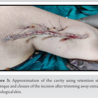 October 10, 2022 Underestimation of a Giant Lipoma on Magnetic Resonance Imaging: A Rare Case Report
 May 10, 2021 Trapezium and Bennett Fracture – A Rare Base Thumb Fracture Pattern:A Case Report
 April 10, 2021 Acute Septic Carpal Tunnel Syndrome in a Rock Climber