

An acute injury superimposed on congenital pseudoarthrosis of the clavicle stimulated spontaneous ossification, suggesting conservative management as a viable alternative to surgery in select pediatric patients.
Dr. Joel P White, School of Medicine, M.S., Texas Tech University Health Sciences Center, Texas, USA. E-mail: joel.p.white@ttuhsc.edu
Abstract
Introduction: Congenital pseudoarthrosis of the clavicle (CPC) is a rare pathology first described by Fitzwilliams in 1910, with fewer than 300 cases reported worldwide. It arises from failure of the primary ossification centers to coalesce, resulting in a false joint connected by periosteum and a synovial membrane. The pathology classically presents in early childhood as a painless midshaft clavicle deformity with no history of trauma, most commonly occurring unilaterally on the right side and more frequently in females. While conservative management is an option in asymptomatic patients, surgical intervention is generally recommended as the deformity progresses in functional and cosmetic significance with age. To our knowledge, spontaneous ossification with conservative treatment has not been previously reported.
Case Report: A 5-year-old boy was referred for a possible non-union clavicle fracture. After assessment, a CPC with an acute fracture was diagnosed. This was treated conservatively and followed an acceptable healing process. Radiographs 2 years later, after conservatively treating a separate acute fracture to the same clavicle, show no evidence of the prior pseudoarthrosis.
Conclusion: The aim of this study was to present radiological outcomes of a CPC that spontaneously healed without intervention. The results of this study show the possibility of less invasive treatment for a CPC.
Keywords: Congenital pseudoarthrosis of clavicle, pediatric orthopedics, non-operative.

Congenital pseudoarthrosis of the clavicle (CPC) is a rare pathology with a poorly understood etiology. CPC was first recognized as a pathology by Fitzwilliams in 1910, distinct from more common conditions such as pediatric cleidocranial dysostosis, obstetric complicated fractures or neurofibromatosis [1]. Despite the failure of the primary ossification centers to coalesce, the clavicle is connected by a periosteum that allows a synovial membrane to develop. Mechanically, there is little restriction until later in life [2].
Classically, CPC presents with a painless midshaft clavicle deformity characteristic of a fracture with no history of trauma and normal upper extremity mechanics in the first few years of life [2]. The non-ossified ends of the clavicle appear enlarged with no callus formation and show a degree of motion between them due to muscular forces on the proximal and distal portions [2,3]. CPC most commonly occurs unilaterally on the right but may occur bilaterally or unilaterally on the left [3]. The pathology is 25–80% more common in females than in males [2,4]. Right-sided CPC may present with a cervical rib, while left-sided CPC may present with dextrocardia [4]. There have been only 200–300 cases reported worldwide since the pathology was first recognized [5].
Treatment may be done conservatively in patients with no pain or restrictions, but may be complicated with upper extremity deficits or vascular complications upon maturity [5]. Naturally, as the patient matures, the deformity becomes increasingly noticeable both functionally and cosmetically. Surgical intervention is recommended as treatment due to pain, activity restrictions, and cosmetic concerns, and this method is customary [5].
To our knowledge, there has not been a case of CPC that has spontaneously healed with conservative treatment and observation. We report a 5-year-old boy referred to us for a possible right clavicle fracture with non-union, which was appropriately diagnosed given radiographs and patient history as a CPC with an acute injury.
A previously healthy 5-year-old boy was referred to our practice by an orthopedist after an acute clavicle injury 11 weeks before the current presentation. He was immobilized in a sling with activity restrictions following the initial outside clinic visit. The patient’s mother shared that he only experienced pain for 3 weeks following the initial incident and was back to baseline at present with no pain, tenderness, or functional limitation. The initial fracture was found to have no radiographic evidence of callus formation by the referring orthopedist, who was seeking a second opinion.
Radiographs show an angulated, right-sided fracture with very mild callus formation over the distal aspect of the clavicle. No callus formation was present over the proximal segment of the supposed fracture, as shown in Fig. 1. In addition, initial images appeared to have well-corticated edges at the level of the aperture, which would be more suggestive of pseudoarthrosis. Due to the unusual history of his presentation, location, and appearance of his radiographs, we leaned toward an undiagnosed pseudoarthrosis of the clavicle with an acutely superimposed injury.
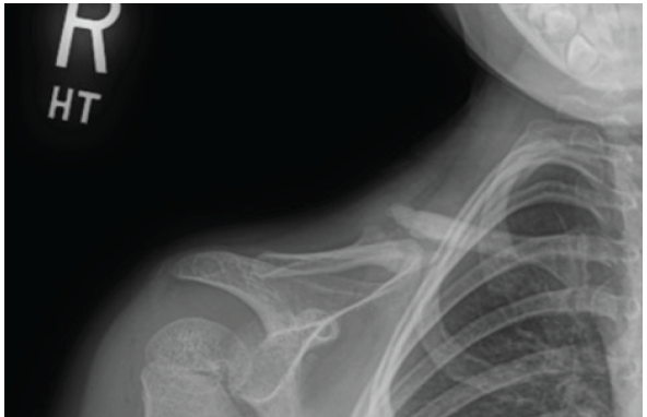
Figure 1: Radiograph demonstrating a pseudarthrosis of the midshaft of the right clavicle.
Treatment options were discussed in detail, including operative versus conservative interventions. As the patient was still in the acute phase of his injury and there was some distal callus formation of note, we chose to continue to monitor the pathology radiographically. We had the patient follow up every 3 months with repeat images of the right clavicle to further evaluate for changes in the appearance.
At 3 months follow up, the patient’s mother was primarily concerned by the increased cosmetic deformity of his clavicle. Radiographs at this visit showed the patient has significant callus formation, as shown in Fig. 2, which accounts for the cosmetic change. At this point, the patient was released to participate in sports safely, as the mother felt comfortable with the understanding that the right clavicle has an increased risk of injury.
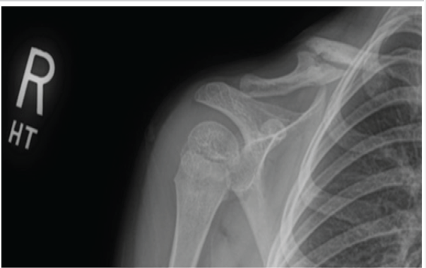
Figure 2: Radiograph demonstrating a pseudarthrosis of the midshaft of the right clavicle with periosteal reaction.
At 6-month follow-up, radiographs showed continued callus formation bridging the previously seen area of pseudoarthrosis with near obliteration of the previously visualized space between the distal and proximal fragments, which may be visualized in Fig. 3. At this point in care, the patient was to follow up pro re nata.
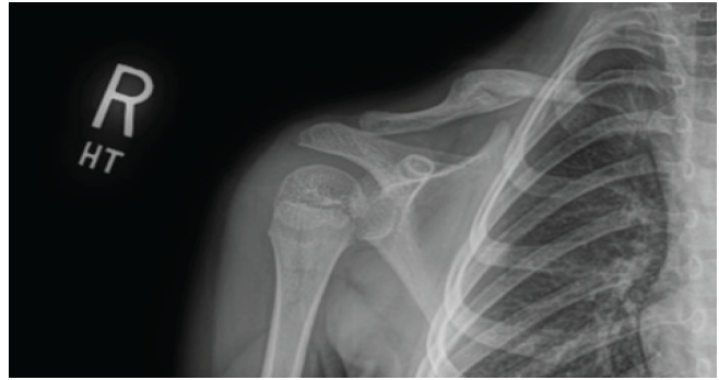
Figure 3: Radiograph demonstrating a pseudarthrosis of the midshaft of the right clavicle healing.
Two years after this patient was released, he presented back in clinic with a right transverse acute diaphyseal clavicle fracture following a sports injury, as shown in Fig. 4. It was notable that the bony characteristics of CPC were no longer able to be appreciated in this radiograph. The fracture was minimally displaced and treated nonoperatively. At follow-up for this acute fracture, radiographs showed a healing, well-aligned clavicle consistent with the physiology of a normal clavicle fracture, as shown in Fig. 5. The patient was released to safe return to activities and to follow up pro re nata.
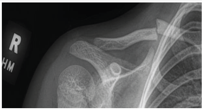
Figure 4: Radiograph demonstrating a right transverse acute diaphyseal clavicle fracture.
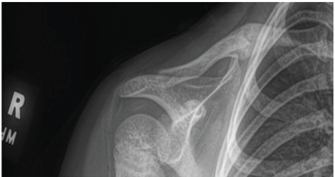
Figure 5: Radiograph demonstrating a healed transverse diaphyseal fracture of the midshaft of the right clavicle.
Typically, a CPC is discovered after a patient complaint of a prominence on the middle to distal third of the clavicle in the first few years of life [3,4]. While the prominence is often grossly identifiable, the varying anatomy of patients may allow the deformity caused by CPC to go unnoticed, especially considering this pathology occurs in otherwise healthy patients with no obstetric complications, other deformities, or clinical signs [6]. In this case, the diagnosis was made coincidentally after the injury occurred to the patient.
Given the limited history of the patient, it was important to include a non-union midshaft fracture of the clavicle in the differential diagnosis. Non-union clavicle fractures are extremely rare in the pediatric population and typically occur in patients around 13 years old who previously underwent surgical fixation of a clavicle fracture [7]. In the initial radiograph, there was evidence of distal callus formation, which indicates that an injury did occur. Still, this formation was localized exclusively to the distal portion of the clavicle. The lateral portion of the clavicle featured no periosteal reaction or callus formation. Aside from minimal callus formation, the radiograph is consistent with previous descriptions and illustrations of CPC, where the sternal half is larger, protruding upward, and usually above the acromial half [8,9]. Considering the patient’s age, there is typically reliable healing of traumatic fractures to the diaphysis of the clavicle even with displacement [7]. Considering the patient’s age, lack of pain or tenderness, functional normalcy, and radiographic findings allowed us to firmly rule non-union of the clavicle out of our differential, as the presentation is more suspicious of CPC with an acute injury to the distal end of the deformity [10]. This case highlights the importance of taking a meaningful patient history; this, along with careful radiographic introspection, could have prevented the initial misdiagnosis.
This incidence of CPC is unique in that it was not treated surgically, while still achieving adequate bone union as a result. It is well understood that while there is no complete closure of ossification centers in the presentation of CPC, there is some level of fibrous connection between the medial and lateral aspects of his pseudoarthrosis [2,6]. We speculate that the injury to the distal portion of the pseudoarthrosis initiated a periosteal reaction in this tissue, allowing for the complete ossification of the clavicle. Unfortunately, the patient was lost to follow-up due to reinjury; however, there was a normal display of physiology 2 years later after the clavicle was refractured. The CPC resolved impressively with little radiographic evidence of its occurrence and a normal healing process during the reinjury. This sheds light on other possible treatments to elicit periosteal reactions, such as electrical, mechanical, ultrasound stimulation or a less invasive surgical procedure than the current accepted standard. This would be challenging to test, though, given the rarity of CPC and the already accepted surgical treatments.
To our knowledge, this is the first diagnosis of CPC that has completed ossification without surgical intervention. This case demonstrates an acute injury to the distal portion of a congenital pseudoarthrosis of the clavicle in a pediatric patient. The pathology was observed radiographically over a period of 6 months. This case may shed light on future possibilities to treat CPC non-invasively.
Congenital pseudoarthrosis of the clavicle is a rare diagnosis that can be easily mistaken for a pediatric clavicle fracture non-union. A careful patient history and close radiographic evaluation are essential to an accurate diagnosis. This case demonstrates that an acute superimposed injury may stimulate a periosteal reaction sufficient to drive spontaneous ossification of the pseudoarthrosis, suggesting that conservative, non-operative management may be a viable consideration in select pediatric patients, particularly in the setting of an acute injury.
References
- 1. Fitzwilliams D. Hereditary cranio – cleido-dysostosis. Lancet 1910;176:1466-75. [Google Scholar] [PubMed]
- 2. Galanopoulos I, Ashwood N, Garlapati AK, Fogg Q. Congenital pseudarthrosis of clavicle: Illustrated operative technique and histological findings. BMJ Case Rep 2012;2012:bcr2012006908. [Google Scholar] [PubMed]
- 3. Beslikas TA, Dadoukis DJ, Gigis IP, Nenopoulos SP, Christoforides JE. Congenital pseudarthrosis of the clavicle: A case report. J Orthop Surg (Hong Kong) 2007;15:87-90. [Google Scholar] [PubMed]
- 4. Lloyd-Roberts GC, Graham Apley A, Owen R. Reflections upon the aetiology of congenital pseudarthrosis of the clavicle. J Bone Joint Surg Br Volume 1975;57-B:24-9. [Google Scholar] [PubMed]
- 5. Alsaeed AA. Surgical management of congenital pseudoarthrosis of the clavicle: A review of current concepts. Cureus 2021;13:e18482. [Google Scholar] [PubMed]
- 6. De Figueiredo MJ, Dos Reis Braga S, Akkari M, Prado JC, Santili C. Congenital pseudarthrosis of the clavicle. Rev Brasil Ortop 2015;47:21-6. [Google Scholar] [PubMed]
- 7. Hughes K, Kimpton J, Wei R, Williamson M, Yeo A, Arnander M, et al. Clavicle fracture nonunion in the paediatric population: A systematic review of the literature. J Hildren’s Orthop 2018;12:2-8. [Google Scholar] [PubMed]
- 8. Currarino G, Herring JA. Congenital pseudarthrosis of the clavicle. Pediatr Radiol 2009;39:1343-9. [Google Scholar] [PubMed]
- 9. Owen R. Congenital pseudarthrosis of the clavicle. J Bone Joint Surg Br 1970;52:644-52. [Google Scholar] [PubMed]
- 10. Chalfant JS, Sanchez TR. Congenital clavicular pseudoarthosis-how to differentiate it from the more common clavicular fractures. Pediatr Emerg Care 2019;35:e37-9. [Google Scholar] [PubMed]


Related Articles in Journal of Orthopaedic Case Reports
 August 1, 2026 Surgical Excision of a Rare Ventral Scapular Osteochondroma using the Prone “Chicken-Wing” Position: A Case Report
 July 1, 2026 Step-cut and Butterfly Femoral Osteotomies Following Intraoperative Fracture Propagation during Distraction Osteogenesis: Report of Two Cases
 July 1, 2026 Beyond the Biopsy: Macrodystrophia Lipomatosa Unmasked After False Osteochondroma Diagnosis
 June 1, 2026 Functional Outcomes of All-Inside Arthroscopic Repair for Chronic Ankle Instability in Adolescents: A Case Series