

Navicular agenesis is a rare cause of flatfoot deformity and subtalar arthritis; accurate imaging-based diagnosis allows effective management with arthrodesis to achieve a stable, plantigrade foot.
Dr. Sanjey Sivakumar, Department of Orthopaedics, Jawaharlal Institute of Postgraduate Medical Education and Research, Puducherry - 605006, India. E-mail: sanjeysivakumar@gmail.com
Abstract
Introduction: Anatomical variations of the navicular bone, including accessory ossicles, hypoplasia, and complete absence, can influence foot biomechanics and clinical presentation. Complete absence of the navicular bone is exceptionally rare and may not be readily recognized.
Case Report: We report a rare case of bilateral navicular agenesis in a 34-year-old male presenting with chronic foot pain and deformity. Imaging revealed a complete absence of the navicular bone with altered midfoot biomechanics and secondary degenerative changes. The patient underwent subtalar arthrodesis with midfoot correction, achieving a stable plantigrade foot. At 12 months follow-up, there was significant pain relief and a satisfactory functional outcome.
Conclusion: Navicular agenesis should be considered in atypical cases of flatfoot deformity. Accurate diagnosis using advanced imaging is essential, and surgical management can provide good functional outcomes.
Keywords: Navicular agenesis, flatfoot, subtalar arthrodesis, midfoot deformity, subtalar arthritis, planovalgus deformity, tarsal bone agenesis.

The navicular bone plays a central role in maintaining the medial longitudinal arch and overall biomechanics of the foot. It functions as a key link between the talus and the cuneiforms, helping to distribute forces during weight-bearing and enabling efficient gait. It also serves as the primary insertion site of the posterior tibial tendon, which is an important dynamic stabilizer of the arch [1]. Ossification of the navicular occurs relatively late compared to other tarsal bones, making it particularly vulnerable to developmental disturbances [2].
While accessory navicular and other minor variations are relatively common, the complete absence of the navicular bone is extremely rare. Most reported cases describe unilateral involvement, often identified incidentally or during evaluation for foot pain or deformity. Bilateral absence is far less frequently encountered and may not be readily recognized in clinical practice [3,4,5,6].
Failure to identify this anomaly can lead to diagnostic confusion, as it may mimic more common conditions such as tarsal coalition, congenital deformities, or post-traumatic changes. We present a case of bilateral navicular agenesis in an adult presenting with midfoot pain and functional limitation, highlighting the diagnostic challenges, biomechanical implications, and considerations for appropriate management of this rare condition.
A 34-year-old male presented with a 4-year history of right foot pain and stiffness, insidious in onset and progressively worsening. Pain was aggravated by prolonged walking, particularly on uneven ground, and was partially relieved with oral analgesics. He was unable to walk beyond 200 m without discomfort. Pre-operative Visual Analog Scale (VAS) score was 9/10.
There was no history of trauma, fever, or inflammatory joint symptoms, or significant medical or family history.
He had bilateral finger contractures and plantar nodules, previously diagnosed as plantar fibromatosis (Ledderhose disease) (Fig. 1), for which he underwent dermatofasciotomy and finger fasciotomy. Three months earlier, he had undergone left subtalar arthrodesis with hallux varus correction for subtalar arthritis with cavus deformity.
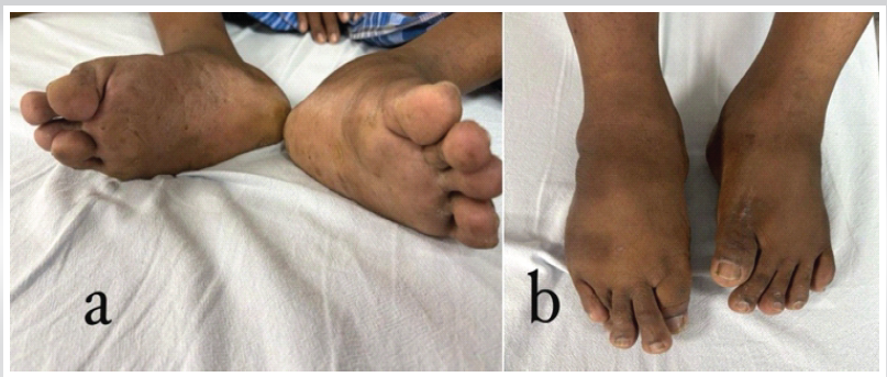
Figure 1: Clinical photographs of the right foot showing plantar fibromatosis with nodular thickening over the plantar aspect (a) and planovalgus deformity on dorsal view (b).
On examination, he had an antalgic gait. The right foot revealed nodular thickening along the central plantar fascia with associated skin dimpling and puckering. Both feet demonstrated a mobile planovalgus alignment. Contractures of the first and fourth toes of the right foot were present, with overriding of the fifth over the fourth toe. The ankle had a full painless range of motion, whereas subtalar movements were restricted and painful, with no instability. Posterior tibial tendon function was clinically intact, with no evidence of tendon insufficiency.
Investigations revealed normal laboratory parameters, including erythrocyte sedimentation rate and C-reactive protein, supporting a non-inflammatory etiology.
Plain radiographs of both feet (Fig. 2) demonstrated a complete absence of the navicular, with direct articulation between the talus and the cuneiforms. Features of flatfoot deformity and subtalar arthritic changes were noted, with no fracture, infection, or other osseous abnormalities.
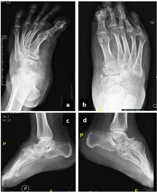
Figure 2: Plain radiographs of the foot showing anteroposterior (a and b) and lateral (c and d) views of the bilateral foot showing a complete absence of the navicular with altered talo-cuneiform articulation.
Alignment assessment revealed midfoot and hindfoot flatfoot, with an absent talonavicular joint. Computed tomography (CT) confirmed complete agenesis of the navicular (Fig. 3) and excluded hypoplasia and post-traumatic causes.
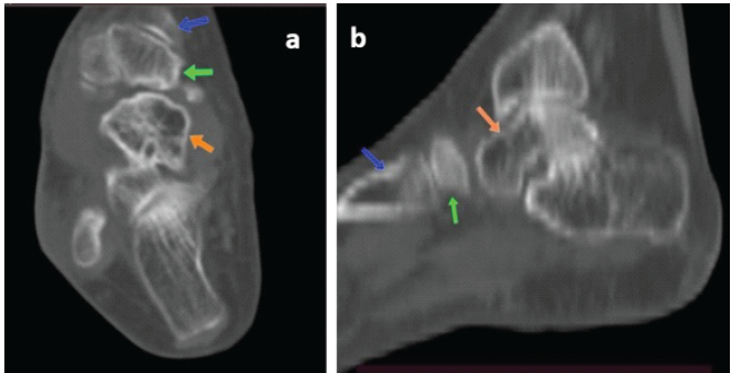
Figure 3: Computed tomography images of the right foot. (a) Axial section and (b) sagittal section demonstrating complete absence of the navicular bone with direct articulation between the talus (orange arrow) and cuneiform (green arrow); first metatarsal bone (blue arrow).
Diagnosis was established based on radiographic findings and confirmed with CT, which clearly demonstrated the absence of the navicular and altered midfoot articulation.
Differential diagnosis
The differential diagnosis of apparent navicular absence includes conditions in which the bone is present but altered, displaced, or poorly visualized.
Tarsal coalition is a fibrous or osseous union(commonly talocalcaneal or calcaneonavicular), presenting with restricted subtalar motion, pain, and rigid flatfoot. CT typically demonstrates the presence of the navicular bone with abnormal fusion rather than complete absence [2].
Congenital vertical talus is a rare deformity characterized by dorsal dislocation of the navicular on the talar head, causing a rocker-bottom foot. In early childhood, the navicular may appear poorly visualized due to delayed ossification. However, imaging typically shows abnormal alignment rather than absence of the bone, and this condition typically presents in infancy [7].
Navicular hypoplasia is a small or underdeveloped navicular that may resemble agenesis on plain radiographs. Advanced imaging, particularly CT, helps distinguish hypoplasia from true agenesis by identifying residual navicular bone [6].
Post-traumatic navicular resorption can occur following severe comminution or surgical excision and may mimic congenital absence on imaging. Clinical history and radiological features, such as irregular bone margins, help differentiate this from a congenital anomaly. In our present case, this was unlikely due to no trauma history [8].
Kohler disease is a childhood osteochondrosis of the navicular, causing sclerosis and fragmentation. Despite radiographic changes, the bone remains identifiable and typically re-ossifies over time [9].
Treatment
Management aimed to achieve a stable, plantigrade, and pain-free foot while correcting the underlying deformity. The patient was counseled regarding the expected trade-off between pain relief and loss of subtalar joint motion following arthrodesis. Subtalar arthrodesis was performed through a lateral approach. Joint surfaces were prepared by removing articular cartilage and exposing bleeding subchondral bone. Fixation was achieved using two cannulated cancellous screws placed across the subtalar joint under fluoroscopic guidance [10]. Calcaneocuboid shortening was performed to correct the varus alignment. No structural bone graft was used (Fig. 4).
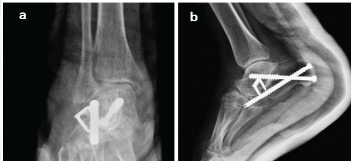
Figure 4: Immediate post-operative radiographs of right foot anteroposterior (a) and lateral (b) views showing subtalar arthrodesis with two cannulated cancellous screws and calcaneocuboid shortening stabilized with a bone staple.
Postoperatively, the limb was immobilized in a below-knee cast, with non-weight-bearing ambulation for 1 month. Gradual weight-bearing was then initiated over the next 6–8 weeks, based on clinical and radiological assessment.
Outcome
The patient was followed up with clinical and radiographic assessment for fusion and functional recovery. At the 12-month follow-up, complete arthrodesis was achieved radiologically (Fig. 5). Clinically, the patient reported a significant reduction in pain and improvement in functional capacity. He was able to ambulate longer distances without analgesics. On examination, the foot was plantigrade with satisfactory correction of the valgus deformity without implant complications, wound issues, or recurrence of deformity.
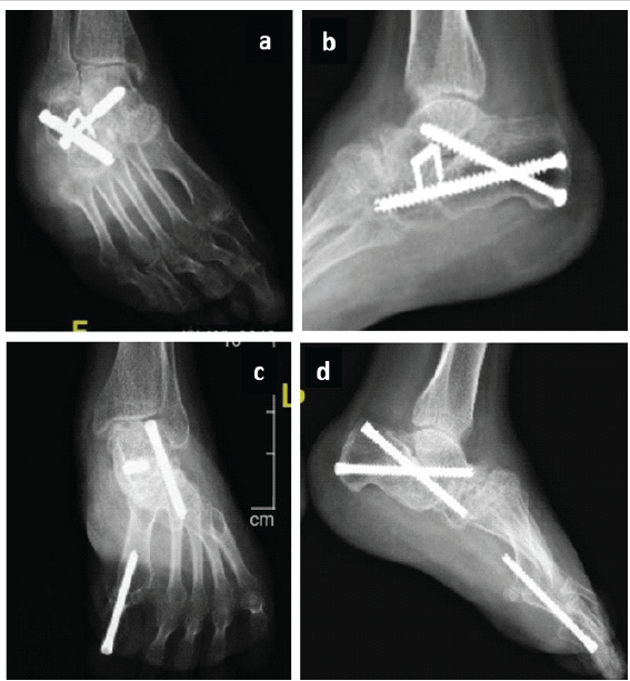
Figure 5: Follow-up radiographs (a) at 12 months of right foot showing complete subtalar arthrodesis with maintained alignment and no evidence of implant-related complications and (b) at 15 months of left foot (c and d) showing complete subtalar arthrodesis and with maintained alignment and corrected hallux varus with no evidence of implant-related complications.
The VAS score improved from 9 preoperatively to 1 at 3 months postoperatively. Overall, the surgical intervention resulted in a stable, pain-free foot with improved gait and functional outcome at short-term follow-up.
Congenital absence of the navicular is a rare anomaly that alters foot biomechanics by disrupting the medial arch and load transmission. Its absence leads to direct talus–cuneiform articulation, resulting in abnormal midfoot loading, deformity, instability, and pain [2,3]. In this case, these changes were evident and associated with secondary subtalar arthritis due to chronic overload.
The exact etiology of navicular agenesis remains unclear, but it is thought to arise from abnormal embryological chondrification and ossification. From a clinical perspective, it is important to distinguish true agenesis from other conditions such as hypoplasia, post-traumatic bone loss, or avascular necrosis, as these entities may have similar radiographic appearances but require different management [4,5,6,7].
Diagnosis is primarily based on imaging. While plain radiographs may suggest the absence of the navicular, cross-sectional imaging is often necessary to confirm the diagnosis and to evaluate associated structural changes. CT is particularly useful in defining bony anatomy and confirming the diagnosis. In this case, the combination of imaging modalities was essential in establishing the diagnosis and excluding other potential causes of midfoot pathology [4,5].
Clinically, navicular agenesis may present with foot pain, deformity, or gait disturbance, although some cases may remain asymptomatic and be detected incidentally. Importantly, the presentation may mimic more common conditions such as posterior tibial tendon dysfunction or tarsal coalition, which can lead to misdiagnosis. Failure to recognize this anomaly preoperatively may result in inappropriate surgical planning and suboptimal outcomes [4,5].
Management depends on symptom severity. Asymptomatic patients can be managed conservatively, whereas symptomatic cases may require intervention. In the presence of deformity or arthritis, arthrodesis provides a reliable option for pain relief and functional restoration. In our patient, subtalar arthrodesis with midfoot correction achieved a stable, plantigrade, and pain-free foot. Previous reports have described both conservative and reconstructive approaches; however, arthrodesis offers more predictable outcomes in cases with secondary arthritis [4,5].
This case underscores the importance of considering rare structural anomalies in patients presenting with chronic foot pain and atypical radiographic findings. Early recognition is essential not only for accurate diagnosis but also for appropriate surgical planning and optimal functional outcomes. This report is limited by the single-case design, and a longer follow-up is required to assess long-term functional outcomes.
Bilateral congenital navicular agenesis is an exceptionally uncommon developmental anomaly with the potential to cause progressive biomechanical dysfunction and secondary degenerative changes in the hindfoot. This case highlights the importance of recognising rare congenital osseous abnormalities when evaluating atypical foot deformities in adults. Awareness of this entity and timely diagnosis facilitate appropriate treatment planning, while documentation of such rare presentations contributes to the limited literature and improves understanding of their clinical behaviour.
Navicular agenesis should be considered in adults presenting with atypical flatfoot deformity and chronic foot pain. Advanced imaging is essential for accurate diagnosis, and appropriate surgical management can provide a stable, plantigrade, and pain-free functional outcome.
References
- 1. Prapto D, Dreyer MA. Anatomy, bony pelvis and lower limb: Navicular bone. In: StatPearls [Internet]. Treasure Island (FL): StatPearls Publishing; 2025 [cited 2026 Jul 24]. Available from: https://www.ncbi.nlm.nih.gov/books/NBK547675/ [Google Scholar] [PubMed]
- 2. Mehdi N, Bernasconi A, Lintz F. Tarsal coalition in adults. Orthop Traumatol Surg Res 2024;110:103761. [Google Scholar] [PubMed]
- 3. Stolarz K, Osiowski A, Preinl M, Osiowski M, Jasiewicz B, Taterra D. The prevalence and anatomy of accessory navicular bone: A meta-analysis. Surg Radiol Anat 2024;46:1731-43. [Google Scholar] [PubMed]
- 4. Buckingham RA, Winson IG. Bilateral agenesis of the tarsal navicular. Foot Ankle Surg 2000;6:185-8. [Google Scholar] [PubMed]
- 5. Alhashimi I, Al-Taie A, Alam S, Ibrahem Adam R. Congenital absence of the navicular bone: A rare cause of adult-acquired flatfoot and posterior tibial tendon dysfunction. Cureus 2025;17:e85151. [Google Scholar] [PubMed]
- 6. Kessomtini W, Laajili H, Said W, Jerbi S. Club foot revealing a hypoplasia of the navicular bone: The first case report. Ann Phys Rehabil Med 2014;57:210-2. [Google Scholar] [PubMed]
- 7. Wirth T. Congenital vertical talus. Foot Ankle Clin 2021;26:903-13. [Google Scholar] [PubMed]
- 8. Swords M, Schepers T. Post traumatic complications after navicular fractures. Foot Ankle Clin 2025;30:127-40. [Google Scholar] [PubMed]
- 9. Chan JY, Young JL. Köhler disease: Avascular necrosis in the child. Foot Ankle Clin 2019;24:83-8. [Google Scholar] [PubMed]
- 10. Easley ME, Trnka HJ, Schon LC, Myerson MS. Isolated subtalar arthrodesis. J Bone Joint Surg Am 2000;82:613-24. [Google Scholar] [PubMed]






Related Articles in Journal of Orthopaedic Case Reports
 July 1, 2025 Fusion under the Scope: Minimally Invasive Arthroscopic Tibiotalocalcaneal Arthrodesis in the Setting of Post-traumatic Fibular Loss: A Case Report
 June 1, 2026 Cortical Lytic Lesion of the Proximal Tibia in an 8-Year-Old Child: A Pediatric Diagnostic Conundrum
 September 1, 2026 A Prospective Study Showing Functional Outcome of the Modified Boytchev Procedure for Recurrent Anterior Shoulder Dislocation
 December 4, 2020 TFNA Implant Failure with Helical Blade Cut-out