

Accurate anatomical placement of femoral and tibial tunnels best assessed by CT is critical for successful ACL reconstruction, as malposition (especially femoral) leads to residual instability and poorer functional outcomes.
Dr. Shashank Prakash, Sport Injury Center, VMMC and Safdarjung Hospital, New Delhi, India. E mail: dr.sps.gmc@gmail.com
Abstract
Introduction: Anterior cruciate ligament (ACL) reconstruction aims to restore knee stability, but improper femoral and tibial tunnel placement can lead to persistent instability. Computed tomography (CT)-based 3D assessment helps correlate tunnel position with outcomes.
Materials and Methods: A study of 50 patients undergoing primary arthroscopic ACL reconstruction used post-operative CT to assess tunnel placement relative to anatomical landmarks. Knee stability was evaluated using Lachman and pivot shift tests, and patients were grouped by residual instability.
Results: Residual instability occurred in 36% of patients. Anatomical femoral and tibial tunnel placement was achieved in 62% and 68% of cases, respectively. Non-anatomical femoral (66.7%) and tibial (55.6%) tunnels were more common in unstable knees, with combined malposition showing the highest instability rates.
Conclusion: Non-anatomical tunnel placement, especially femoral, is strongly associated with residual instability, emphasizing the importance of precise anatomical positioning in ACL reconstruction.
Keywords: Anterior cruciate ligament reconstruction, computed tomography, femoral tunnel position, residual knee instability, tibial tunnel position.

The anterior cruciate ligament (ACL) injuries are one of the most common knee injuries that leave individuals who are involved in active lifestyles and the performance of sports activities with untreated injuries experiencing dysfunctional instability and an inability to continue their activities at normal levels, and the onset of joint degeneration which occurs at an early age [1]. Arthroscopic ACL reconstruction has become the gold standard of ACL reconstruction surgery because it has demonstrated the best restoration of knee stability and functional outcome and allowing patients to resume their activity at their pre-injury level [2]. Although surgical techniques, fixation devices and graft choices have advanced, “ACL reconstruction patients who experience knee instability following their surgery remain a significant issue since their instability manifests itself in frequent giving-way episodes and rotational laxity of their knees and poor functional development [3]. The most prevalent modifiable cause that causes graft failure and long-term instability is caused by surgeons during the operation that make incorrect tunnel placements, as they form non-anatomic tunnel placements on both the femoral and tibial tunnels [4]. The native ACL is a complex three-dimensional structure that is important in regulating anterior tibial translation and rotational stability of the knee and, thus, any minor distortion of the anatomic footprint on the creation of the tunnel may affect the graft biomechanics dramatically [5]. Misaligned tunnels could cause excessive graft tension, graft strain, contact with the intercondylar roof, and loss of adequate rotational stability, leading to chronic instability and poor clinical results [6]. Historically, plain radiographs and intraoperative arthroscopic landmarks have been used to assess the position of tunnels, but they are poor due to some projectional errors and lack of three-dimensional visualization [7]. Computed tomography (CT) has proven to be a highly reliable and reproducible imaging technique to evaluate the tunnel position post-operatively to enable the accurate measurement of tunnel orientation, depth, and relative position to anatomic landmarks using standardized methods of measurement, such as the Bernard-Hertel grid and three-dimensional reconstructions [8]. Tunnel analysis by CT has been demonstrated to be better than traditional radiography to identify subtle malpositions that cannot be seen on the standard imaging but can be clinically important [9]. A number of studies have shown a close connection between non-anatomic tunnel positioning as seen on CT scans and higher rates of residual instability, graft failure, and revision surgery [10]. Specifically, there is evidence that anterior or vertical placement of the femoral tunnel can result in chronic rotational instability, with a positive pivot shift test, whereas posterior or overly vertical tibial tunnels can impair graft function and knee kinematics [11]. As the present focus on anatomic ACL reconstruction, the correlation of CT-confirmed tunnel positions and post-operative knee stability has become a key issue in streamlining surgical procedures and enhancing patient outcomes. Moreover, objective analysis of the place of the tunnel by means of CT scans can be of great benefit to the surgeon, helping to detect technical flaws and support the clinical decision-making process, in particular, when the results are unsatisfactory or when it is necessary to consider revision ACL reconstruction. Thus, the CT-based tunnel position analysis and its association with the residual instability following ACL reconstruction should be thoroughly studied to contribute to a better understanding of the impact of surgical accuracy on post-operative knee stability, improve the reconstructive approach, and eventually decrease the rate of persistent instability and graft failure.
Study design
This study was conducted as a hospital-based observational analytical study with a cross-sectional design.
Study setting
The study was carried out in the Department of Orthopedics of a tertiary care teaching hospital with facilities for arthroscopic knee surgery and advanced imaging. The study protocol was approved by the Institutional Ethics Committee, and written informed consent was obtained from all patients before inclusion in the study.
Study duration
The study was conducted over a period of 18 months.
Participants
Enrolment was done by a certain set of criteria for the performance of arthroscopic ACL reconstruction.
Inclusion criteria
- Patients aged between 18 and 45 years
- Patients who had undergone primary arthroscopic ACL reconstruction
- Patients with a minimum post-operative duration of 6 months
- Patients who consented to undergo post-operative CT evaluation
- Patients willing to participate and provide written informed consent.
Exclusion criteria
- Patients with revision ACL reconstruction
- Patients with associated multi-ligament knee injuries
- Patients with significant osteoarthritis of the knee
- Patients with previous fractures around the knee joint
- Patients with post-operative infection or graft rupture
- Patients with contraindications to CT imaging.
Study sampling
The researchers used purposive sampling to select their study participants. The research team enrolled all eligible patients who arrived during the study period and met both inclusion and exclusion criteria until they reached their target sample size. The research method achieved both operational requirements and sufficient representation of patients who underwent post-operative ACL reconstruction.
Study sample size
The total sample size for the study was 50 patients. The sample size was selected based on feasibility, availability of eligible patients during the study period, and consistency with similar studies published in the literature evaluating CT-based tunnel position and post-operative instability following ACL reconstruction.
Study parameters
The following parameters were assessed:
- Demographic variables, such as age and sex
- Side of injury
- Time interval since surgery
- Femoral tunnel position on CT scan
- Tibial tunnel position on CT scan
- Tunnel orientation and location based on standardized anatomical grids
- Clinical instability parameters, including Lachman test and pivot shift test
- Presence or absence of residual instability.
Patient-reported outcome measures, instrumented laxity testing, graft maturation assessment, and return-to-sport evaluation were beyond the scope of the present study and were therefore not included in the analysis.
Study procedure
The post-operative records to find patients who met their criteria for inclusion. Medical professionals conducted a complete knee examination after they received informed consent from the patient. An orthopedic surgeon who had experience conducted clinical instability tests, which included the Lachman test and pivot shift test. The patients received CT scanning of their surgically treated knee. The team conducted CT scans according to standard procedures which required specific knee positions. CT measurements were performed according to standardized protocols by experienced observers using pre-defined anatomical landmarks. However, formal assessment of interobserver and intraobserver reliability was not performed and should be considered in future studies.
Study data collection
The researchers gathered information through their use of a validated data collection instrument. The research team recorded patient data, which included clinical observations and radiological findings and demographic information. CT measurements were performed using digital imaging software, and tunnel positions were documented in percentage values relative to the anatomical reference grids. All collected data were verified for completeness and accuracy before analysis.
Data analysis
The study input the gathered data into Microsoft Excel before conducting their analysis with suitable statistical analysis software. The researchers used descriptive statistics, which included mean values, standard deviation measurements, and frequency plus percentage data to create a summary of the collected information. The researchers used inferential statistical tests to determine the relationship between tunnel position parameters and residual instability in their study. The researchers considered a P-value below 0.05 to indicate statistical significance. The results were displayed in tables and figures, which the researchers used to present their findings whenever it was suitable to do so.
Radiological (CT-based) and clinical evaluations were performed on 50 patients who underwent arthroscopic ACL reconstruction to analyze femoral and tibial tunnel positions and their association with post-operative knee instability. The majority of patients were aged 26–35 years (42%), followed by 36–45 years (30%) and 18–25 years (28%), reflecting the typical demographic prone to ACL injuries. There was a clear male predominance (76% males vs. 24% females), likely due to higher participation in high-risk physical activities. Regarding laterality, the right knee was more commonly involved (56%) compared to the left (44%), although the distribution was relatively balanced without a strong side predilection.
The study found that 36% of patients showed residual knee instability while 64% of patients exhibited stable knees after their surgical procedure. The finding shows that many patients continue to suffer from unstable knee conditions after they undergo ACL reconstruction surgery (Table 1).
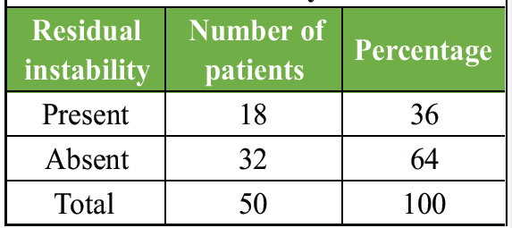
Table 1: Post-operative residual knee instability
CT-based assessment showed that anatomical femoral tunnel placement was achieved in 31 patients (62%), while 19 patients (38%) had non-anatomical positioning, highlighting the technical difficulty of accurate femoral tunnel placement. In comparison, tibial tunnel positioning was more precise, with 34 patients (68%) demonstrating anatomical placement and 16 patients (32%) showing non-anatomical positioning, indicating relatively better accuracy for tibial tunnels than femoral tunnels.
Patients who had their femoral tunnels placed incorrectly through non-anatomical methods showed higher rates of residual instability. The study found that 66.7% of patients with instability used non-anatomical femoral tunnels, which demonstrated a strong link between femoral tunnel malposition and post-operative instability (Table 2 and Fig. 1).
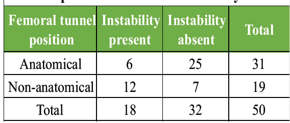
Table 2: Association between femoral tunnel position and residual instability
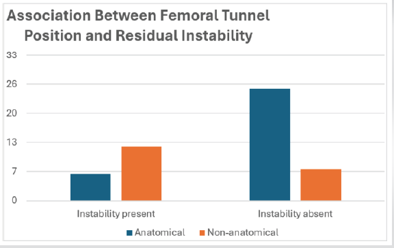
Figure 1: Association between femoral tunnel position and residual instability.
The study results showed that patients who received non-anatomical tibial tunnel placement experienced more residual instability than patients who received anatomical placement, which demonstrates that incorrect tibial tunnel placement leads to post-operative instability issues (Table 3 and Fig. 2).
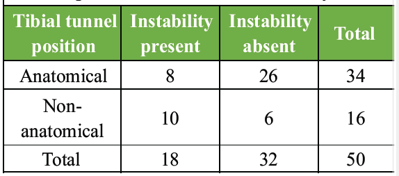
Table 3: Association between tibial tunnel position and residual instability
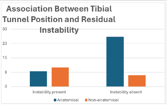
Figure 2: Association between tibial tunnel position and residual instability.
The patients who underwent non-anatomical femoral tunnel placement showed increased positive pivot shift test results. The research findings show that incorrect femoral tunnel placement results in continuous rotational instability issues that impact knee joint function (Table 4 and Fig. 3).
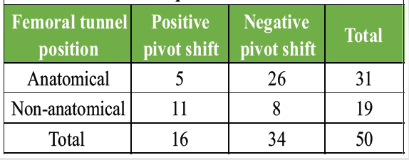
Table 4: Pivot shift test findings and femoral tunnel position
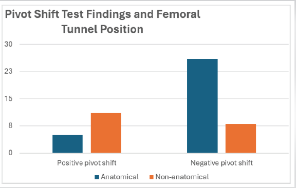
Figure 3: Pivot shift test findings and femoral tunnel position.
Combined analysis showed that residual instability was lowest when both tunnels were anatomical (4/26; 15.4%) compared to cases with either tunnel non-anatomical (7/15; 46.7%) and highest when both tunnels were non-anatomical (7/9; 77.8%), demonstrating a strong association between tunnel malposition and post-operative instability.
The present study evaluated CT-based tunnel positioning of the femur and tibia after ACL reconstruction and compared it with residual knee instability in 50 patients. The demographic analysis revealed that the majority of patients aged between 26 and 35 years (42%), with a majority being male (76), who constitute the most at-risk population, were the most prevalent. Although they had the standard arthroscopic ACL reconstruction, 36% of the patients showed residual knee instability, and they have not reached the goal of restoring clinical stability. CT analysis revealed that the anatomical location of femoral tunnels was observed in 62% of patients, non-anatomical in 38%, and tibial tunnels were anatomically located in 68 and non-anatomically located in 32 cases. These results highlight the ongoing technical difficulties that have been linked with proper placement of tunnels, especially on the femoral side. The close correlation between tunnel malposition and residual instability was also observed, where 66.7% of patients with post-operative instability had non- anatomical femur tunnels, whereas only 33.3% had anatomically placed femur tunnels. On the same note, tibial tunnel malposition was evident in 55.6% of the instable patients. Rotational instability, measured by the pivot shift test, was much higher in patients with non-anatomic position of femoral tunnels (57.9%) than with anatomical placement (16.1%). In addition, a concomitant malposition of the two tunnels led to the most frequent instability rate (77.8%), and the least instability occurred when the two tunnels were anatomically positioned (15.4%). All these findings substantiate the conclusion that anatomic tunnel positioning is an essential factor of post-operative knee stability.
The results of the present research are highly consistent with the results of the article by Shafizadeh et al., [12] which evidenced that in experienced surgeons, ACL tunnel positioning has a significant inter- and intra-observer variability. In a cadaveric fluoroscopy-based navigation study, the authors found that intraobserver variability was 3.3 mm in tibial tunnels and 2.0 mm in femoral tunnels, and interobserver variability of 3.7 mm in femoral tunnels with a maximum of 13.2 mm and 18.3 mm in tibial tunnels. These are minor deviations that were pointed out as possible causes of graft failure. The comparatively large percentage of non-anatomic tunnel placements in the present study (38% femoral, 32% tibial) can thus be attributed to the natural variability in the technique of tunnel placement given, supporting Shafizadeh in his conclusion that better accuracy of the surgical procedure is necessary to achieve less post-operative instability.
Tunnel malposition of the femur is also correlated with rotational instability, as was found in the present study, and this fact is additionally supported by Tashiro et al., [13] assessed in vivo knee stability following reconstruction with bone-patellar tendon-bone bone autografts. They were able to show that femoral tunnel positions were found to be more strongly correlated with anterolateral rotatory translation by a shallower and higher position (R = 0.42, P < 0.01). Patients who were reconstructed with the trans-tibial method had higher rotatory translation (3.2 ± 1.6 mm) than those who were reconstructed with trans-portal (2.0 ± 1.8 mm). These results are quite consistent with the present study that found a positive pivot shift significantly more common among patients who received non-anatomical tunneling of the femur. Even though the anterior-posterior stability in a study conducted by Tashiro did not show significant differences between the techniques, the high degree of rotational laxity with malposition of the femoral tunnel highlights the significance of anatomical positioning of the femoral tunnel, which is also evident in the present outcomes.
Conversely, research on refined anatomic reconstruction techniques has shown high accuracy in tunnel positioning. Buscayret et al. [14] compared tunnel positioning during anatomic ACL reconstruction when the single anteromedial bundle biological augmentation technique was performed, and the remnant was preserved. On 99 patients, they found a mean of 19.4% of notch depth (standard deviation [SD] 2) and 23.1% of lateral wall height (SD 3.5%) at the location of the femoral tunnel and a mean of 36.3% of anteroposterior tibial plateau length (SD 3.8%) at the location of the tibial tunnel by means of The standard deviations were small and the tunnel placement was very consistent and precise; inter and intra-observer consistency as well as inter-observer consistency were all high. In comparison to the present study, where a significant preponderance of tunnels was non-anatomic, the results of Buscayret indicate that refined anatomic methods and uniform guides and remnant conservation can greatly decrease the variability of tunnel placement and, accordingly, residual instability.
Additional evidence to support the presence of advanced instrumentation is based on Kubota et al., [15] compared the accuracy of tunnels in revision ACL reconstruction with a rectangular retro-dilator. They found that all of the femoral revision tunnels were anatomically placed (Type I by the Magnussen classification), and all tibial tunnels were within the native site of ACL attachments. Notably, the difference in the revision tunnel locations was much less as compared to the primary tunnels. These results show that better instrumentation can be more precise in locating the tunnel than primary reconstruction methods that are traditional. The given observation applies to the present work, as the cases where malposition of the tunnel was the most evident in both the case of combined malposition of the femur and tibia, and thus the use of more specific tools to create the tunnel might possibly result in lower instances of instability.
The importance of technology-assisted surgery is also indicated by Zhang et al., [16] in relation to surgical navigation robot-assisted ACL reconstruction versus conventional handheld locator methods [5]. They have found a much better success rate of the femoral tunnel positioning in the robot-assisted group (82.1) than in the control group (50, P = 0.003), with no augmentation in adverse events. The longer surgical time in the robotic group is less important, but the increased accuracy of the femoral tunnel placement is especially pertinent, as the present study found malposition of the femoral tunnel to be the greatest contributive factor to residual instability. The results demonstrate that robotics systems combined with navigation systems will produce better surgical results because they reduce errors during tunnel placement procedures. The results of the present research together with existing literature demonstrate that proper anatomical positioning of tunnels during ACL reconstruction represents the most important factor for success. The high rates of residual instability that occur when both femoral and combined tunnel malposition exist demonstrate the limitations of conventional methods, which need surgical accuracy improvement. The CT-based assessment method provided a precise objective evaluation of tunnel position together with the technical factors that caused post-operative instability. The present study results confirm previous research, which demonstrates that advanced imaging, navigation and instrumentation techniques should be used to achieve accurate tunnel placement, which will enhance long-term knee stability results after ACL reconstruction.
The present study has several limitations that should be considered while interpreting the findings. The relatively small sample size (n = 50) and single-center design may limit the statistical power and generalizability of the results to broader populations. As a cross-sectional observational study, only associations between tunnel position and residual instability could be identified, and causal relationships cannot be established. The minimum follow-up period of 6 months may not adequately reflect long-term graft survival, functional recovery, return-to-sport outcomes, or revision rates. The use of purposive sampling may have introduced selection bias, while the absence of detailed pre-operative instability grading and baseline functional assessments limited direct pre- and post-operative comparisons. Furthermore, patient-reported outcome measures, such as International Knee Documentation Committee, Knee Injury and Osteoarthritis Outcome Score, Lysholm, and Tegner scores were not evaluated. Residual instability was assessed using clinical examination methods, including the Lachman and pivot shift tests, which are subject to examiner variability, and objective instrumented laxity measurements, such as KT-1000 or KT-2000 arthrometry were not performed. Although CT provided accurate assessment of tunnel position, interobserver and intraobserver reliability analyses were not conducted. In addition, potential confounding factors, including graft type, fixation method, surgeon experience, rehabilitation compliance, associated meniscal or chondral injuries, body mass index, activity level, and time from injury to surgery were not analyzed. CT evaluation was restricted to tunnel positioning and did not assess graft maturation, graft tension, tunnel widening, biological incorporation, or other contributors to post-operative instability. Therefore, larger multicenter prospective studies with longer follow-up and comprehensive functional and radiological assessments are warranted to validate and expand upon the present findings.
Future research should focus on multicenter prospective cohort studies and randomized investigations with larger patient populations and longer follow-up durations. Incorporation of validated patient-reported outcome measures, objective instrumented laxity assessment, evaluation of graft maturation and tunnel widening, and analysis of return-to-sport outcomes would provide a more comprehensive understanding of factors contributing to post-operative instability. Standardization of surgical techniques and rehabilitation protocols may further clarify the independent influence of tunnel position on clinical outcomes following ACL reconstruction.
Optimal anatomical placement of femoral and tibial tunnels is critical for post-operative knee stability after ACL reconstruction; CT evaluation shows femoral malposition – especially combined with tibial errors is strongly associated with residual instability, highlighting the need for precise anatomic reconstruction to improve long-term outcomes.
Precise anatomical tunnel placement in ACL reconstruction is essential to ensure knee stability and prevent persistent symptoms, such as giving way and rotational instability. Femoral tunnel malposition is particularly associated with poorer functional outcomes. CT imaging aids in early detection of technical errors, enabling timely intervention and improved long-term results.
References
- 1. Kondo E, Merican AM, Yasuda K, Amis AA. Biomechanical comparison of anatomic double-bundle, anatomic single-bundle, and nonanatomic single-bundle anterior cruciate ligament reconstructions. Am J Sports Med 2011;39:279-88. [Google Scholar] [PubMed]
- 2. Zantop T, Diermann N, Schumacher T, Schanz S, Fu FH, Petersen W. Anatomical and nonanatomical double-bundle anterior cruciate ligament reconstruction: Importance of femoral tunnel location on knee kinematics. Am J Sports Med 2008;36:678-85. [Google Scholar] [PubMed]
- 3. Forsythe B, Kopf S, Wong AK, Martins CA, Anderst W, Tashman S, et al. The location of femoral and tibial tunnels in anatomic double-bundle anterior cruciate ligament reconstruction analyzed by three-dimensional computed tomography models. J Bone Joint Surg Am 2010;92:1418-26. [Google Scholar] [PubMed]
- 4. Rayan F, Nanjayan SK, Quah C, Ramoutar D, Konan S, Haddad FS. Review of evolution of tunnel position in anterior cruciate ligament reconstruction. World J Orthop 2015;6:252-62. [Google Scholar] [PubMed]
- 5. Loh JC, Fukuda Y, Tsuda E, Steadman RJ, Fu FH, Woo SL. Knee stability and graft function following anterior cruciate ligament reconstruction: Comparison between 11 o’clock and 10 o’clock femoral tunnel placement. 2002 Richard O’Connor award paper. Arthroscopy 2003;19:297-304. [Google Scholar] [PubMed]
- 6. Suzuki T, Shino K, Otsubo H, Suzuki D, Mae T, Fujimiya M, et al. Biomechanical comparison between the rectangular-tunnel and the round-tunnel anterior cruciate ligament reconstruction procedures with a bone-patellar tendon-bone graft. Arthroscopy 2014;30:1294-302. [Google Scholar] [PubMed]
- 7. Abebe ES, Kim JP, Utturkar GM, Taylor DC, Spritzer CE, Moorman CT 3rd, et al. The effect of femoral tunnel placement on ACL graft orientation and length during in vivo knee flexion. J Biomech 2011;44:1914-20. [Google Scholar] [PubMed]
- 8. Bedi A, Musahl V, Steuber V, Kendoff D, Choi D, Allen AA, et al. Transtibial versus anteromedial portal reaming in anterior cruciate ligament reconstruction: An anatomic and biomechanical evaluation of surgical technique. Arthroscopy 2011;27:380-90. [Google Scholar] [PubMed]
- 9. Wang H, Fleischli JE, Zheng NN. Transtibial versus anteromedial portal technique in single-bundle anterior cruciate ligament reconstruction: outcomes of knee joint kinematics during walking. Am J Sports Med 2013;41:1847-56. [Google Scholar] [PubMed]
- 10. Shen W, Forsythe B, Ingham SM, Honkamp NJ, Fu FH. Application of the anatomic double-bundle reconstruction concept to revision and augmentation anterior cruciate ligament surgeries. J Bone Joint Surg Am 2008;90 Suppl 4:20-34. [Google Scholar] [PubMed]
- 11. Shaerf DA, Pastides PS, Sarraf KM, Willis-Owen CA. Anterior cruciate ligament reconstruction best practice: A review of graft choice. World J Orthop 2014;5:23-9. [Google Scholar] [PubMed]
- 12. Shafizadeh S, Balke M, Hagn U, Hoeher J, Banerjee M. Variability of tunnel positioning in ACL reconstruction. Arch Orthop Trauma Surg 2014;134:1429-36. [Google Scholar] [PubMed]
- 13. Tashiro Y, Okazaki K, Murakami K, Matsubara H, Osaki K, Iwamoto Y, et al. Anterolateral rotatory instability in vivo correlates tunnel position after anterior cruciate ligament reconstruction using bone-patellar tendon-bone graft. World J Orthop 2017;8:913-21. [Google Scholar] [PubMed]
- 14. Buscayret F, Temponi EF, Saithna A, Thaunat M, Sonnery-Cottet B. Three-dimensional CT evaluation of tunnel positioning in ACL reconstruction using the single anteromedial bundle biological augmentation (SAMBBA) technique. Orthop J Sports Med 2017;5:2325967117706511. [Google Scholar] [PubMed]
- 15. Kubota D, Hayashi H, Kurosaka D, Ikeda R, Kayama T, Omori T, et al. Anatomical evaluation of the positions of bone tunnels created using a rectangular retro-dilator in revision anterior cruciate ligament reconstruction. BMC Musculoskelet Disord 2025;26:627. [Google Scholar] [PubMed]
- 16. Zhang L, Hu H, Huang W, Xu J, Zhao J, Fei W, et al. Superior accuracy of femoral bone tunnel drilling in robot-assisted anterior cruciate ligament reconstruction: A multicenter, randomized, controlled trial. Int J Surg 2025;111:3886-94. [Google Scholar] [PubMed]






Related Articles in Journal of Orthopaedic Case Reports
 September 1, 2026 From Ankle to Knee: Donor-Site Morbidity after Peroneus Longus Tendon Graft for ACL Reconstruction Assessed by the Kitaoka Ankle-Hindfoot Score – A Prospective Cohort Study
 September 1, 2026 Serial Magnetic Resonance Imaging Graft Signal Intensity and its Correlation with Functional Recovery Following Primary Anterior Cruciate Ligament Reconstruction
 September 1, 2026 Comparative Analysis of Functional Outcomes of Accelerated versus Conventional Rehabilitation after Anterior Cruciate Ligament Reconstruction Surgery
 August 1, 2026 Calcific Tendinitis of the Linea Aspera with Bilateral Deposits and Inflammatory Asymmetry Presenting as Unilateral Pain: A Case Report and Review of Literature