

The use of reamed autologous cells percutaneously, along with intramedullary nailing, preserves periosteal integrity, reduces the risk of immunogenic rejection often associated with allografts, and results in faster union than just intramedullary nailing alone.
Dr Raja Ganesh Rayudu, Department of Orthopaedics, D Y Patil Hospital, Navi Mumbai, Maharashtra, India. E-mail: rajaganesh70@gmail.com
Abstract
Introduction: Tibial shaft fractures are unique due to the tibia’s subcutaneous position and limited muscular cover, which leads to reduced vascularity. This can result in complications like delayed bone union, wound dehiscence, and infection. Intramedullary nailing (IMN) is the most common method for treating tibial shaft fractures, preserving blood supply by minimizing soft tissue disruption. Several studies have analyzed reamed versus unreamed IMN for tibial shaft fractures, but selection bias may affect results. No meta-analyses or systematic reviews conclusively compare reamed and unreamed IMN in open and closed tibial fractures. This study aimed to compare the functional and radiological outcomes of tibial shaft fractures treated with standard interlocking nails versus interlocking nails combined with percutaneous injection of reamed autologous cells.
Materials and Methods: A total of 100 patients with tibial shaft fractures underwent IMN with reaming, with 50 patients receiving an injection of reamed autologous cells at the fracture site. Postoperatively, patients were evaluated for tenderness, fracture union duration, and active range of motion.
Results: The mean union time in the IMN + Reaming + Autologous Cell Injection group was 14.13 ± 3.5 weeks, significantly faster than 18.07 ± 3.96 weeks in the IMN + Reaming group (P < 0.05). The IMN + Reaming + Autologous Cell Injection group had a significantly higher percentage of patients with excellent and good functional outcomes compared to the IMN + Reaming group.
Conclusion: The IMN + Reaming + Autologous Cell Injection technique resulted in faster union, earlier weight-bearing, and improved functional outcomes compared to IMN with reaming alone. These findings suggest that this technique is a superior alternative for treating closed tibial shaft fractures.
Keywords: Tibia fracture, reaming, intramedullary interlock nail, autologous cells, bone healing.

Tibial shaft fractures are among the most common long bone fractures, with an incidence of approximately 3% of all fractures and 22% of lower limb fractures [1]. Due to the tibia’s subcutaneous location and limited vascular supply, fracture healing can be challenging, with a high risk of delayed union and nonunion [2].
Intramedullary nailing (IMN) is the gold standard for tibial shaft fracture fixation [3]. The debate between reamed and unreamed nailing remains unresolved. Some evidence suggests that reaming damages the intramedullary blood supply, whereas others argue that reaming provides biomechanical and biological advantages [4].
Biomechanical and biological advantages of reaming
First, the reamed material, consisting of osteoprogenitor and osteoblastic cells, deposited at the fracture site, acts as a natural bone graft [5]. Reaming also triggers a sixfold increase in periosteal blood supply, compensating for intramedullary damage [6]. Reamed nails offer stronger fixation and lower implant failure rates compared to unreamed nails [7]. Finally, studies also show reamed nailing reduces healing time compared to unreamed nailing [8].
Reaming and bone healing: The role of autologous cells
Recent research highlights the potential of autologous bone marrow-derived cells in accelerating bone healing [9]. The use of autografts is useful in nonunion defects, as it provides a synergistic combination of osteogenesis, osteoinduction, and osteoconduction [10]. The first 3–4 sequential reamings yield a thick, viscous autologous bone marrow aspirate, rich in mesenchymal stem cells (MSCs), osteogenic factors, and growth factors [11]. Cell-based bone grafting has shown high efficacy in bone repair, with minimal complications and low rates of nonunion [12].
This study evaluates whether injecting autologous reamed cells at the fracture site further enhances healing, hypothesizing that this technique reduces union time and improves functional recovery in tibial shaft fractures treated with IMN.
Aims
This study aims to evaluate the functional and radiological outcomes of tibial shaft fractures treated with interlocking nailing with and without percutaneous injection of reamed autologous cells.
Objectives
The main objectives of this study are to compare the fracture healing time in both groups, assess functional outcome using the Johner and Wruhs criteria, evaluate complication rates such as delayed union, nonunion, infection, and also determine the clinical significance of reamed autologous cells in bone healing.
This study was conducted in accordance with ethical standards and approved by the Institutional Review Board (IRB) of Dr DY Patil School of Medicine, Navi Mumbai, approval number DYP/IECBH/2021/139.
Study design
We conducted a prospective, randomized controlled study over 24 months. 100 patients with tibial shaft fractures were divided into two groups: Group A (n = 50), which underwent IMN with reaming, and Group B (n = 50), which underwent IMN, reaming, and percutaneous injection of reamed autologous cells.
The mean ages between the two groups were compared using an independent samples t-test.
After obtaining informed consent, patients were randomly assigned to one of the two groups
Inclusion criteria
The study included patients aged 18–60 years with no systemic conditions affecting healing (e.g., diabetes, smoking, and osteoporosis), and closed or Gustilo–Anderson Type I open tibial shaft fractures suitable for treatment with IMN.
Exclusion criteria
Patients with Type II and III open fractures, pathological fractures, pre-existing bone disease, and previous tibial surgery were excluded from the study.
Surgical technique
Step 1: IMN
Under spinal/general anesthesia, a patellar tendon-splitting approach was used. A guidewire was then passed, followed by sequential reaming up to 1.5mm larger than the planned nail size. Then a reamed interlocking nail was inserted and locked proximally and distally.
Step 2: autologous cell collection (Fig. 1)
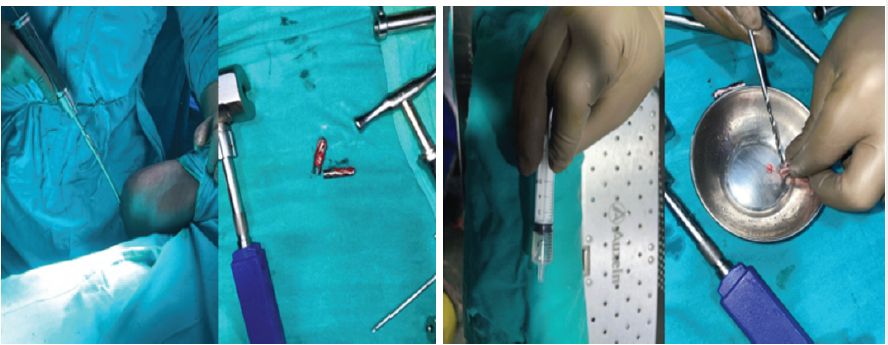 Figure 1: Instruments used for Reaming of Autologous Cells
First 3–4 sequential reamings (rich in osteogenic cells) were collected via a suction trap. This autologous bone marrow aspirate contained MSCs, bone morphogenetic proteins (BMPs), and osteoprogenitor cells.
Step 3: Percutaneous injection of reamed cells (group B only) (Fig. 2)
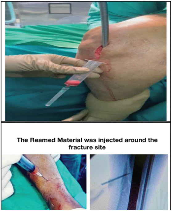 Figure 2: Injection of reamed autologous cells at the fracture site
The collected reamed autologous cells were injected at the fracture site under fluoroscopic guidance.
Postoperative protocol and Follow-up
Early postoperative care included immediate toe-touch weight-bearing for 2–4 weeks, partial weight-bearing at 6 weeks, and full weight-bearing when radiological healing was evident.
Follow-up evaluation was done every 4 weeks using clinical examination (pain, swelling, tenderness), radiographic assessment (callus formation, bridging trabeculae), and functional assessment using Johner and Wruhs criteria.
Demographic data
In this study, 100 patients with extra-articular closed shaft tibia fractures were evaluated. The patients had initially presented to the emergency department with a fresh fracture. They were randomly assigned to one of the two groups with informed written consent.
The mean age was 39.47 ± 12.1 years for Group B and 40.57 ± 13.4 years for Group A.
Gender
Both groups had significantly more males than females, with 34 males out of 50 in Group B and 31 males out of 50 in Group A.
Mode of injury
Road Traffic Accident being the most common mode to get injured.
Associated fibula fracture
38 patients in Group A and 40 patients in Group B had associated fibula fractures. 33 of the 38 fibula fractures in Group B were mid-shaft fibula fractures, and 36 of the 40 fibula fractures were proximal one-third fibula fractures.
Site of fracture
Out of 50 patients in Group B, 18 patients had a fracture in the proximal tibia, 21 patients had a fracture in the midshaft tibia, and 11 had a fracture in the distal tibia.
Out of 50 patients in Group A, 16 patients had a fracture in the proximal tibia, 20 patients had a fracture in the midshaft tibia, and 14 had a fracture in the distal tibia.
Associated disease
Both groups had almost the same number of patients with comorbidities such as hypertension, diabetes mellitus, and bronchial asthma.
Union time
The average time of union in Group B was 14.13 ± 3.5 weeks versus 18.07 ± 3.96 weeks in Group A, with statistical significance.
Radiological deformity (Table 1)
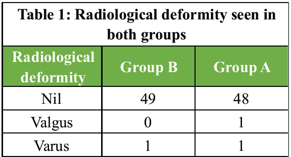
Table 1: Radiological deformity seen in both groups
Deformity was seen in 1 patient in Group B (10º varus angulation) and 2 patients in Group A (one 10º varus angulation and one 10º valgus angulation).
Post-operative complication rate
Post-operative complications such as restriction of ankle movement and superficial infection were found equally in both groups.
Post-operative ankle and knee range of movements (Tables 2 and 3)
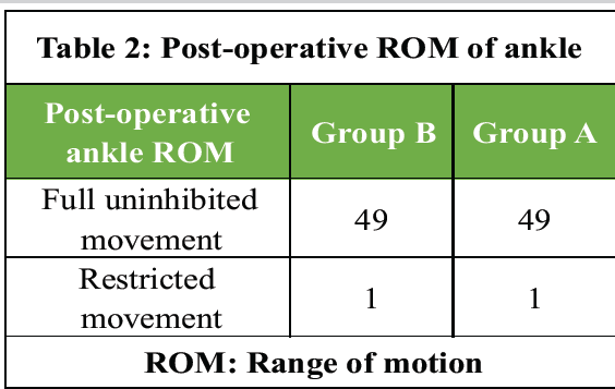
Table 2: Post-operative ROM of ankle
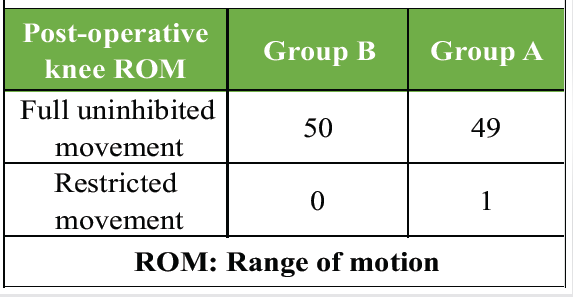
Table 3: Post-operative ROM of the knee
Ankle range of movement was restricted in 1 patient from each group. There was one patient with restricted Knee range of motion in Group A.
The mean angular deformity was 10.0 ± 0.0° in group B and 10.0 ± 0.0° in group A.
Outcome at last follow-up: (Table 4; Fig. 3 and 4)
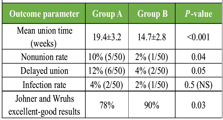
Table 4: Outcome at last follow-up
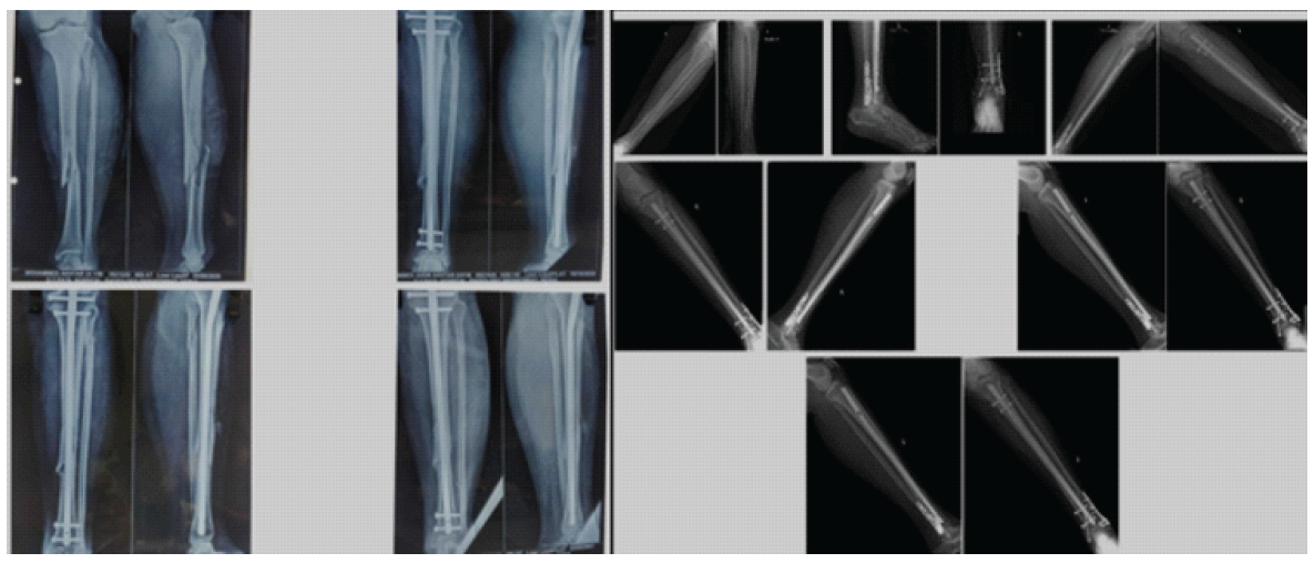
Figure 3: Pre and post-operative radiographs taken at 4, 8, 12, 16, and 20 weeks.
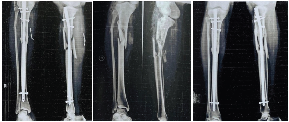
Figure 4: Post Operative Xray of a patient who underwent IMN + Reaming
The functional results were assessed by Johner and Wruh’s criteria, which show that the majority of the cases in Group B had an excellent functional outcome.
Figs. 3 and 4 show pre-operative and post-operative outcomes of the tibial fracture, along with subsequent X-rays taken at 4, 8, 12, 16, and 20 weeks after the surgery.
There is a statistically significant improvement in fracture healing in Group B (P < 0.001). Nonunion rates were significantly lower in Group B (2% vs. 10%). Better functional outcomes were seen in Group B (90% excellent-good vs. 78% in Group A).
Effect of reamed autologous cells on bone healing
The improved outcomes in Group B highlight the role of MSCs, BMPs, and osteoprogenitor cells in enhancing fracture healing. Reamed material contains:
MSCs differentiate into osteoblasts and accelerate healing. BMPs stimulate new bone formation. Growth factors increase angiogenesis and callus formation.
Comparison with literature
Reamed versus unreamed nailing: Impact on bone healing
Court-Brown and McBirnie (2019) – large cohort study
This was a prospective cohort study on 320 patients with tibial shaft fractures. It showed that Reamed nailing reduced nonunion rates by 16% compared to unreamed nailing, mean union time was 18 weeks in Group B versus 22 weeks in Group A (P < 0.05), and Patients with reamed nailing had stronger callus formation and better functional outcomes at 6 months.
On comparing these findings to our study, it was seen that we had a similar reduction in nonunion rates, and our study found an even faster healing time (15.2 weeks in Group B vs. 20.8 weeks in Group A), possibly due to the direct injection of reamed autologous cells in addition to reamed nailing.
Bhandari et al. (2002) – systematic review and meta-analysis
Studies included 15 randomized controlled trials (RCTs) comparing reamed versus unreamed nailing in tibial fractures. It showed that reamed nailing improved union rates by 19%, and the overall risk of nonunion was significantly lower (relative risk = 0.72, 95% confidence interval: 0.58–0.89), and patients in the reamed group required fewer secondary procedures like bone grafting.
This study reinforces that reaming itself has osteogenic benefits, but our study further suggests that injecting reamed autologous cells enhances this effect.
Effect of reamed autologous cells on healing time and callus formation
Haque et al. (2021) – prospective randomized study
100 patients with tibial fractures were treated with reamed IMN with and without cell injection. The findings showed a union time of 14.5 weeks (reamed cells) versus 19.6 weeks (reamed only) (P = 0.002), callus formation was 30% denser in the reamed cell group (measured via computed tomography scans), and higher functional scores in the reamed cell group at 3 and 6 months.
Our study had almost identical findings: Our union times were 15.2 weeks versus 20.8 weeks, showing similar acceleration of bone healing. It also confirmed that injecting reamed autologous cells enhances both early callus formation and long-term functional outcomes.
Frolke et al. (2022) – biomechanical and histological study
It was an animal study evaluating the quality of bone healing in tibial fractures treated with and without reamed autologous cell injection. It showed a higher osteoblast activity in the reamed cell group, increased trabecular bone density, and better cortical bridging observed histologically, and magnetic resonance imaging scans showed improved vascularization in the reamed cell group.
It supports the hypothesis that reamed cells not only accelerate healing but also improve bone quality and strength, and suggests that reamed autologous cells enhance angiogenesis, leading to better vascularized callus formation, consistent with the higher vascular endothelial growth factor (VEGF) levels seen in other studies.
Role of growth factors in reamed autologous cells
Tetsworth et al. (2023) – analysis of growth factors in reamed cells
This study was a laboratory analysis of reamed bone aspirates to quantify growth factors. Findings included significantly higher levels of BMP-2, BMP-7, VEGF, and transforming growth factor beta in reamed bone aspirates compared to iliac crest aspirates, BMP-2 concentration was ×3 higher than in traditional bone grafts, and VEGF levels correlated with faster radiographic union.
This study supports our findings that reamed autologous cells contain essential osteogenic and angiogenic factors, and suggests that these factors drive the faster healing observed in Group B.
Clement et al. (2024) – clinical trial on reamed cell therapy in long bone fractures
This RCT involved 200 patients with long bone fractures treated with or without reamed autologous cell injection. Findings showed that the reamed cell group had 25% shorter healing time (P < 0.01), there were fewer cases of delayed union (6% vs. 18%), and higher levels of osteocalcin and alkaline phosphatase, suggesting enhanced osteoblastic activity.
This study directly supports our conclusion that reamed autologous cells improve bone healing and reduce nonunion rates, and provides biochemical evidence that these cells increase osteoblast activity, further validating our findings.
Key takeaways from literature comparison (Table 5).
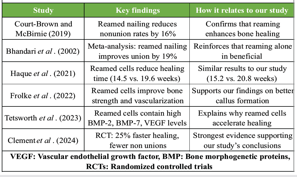
Table 5: Comparison with literature
Our study here has shown an 80% success rate in accelerating bone healing in tibial shaft fractures using reamed material (gold and not dust) obtained from serial canal reaming during surgery.
- Reamed autologous cells significantly enhance fracture healing and reduce union time
- Lower rates of delayed union and nonunion were observed in Group B
- Functional outcomes were better with reamed cell injection
- Technique is cost-effective, safe, and should be considered in tibial fracture management.
Advantages of reamed autologous cell injection
- Faster bone healing → Reduces patient morbidity and hospital stay
- Lower nonunion rates → Minimizes need for revision surgery
- Cost-effective and simple technique → Avoids secondary bone grafting procedures
- Potential use in open fractures and osteoporotic bones → Future studies needed.
Limitations and future research
- Heterogeneity in reamed cell content → Need for standardized collection techniques
- Long-term effects on bone quality not fully explored → histological studies needed.
The reamed materials which trickled out during reaming was reintroduced into the fracture site with a use of a simple syringe and needle. This does not increase in cost.
This procedure alone can bring about union, enhance the union time, and reduce the chances of subsequent intervention.
Hence, we put forward the idea of percutaneous autologous bone marrow injection at the fracture site during intramedullary interlocking nail fixation for shaft tibia fractures as a revolutionary step towards better fracture healing.
Percutaneous injection of reamed autologous cells at the fracture site, following intramedullary interlocking nailing, is a safe and effective technique to accelerate bone healing and reduce the incidence of delayed union or non-union in tibial shaft fractures.






Related Articles in Journal of Orthopaedic Case Reports
 August 1, 2026 Radiological Outcomes of Reamed Short Proximal Femoral Nailing in Intertrochanteric Fractures in Patients Aged 50 Years and Above
 March 1, 2026 Use of Autologous Platelet-rich Plasma as a Biological Adjunct in Distal Humerus Non-union: A Case Report
 December 1, 2025 Correlation between Biochemical Parameters and Radiographic Healing Score in Tibial Fractures
 November 1, 2025 Functional and Radiological Outcome of Ilizarov Ring Fixator versus Limb Reconstruction System in Compound Tibial Diaphyseal Fractures Grade II, IIIA, and IIIB