

Dual fixation should be considered in long bone fractures where single-implant constructs are predicted to be biomechanically insufficient, including highly comminuted diaphyseal fractures, periarticular injuries, osteoporotic bone, and established non-union
Dr. Priyank Sundriyal, Department of Orthopedics, Government Medical College, Chhatrapati Sambhajinagar, Maharashtra, India. E-mail: priyank.sl17@gmail.com
Abstract
Introduction: Complex long bone fractures characterized by comminution, segmental patterns, metaphyseal extension, or associated joint instability pose significant challenges to stable fixation. While intramedullary nailing remains the standard treatment for diaphyseal fractures, certain fracture configurations are prone to mechanical failure, delayed union, or non-union when treated with a single fixation modality. Dual fixation using a combination of intramedullary nailing and plate augmentation may provide superior biomechanical stability and improve healing outcomes.
Objectives: To evaluate the clinical and radiological outcomes of primary dual fixation in complex long bone fractures and to assess its role as a proactive strategy for preventing fixation failure and non-union.
Materials and Methods: A prospective case series was conducted at a tertiary care center over a 24-month period. Thirty adult patients (24 males, 6 females; mean age 36.9 years) with complex long bone fractures were treated using primary dual fixation consisting of a non-locking intramedullary nail supplemented with plate augmentation. Fracture locations included the femur (n = 14), humerus (n = 14), and ulna (n = 2). Patients were followed for a minimum of 12 months. Outcome measures included radiological union, functional scores using the Disabilities of the Arm, Shoulder, and Hand (DASH) and lower extremity functional scale (LEFS), time to mobilization, and complications.
Results: Union was achieved in all 30 patients (100%). Mean time to radiological union was 16.5 weeks (range 13–22 weeks). Delayed union occurred in 3 patients (10%) but resolved without additional intervention by 24 weeks. No cases of non-union or construct failure were observed. Mean DASH score for upper-limb fractures was 12.8, while the mean LEFS score for lower-limb fractures was 75.6. Excellent-to-good functional outcomes were achieved in 26 patients (86.7%). Partial weight-bearing commenced at a mean of 4.5 weeks, and full weight-bearing was achieved at a mean of 10.8 weeks in lower-limb fractures. Complications included superficial infection in 2 patients (6.7%), implant irritation in 3 patients (10%), and migration of a titanium elastic nail in 2 patients (6.7%). No deep infections occurred.
Conclusion: Primary dual fixation provides reliable union, excellent functional outcomes, and a low complication rate in complex long bone fractures. By combining the load-sharing advantages of intramedullary nailing with the rotational and axial stability of plate augmentation, this strategy may prevent the delayed union, non-union, and fixation failures traditionally associated with mechanically unstable fracture patterns. These findings support consideration of dual fixation as a definitive primary treatment option in selected complex fractures and warrant further evaluation through prospective comparative studies.
Keywords: Dual fixation, nail-plate construct, complex long bone fractures, intramedullary nailing, plate augmentation, fracture union, orthopedic trauma.

Complex long bone fractures, commonly resulting from high-energy trauma, such as road traffic accidents (RTA) and falls from height, present significant challenges in orthopedic practice. These injuries are often associated with comminution, segmental fracture patterns, and metaphyseal-diaphyseal extension, making stable fixation difficult to achieve with a single modality.
Intramedullary nailing is widely regarded as the gold standard for diaphyseal fractures due to its load-sharing properties and minimally invasive nature [1]. However, in complex fracture configurations, nailing alone may not provide adequate rotational or axial stability. Plate augmentation, when combined with intramedullary nailing, enhances construct rigidity and helps maintain alignment, especially in comminuted or segmental fractures [2].
Nail-plate combination fixation has been used traditionally as a revision strategy for long bone diaphyseal fractures in which primary fixation has failed. Whether the index procedure was an intramedullary nail or a plate, the resulting non-union or delayed union has historically been addressed by combining both implant types at the revision stage. A substantial body of published evidence supports this revisional nail-plate approach, confirming its efficacy in salvaging failed primary fixation and restoring the mechanical environment required for healing [3].
However, the clinical and economic burden of a second operative procedure with its attendant’s anesthetic risk, prolonged rehabilitation, and increased cost is considerable. Humeral shaft fractures, for instance, carry a well-documented predisposition to non-union even after technically satisfactory primary fixation, with reported rates ranging from 5% to 15% [4]. Comminuted distal femoral fractures fixed by intramedullary nailing alone are prone to delayed union secondary to the so-called “windshield wiper” effect, that is, cyclical toggling of the distal nail within the widening medullary canal of the metaphysis generates interfragmentary motion that impedes callus consolidation [5]. Monteggia and Galeazzi fracture-dislocations with long segmental comminution similarly present biomechanical environments in which a single fixation modality struggles to maintain adequate stability throughout the healing period [6].
Recognizing these failure patterns, we propose a paradigm shift in the management of selected unstable long bone diaphyseal fractures. The proactive, primary application of dual fixation combining non-locking intramedullary nailing with supplemental plate fixation, or employing dual-plate constructs at the index procedure, before the patient has had the opportunity to develop non-union or delayed union. The target population for this approach comprises closed fractures, or Grade I open fractures amenable to debridement and primary internal fixation [7], that the operating surgeon judges to be mechanically unstable by virtue of comminution, displacement, segmental bone loss, or association with adjacent joint instability. These fractures, under physiological muscular loads, cannot maintain their reduction with a single implant alone.
By performing dual fixation as a primary procedure, we aim to provide simultaneous load sharing and load bearing, superior rotational stability, enhanced callus formation through micromotion control, and earlier safe mobilization, thereby pre-empting the complications that would otherwise necessitate a second surgery over the course of the 9-month healing window.
We present a series of 30 patients managed with primary dual fixation over a 3-year period, few of whom are described in detail as representative cases. We discuss the biomechanical rationale for implant selection, the operative technique principles, and clinical and radiological outcomes at a minimum follow-up of 12 months.
This prospective case series was conducted at a tertiary care center over a period of 24 months, including 30 patients with complex long bone fractures treated using a nail-and-plate combination technique.
Inclusion criteria
- Patients aged 18–65 years
- Complex long bone fractures (comminuted, segmental, or with metaphyseal extension)
- Closed fractures and open fractures up to Gustilo-Anderson Grade III B
- Patients treated with intramedullary nail combined with plate augmentation.
Exclusion criteria
- Gustilo-Anderson Grade III C fractures
- Patients medically unfit for surgery
- Loss to follow-up.
Patient demographics (Table 1)
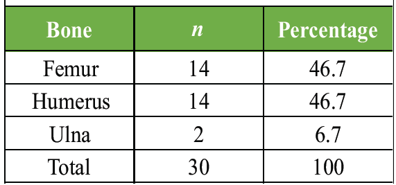
Table 1: Patient demographics (n=30)
Fracture distribution (Table 2).
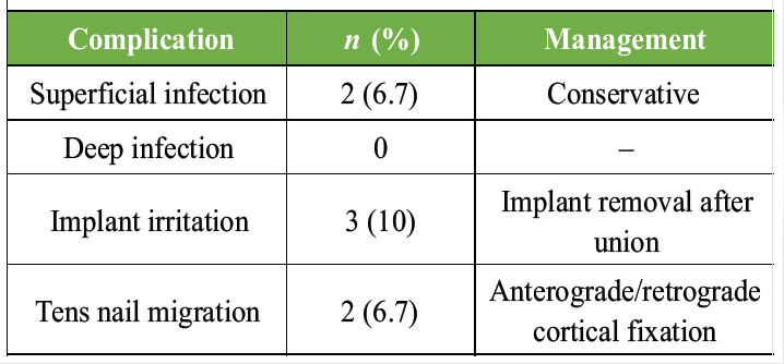
Table 2: Distribution of fractures by bone
Surgical technique
All patients were treated using a standardized dual fixation approach: Non-locking intramedullary nailing followed by supplementary plate augmentation.
- Closed or minimally invasive non-locking intramedullary nailing was performed first to restore length and alignment
- A locking compression plate was then applied to address residual instability, especially in comminuted or metaphyseal regions
- Care was taken to minimize soft tissue dissection and preserve periosteal blood supply (Fig. 1).
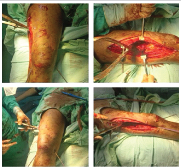
Figure 1: Intra op photo showing surgical technique of tens with plate augmentation in a distal femur fracture patient.
Post-operative protocol
- Early range of motion exercises were initiated within 48 h
- Partial weight-bearing was started between 4 and 6 weeks depending on fracture stability
- Full weight-bearing was allowed after evidence of radiological union.
Follow-up and outcome assessment
Patients were followed at regular intervals up to 12 months.
Outcome measures included:
- Time to radiological union
Functional outcome scores:
- Disabilities of the arm, shoulder and hand (DASH) (upper limb)
- Lower extremity functional scale (LEFS) (lower limb)
- Complications, including infection, implant failure, delayed union, and non-union.
Statistical analysis
Descriptive statistical analysis was performed, with mean and range calculated for continuous variables.
Illustrative cases
- A 55-year-old male with a/h/o RTA with Vancouver type C peri-prosthetic femur fracture of the left side (Fig. 2)
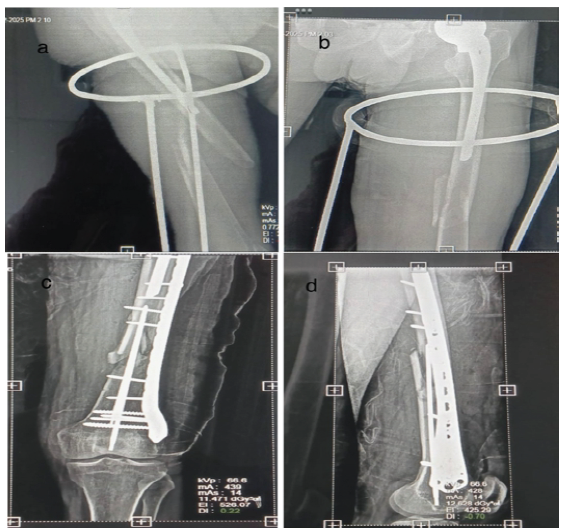
Figure 2: (a and b) pre-operative X-rays; (c and d) post-operative X-rays.
- 18-year-male with a/h/o self-fall from height with distal femur fracture managed by tens nail with fibular strut graft with distal femur plate (Fig. 3)
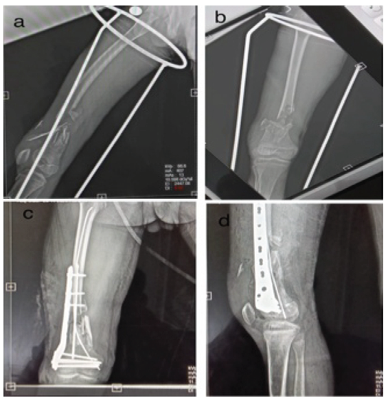
Figure 3: (a and b) pre-operative X-rays; (c and d) – post-operative X-rays.
- 42-year-old female with type 12-A3 shaft humerus fracture (Fig. 4)
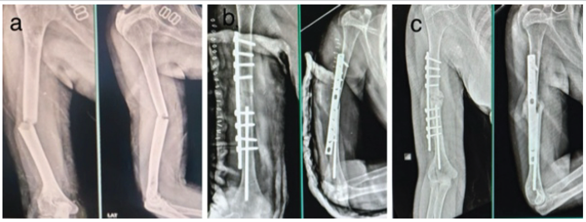
Figure 4: (a) pre-operative X-rays; (b) post-operative X-rays; (c) 3 month follow-up X-rays.
- A 19-year-old female with type 12-A1 shaft humerus fracture (Fig. 5).
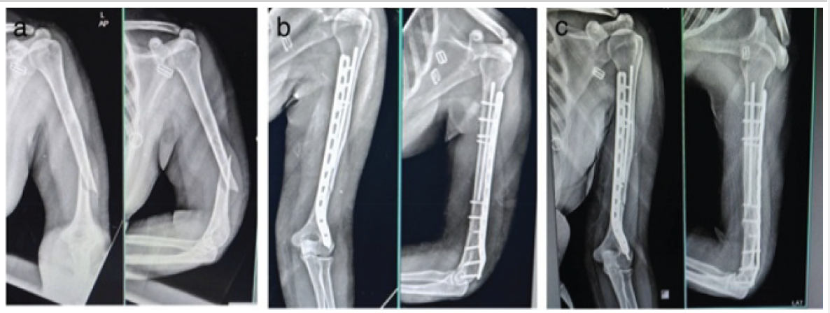
Figure 5: (a) pre-operative X-rays; (b) post-operative X-rays; (c) 2 month follow-up X-rays.
In case number 3rd and 4th, tens nail was inserted through the fracture site to maintain alignment, which was augmented with a plate. In this approach, migration of the tens nail was seen in post-operative X-rays as the tens nail was not fixed in the canal, so we fixed this problem by inserting the tens nail in anterograde or retrograde fashion through the bony cortex so that it gets a fixation point in bone.
- A 76-year-old male with type P2A shaft humerus fracture managed with anterograde tens nail and 4.5 dcp plate (Fig. 6)
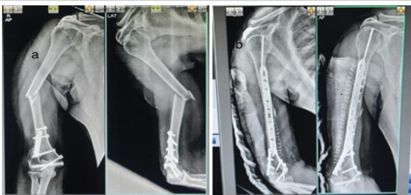
Figure 6: (a) pre-operative X-ray; (b) post-operative X-ray showing tens nail inserted in anterograde fashion.
- 49-year-old male with non-union distal third humerus, plating done 6 months back, managed by long plate, retrograde tens nail, and vascularized pedicular fibular bone graft (Fig. 7)
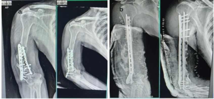
Figure 7: (a) pre-operative X-rays; (b) post-operative X-rays.
- A 46-year-old male presented with a/h/o RTA with a type 3 comminuted fracture of the olecranon managed with tens and olecranon hook plate (Fig. 8).
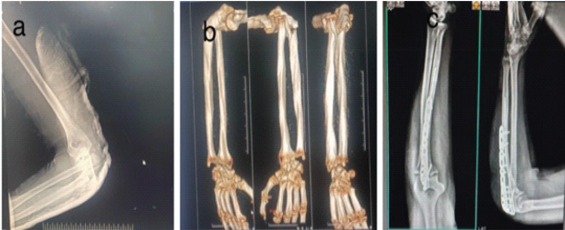
Figure 8: (a and b) pre-operative X-rays and 3D computed tomography; (c) post-operative X-rays.
The mechanical behavior of a fixation construct is determined by its bending stiffness, torsional rigidity, axial stability, and resistance to fatigue failure. In simple fracture patterns, a single well-positioned implant can adequately address all four parameters. However, in comminuted, segmental, or periarticular fractures, the loss of cortical continuity generates high bending moments at the implant-bone interface, and no single intramedullary or extramedullary device achieves sufficient stability across all loading planes simultaneously.
Laboratory biomechanical studies have consistently demonstrated that nail-plate constructs exhibit superior torsional rigidity compared to nailing alone, while orthogonal dual-plate configurations provide the highest resistance to varus-valgus loading and axial collapse in metaphyseal comminution [8,9]. Previous studies have demonstrated a 34% reduction in implant strain and a 28% increase in construct stiffness when a supplemental plate was added to a locked intramedullary nail (IMN) in a simulated segmental femoral fracture model [10]. These mechanical advantages translate clinically to lower rates of construct failure, malunion, and non-union [11].
Conventionally, nail-plate combinations were reserved for the revision setting and were deployed only after a primary fixation construct had demonstrably failed. Our series challenges this reactive approach. Based on our 3-year, 30-patient experience and a review of the published literature, we propose that the following fracture types warrant primary dual fixation at the index procedure, because the surgeon can predict with sufficient confidence from the initial radiographs and computed tomography (CT) that single-implant fixation will be mechanically inadequate:
- Humeral shaft fractures: Notorious for non-union even after technically satisfactory single-modality fixation. The combination of poor cortical contact at the fracture site, distraction by the weight of the limb, and unpredictable periosteal vascularity justifies primary dual fixation with IMN plus anterolateral plate in comminuted or displaced patterns.
- Distal femoral fractures with significant metaphyseal comminution: The diverging medullary canal of the distal femur allows a locked nail to toggle cyclically under physiological loading – the so-called windshield wiper effect – generating interfragmentary motion that impedes union. A supplemental medial or lateral plate neutralizes this rotational toggle and protects the healing callus.
- Monteggia fracture-dislocations and Galeazzi fracture-dislocations with long segmental or comminuted forearm fracture components: The combination of axial, rotational, and ligamentous instability in these injury patterns renders single-implant fixation prone to loss of reduction and radio-ulnar joint re-dislocation.
- Segmental long bone fractures (intercalary fragment >5 cm): The loss of a continuous cortical strut renders a single intramedullary nail unable to control torsion across both fracture levels simultaneously.
- Comminuted diaphyseal fractures under high muscular deforming forces (e.g., mid-shaft femur, proximal tibia), where physiological loads cannot be resisted by a single implant; dual fixation provides load sharing and reduces cyclic implant strain.
All 30 patients completed a minimum follow-up of 12 months.
Fracture Union:
- Union was achieved in all 30 patients (100%)
- The mean time to union was 16.5 weeks (range: 13–22 weeks)
- Delayed union was observed in 3 cases (10%), which united by 24 weeks without additional intervention
- No cases of non-union were recorded.
Functional outcomes:
- Mean DASH score (upper limb cases): 12.8
- Mean LEFS score (lower limb cases): 75.6
- Excellent to good outcomes were observed in 26 patients (86.7%)
- Fair outcomes in 4 patients (13.3%), primarily due to joint stiffness.
Mobilization:
- Partial weight-bearing initiated at a mean of 4.5 weeks
- Full weight-bearing achieved at a mean of 10.8 weeks in lower limb fractures.
Complications:
- Superficial infection: 2 cases (6.7%), managed conservatively
- Deep infection: None
- Implant irritation: 3 cases (10%), managed with implant removal after union
- Migration of the tens nail was seen (Table 3 and 4).
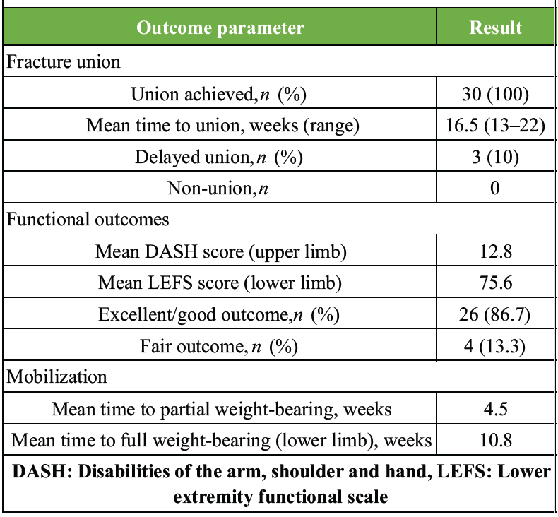
Table 3: Functional and union outcomes (n=30)
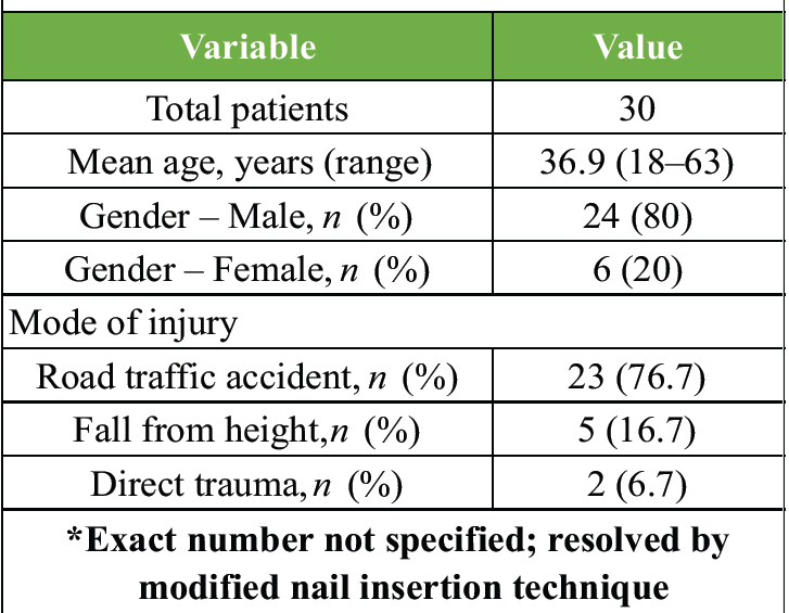
Table 4: Complications and their management
Nail-plate combination fixation has long been validated as a salvage strategy for failed primary fixation of long bone diaphyseal fractures. This series advances that concept by demonstrating that the same dual fixation principle applied proactively at the index procedure can prevent the non-union and delayed union that would otherwise mandate a second surgery in mechanically unstable fractures.
When the treating surgeon assesses, on plain radiographs and CT, that a fracture is comminuted, displaced, or located in a biomechanically vulnerable zone, such as the humeral shaft, distal femoral metaphysis, or the forearm in the context of a Monteggia or Galeazzi dislocation, primary dual fixation with an intramedullary nail supplemented by a long plate at appropriate screw density should be considered as the definitive fixation strategy. This approach simultaneously provides load sharing, load bearing, and rotational stability; promotes robust callus formation; enables earlier mobilization; reduces joint stiffness; and eliminates the morbidity, cost, and psychological burden of a second operative procedure.
Over a 3-year period and 30 patients, this strategy produced no non-unions, no construct failures, and no second fixation procedures in a cohort of fractures where published single-implant revision rates of 5–12% would have been expected. These results, while preliminary, support the broader adoption of primary dual fixation in prospectively identified unstable long bone diaphyseal fractures and justify the design of prospective randomized trials to confirm these findings.
Limitations
This study has several limitations, including its relatively small sample size, single-center design, lack of a control group as it is a case series without any comparative group, non-randomized observational methodology, heterogeneous fracture patterns and fixation constructs, and minimum follow-up of 12 months. Implant selection was based on surgeon judgment rather than standardized objective criteria, and only descriptive statistical analyses were performed. Functional assessment was limited to DASH and LEFS without evaluation of quality of life or patient satisfaction. Economic outcomes, operative variables, and biomechanical validation were not assessed. Consequently, while the present findings demonstrate encouraging clinical outcomes, they should be interpreted as preliminary evidence supporting the feasibility of primary dual fixation in selected mechanically unstable fractures rather than definitive evidence of superiority over conventional single-implant fixation. Future multicenter randomized comparative studies are necessary to validate these findings and establish standardized treatment indications.
1. Dual fixation should be considered in long bone fractures where single-implant constructs are predicted to be biomechanically insufficient, including highly comminuted diaphyseal fractures, periarticular injuries, osteoporotic bone, and established non-union
2. Patient selection, preoperative planning with CT imaging, and attention to implant positioning are critical determinants of outcome in dual fixation procedures
3. A high union rate and satisfactory functional outcomes can be expected with dual fixation when appropriate surgical principles are followed.
References
- 1. Ricci WM, Gallagher B, Haidukewych GJ. Intramedullary nailing of femoral shaft fractures: Current concepts. J Am Acad Orthop Surg 2009;17:296-305. [Google Scholar] [PubMed]
- 2. Stoffel K, Sommer C, Lee M, Zhu TY, Schwieger K, Finkemeier C. Double fixation for complex distal femoral fractures. EFORT Open Rev 2022;7:274-86. [Google Scholar] [PubMed]
- 3. Attum B, Douleh D, Whiting PS, White-Dzuro GA, Dodd AC, Shen MS, et al. Outcomes of distal femur nonunions treated with a combined nail/plate construct and autogenous bone grafting. J Orthop Trauma 2016;31:e301-4. [Google Scholar] [PubMed]
- 4. Leiblein M, Verboket R, Marzi I, Wagner N, Nau C. Nonunions of the humerus – treatment concepts and results of the last five years. Chin J Traumatol 2019;22:187-95. [Google Scholar] [PubMed]
- 5. Henderson CE, Lujan TJ, Bottlang M, Fitzpatrick DC, Madey SM, Marsh JL. Stabilization of distal femur fractures with intramedullary nails and locking plates: Differences in callus formation. Iowa Orthop J 2010;30:61-8. [Google Scholar] [PubMed]
- 6. Xiao RC, Chan JJ, Cirino CM, Kim JM. Surgical management of complex adult Monteggia fractures. J Hand Surg Am 2021;46:1006-15. [Google Scholar] [PubMed]
- 7. Gustilo RB, Anderson JT. Prevention of infection in the treatment of one thousand and twenty-five open fractures of long bones: Retrospective and prospective analyses. J Bone Joint Surg Am 1976;58:453-8. [Google Scholar] [PubMed]
- 8. Cheung ZB, Nasser P, Iatridis JC, Forsh DA. Orthogonal plating of distal femur fractures: A biomechanical comparison with plate-nail and parallel plating constructs. J Orthop 2023;37:34-40. [Google Scholar] [PubMed]
- 9. Liporace FA, Tang A, Jankowski JM, Yoon RS. Distal femur: Nail plate combination and the linked construct. OTA Int 2022;5:e172. [Google Scholar] [PubMed]
- 10. Sanders R, Swiontkowski M, Rosen H, Helfet D. Double-plating of comminuted, unstable fractures of the distal part of the femur. J Bone Joint Surg Am 1991;73:341-6. [Google Scholar] [PubMed]
- 11. Baumann AN, Uhler MA, Fiechter J, Anastasio AT, Walley KC, Coscia A, et al. Nail-plate combination constructs versus single traditional constructs for distal femur fractures: A systematic review and meta-analysis of comparative studies. Arch Orthop Trauma Surg 2024;145:89. [Google Scholar] [PubMed]






Related Articles in Journal of Orthopaedic Case Reports
 September 1, 2026 Distal Humerus Fracture in a 28-Year-Old Male with Achondroplasia Treated with Reconstruction Plate: Case Report and Review of Literature
 August 1, 2026 A Prospective Analysis of Physeal-Sparing Intramedullary Interlocking Nail for Adolescent Tibial Shaft Fractures
 June 1, 2026 Comparison of Surgical Outcomes and Perioperative Laboratory Parameters Following Minimally Invasive Plate Osteosynthesis versus Conventional Plating in Long Bone Fractures
 June 1, 2026 Role of Tranexamic Acid in Orthopedic Trauma: Current Evidence, Clinical Applications, and Ongoing Controversies