

Early recognition of post-operative C5 palsy following posterior cervical decompression and fusion, with prompt rehabilitation and close follow-up, can optimize neurological recovery and functional outcomes.
Dr. Mlsv Prasad, Department of Orthopaedics, JSS Academy of Higher Education and Research, Mysuru, Karnataka, India. E-mail: prasad11181169@gmail.com
Abstract
Introduction: Posterior cervical decompression and fusion (PCDF) is a well-established surgical procedure for the management of multilevel cervical spinal cord compression. Although generally safe, post-operative C5 palsy is a recognized complication that can significantly affect functional recovery.
Case Report: We report the case of a patient with multilevel cervical compressive myelopathy who underwent PCDF. The immediate post-operative period was complicated by new-onset weakness of shoulder abduction and elbow flexion, consistent with C5 palsy. Post-operative imaging confirmed satisfactory decompression and appropriate implant positioning, with no evidence of hematoma or hardware-related complications. The patient was managed conservatively with close neurological monitoring and a structured rehabilitation program.
Discussion: The exact mechanism of post-operative C5 palsy remains unclear, with proposed causes including posterior shift of the spinal cord, nerve root traction, ischemia, and reperfusion injury. Early recognition, exclusion of compressive causes, and rehabilitation are essential for optimal recovery.
Conclusion: C5 palsy is an uncommon but important complication following PCDF. Prompt diagnosis, appropriate imaging, and multidisciplinary rehabilitation are crucial to maximizing neurological recovery and improving functional outcomes. Categories: Neurology, Neurosurgery, Orthopedics
Keywords: C5 palsy, case report, cervical myelopathy, cervical spine surgery, cervical spondylotic myelopathy, posterior cervical decompression and fusion, posterior cervical fusion, post-operative neurological deficit, rehabilitation, spine surgery.

Degenerative cervical myelopathy (DCM) is the most common cause of spinal cord dysfunction in adults worldwide and results from progressive compression of the cervical spinal cord secondary to degenerative changes such as intervertebral disc degeneration, osteophyte formation, hypertrophy of the ligamentum flavum, and facet joint arthropathy [1,2]. Patients commonly present with gait instability, hand clumsiness, upper limb weakness, paresthesia, and upper motor neuron signs, leading to progressive functional impairment if left untreated [2,3].
Magnetic resonance imaging (MRI) is the gold standard for diagnosing DCM because it accurately demonstrates the level and severity of spinal cord compression, associated intramedullary signal changes, and other pathological abnormalities that guide surgical planning [1,3]. Surgical decompression is recommended for patients with moderate-to-severe or progressive myelopathy, as it prevents further neurological deterioration and improves long-term functional outcomes [1].
Posterior cervical decompression and fusion (PCDF) is an established surgical option for patients with multilevel cervical spondylotic myelopathy, particularly in the presence of preserved cervical lordosis. It provides effective decompression of the spinal cord while maintaining cervical stability through instrumented fusion [4,5]. Although neurological recovery after surgery is generally favorable, post-operative C5 nerve root palsy remains a recognized complication that requires early identification and appropriate rehabilitation [6,7].
We present a case of multilevel cervical spondylotic myelopathy managed with PCDF, highlighting the clinical presentation, radiological findings, surgical management, post-operative C5 palsy, and functional recovery in the context of current literature [8].
A 66-year-old female presented with a 5-month history of progressive gait instability, recurrent falls, bilateral hand paresthesia, and right upper limb weakness. She also complained of difficulty with fine motor activities, including buttoning clothes and writing. There was no history of trauma or bowel and bladder dysfunction. Her medical history was significant for hypertension, type 2 diabetes mellitus, and hypothyroidism.
On neurological examination, the patient had a spastic gait, grade 4/5 power in the right upper limb, hyperreflexia in both upper and lower limbs, bilateral Hoffmann’s sign, and extensor plantar responses, consistent with cervical myelopathy.
Laboratory investigations revealed the following:
- Hemoglobin: 10.5 g/dL
- Erythrocyte sedimentation rate: 150 mm/h
- Hemoglobin A1c: 7.1%
- Thyroid-stimulating hormone: 6.33 μIU/mL.
MRI of the cervical spine demonstrated severe spinal canal stenosis and spinal cord compression at the C5–C6 level, with focal intramedullary T2 hyperintensity suggestive of chronic compressive myelopathy (Fig. 1).
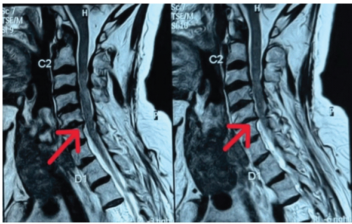
Figure 1: Pre-operative sagittal T2-weighted magnetic resonance image of the cervical spine. Mid-sagittal T2-weighted magnetic resonance imaging demonstrating severe spinal canal stenosis and spinal cord compression at the C5–C6 level, with focal intramedullary T2 hyperintensity consistent with chronic compressive myelopathy (arrows).
Axial T2-weighted imaging at the same level demonstrated a posterior disc-osteophyte complex causing severe central canal stenosis with marked flattening of the spinal cord and near-complete effacement of the surrounding cerebrospinal fluid space (Fig. 2).
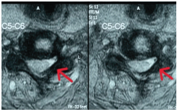
Figure 2: Pre-operative axial T2-weighted magnetic resonance image at the C5–C6 level. Axial T2-weighted magnetic resonance imaging demonstrating a posterior disc-osteophyte complex causing severe central canal stenosis with marked spinal cord compression and near-complete effacement of the surrounding cerebrospinal fluid space (arrows).
Based on the clinical and radiological findings, a diagnosis of DCM secondary to cervical spondylosis was established. After medical optimization, the patient underwent PCDF from C3 to C7 using lateral mass screw-and-rod fixation.
Post-operative radiographs demonstrated satisfactory placement of the instrumentation with maintained cervical alignment and a stable posterior fusion construct (Fig. 3).
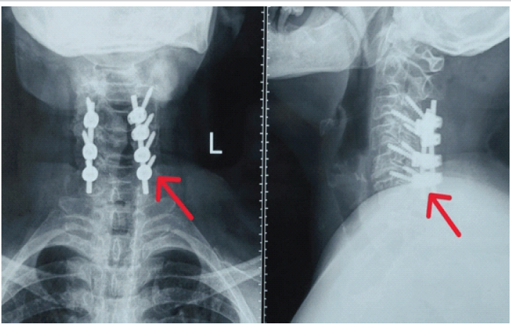
Figure 3: Post-operative radiographs following posterior cervical decompression and fusion (PCDF). Post-operative anteroposterior and lateral radiographs demonstrating satisfactory C3–C7 lateral mass screw–rod fixation (arrows), maintained cervical alignment, and a stable posterior cervical fusion construct following PCDF.
During the post-operative period, the patient developed weakness of shoulder abduction and elbow flexion, consistent with C5 nerve root palsy. She was managed conservatively with physiotherapy and rehabilitation.
The surgical wound healed uneventfully without evidence of infection (Fig. 4).
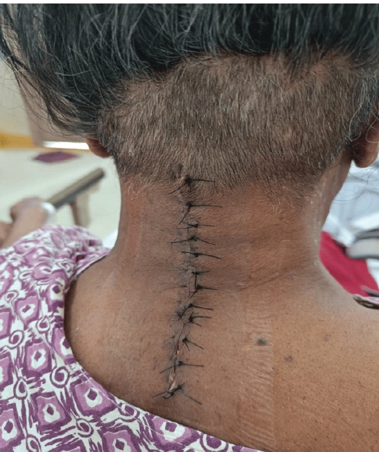
Figure 4: Post-operative wound at follow-up.
Clinical photograph demonstrating a well-healed posterior cervical surgical incision without evidence of wound infection, dehiscence, or implant-related complications during follow-up.
At subsequent follow-up, the patient demonstrated gradual improvement in gait, upper limb strength, and hand function with continued physiotherapy.
DCM is the most common cause of spinal cord dysfunction in adults and results from progressive compression of the cervical spinal cord due to degenerative changes such as intervertebral disc degeneration, osteophyte formation, facet joint arthropathy, and hypertrophy of the ligamentum flavum. The natural history of DCM is characterized by gradual neurological deterioration, making early diagnosis and timely surgical intervention essential to prevent irreversible spinal cord injury and functional disability [1,2].
Our patient presented with progressive gait instability, bilateral hand paresthesia, and upper limb weakness, which are characteristic clinical manifestations of DCM. MRI demonstrated severe spinal cord compression at the C5–C6 level with intramedullary T2 hyperintensity, indicating chronic cord injury. These findings align with previous studies by Kalsi-Ryan et al. and Badhiwala et al., who emphasized that MRI is the imaging modality of choice for confirming the diagnosis, assessing the severity of cord compression, and guiding surgical management [2,3].
The patient underwent PCDF, which is an established surgical procedure for multilevel cervical spondylotic myelopathy, particularly in patients with preserved cervical alignment. Posterior decompression effectively relieves spinal cord compression, while instrumented fusion provides immediate stability, maintains cervical alignment, and reduces the risk of post-operative deformity. Edwards et al. reported that appropriate patient selection and surgical planning are critical determinants of successful neurological recovery following posterior cervical surgery [4]. Similarly, Hirabayashi and Satomi demonstrated favorable long-term neurological improvement following posterior decompression procedures in patients with multilevel cervical myelopathy [5].
A recognized complication following posterior cervical decompression is post-operative C5 nerve root palsy, which typically presents with weakness of shoulder abduction and elbow flexion. The reported incidence ranges from approximately 5% to 10%, although rates vary depending on the surgical technique and patient characteristics [6,7]. The exact pathophysiology remains uncertain, with proposed mechanisms including posterior migration of the spinal cord following decompression, traction injury to the C5 nerve root, reperfusion injury, and pre-existing foraminal stenosis [6,7]. In the present case, the patient developed post-operative C5 palsy but showed gradual neurological improvement with conservative management, including physiotherapy and structured rehabilitation, without requiring revision surgery.
Overall, this case highlights the importance of correlating clinical findings with MRI. These findings agree with current evidence supporting surgical decompression as the standard of care for patients with progressive DCM [1,4,8]. Recent studies have also demonstrated favorable neurological recovery in patients with post-operative C5 palsy managed with structured rehabilitation and close follow-up [9,10].
DCM is a progressive neurological disorder in which early diagnosis and timely surgical intervention are essential to prevent irreversible spinal cord injury and improve functional outcomes. This case demonstrates that PCDF is an effective treatment for multilevel cervical spondylotic myelopathy, providing adequate spinal cord decompression and stable fixation. Although post-operative C5 nerve root palsy remains a recognized complication, prompt diagnosis, appropriate rehabilitation, and close follow-up can result in satisfactory neurological recovery.
Careful patient selection, meticulous surgical technique, and early recognition of complications such as C5 nerve root palsy are critical for achieving favorable neurological and functional outcomes.
Post-operative C5 palsy remains a recognized complication after PCDF, even when surgery is technically satisfactory. Anticipation of this complication, meticulous surgical planning, early identification of post-operative deltoid and biceps weakness, exclusion of structural causes, and timely rehabilitation are critical for favorable neurological and functional outcomes.
References
- 1. Fehlings MG, Tetreault LA, Riew KD, Middleton JW, Wang JC. A clinical practice guideline for the management of degenerative cervical myelopathy: Introduction, rationale, and scope. Global Spine J 2017;7 Suppl 3:21S-7. [Google Scholar] [PubMed]
- 2. Kalsi-Ryan S, Wilson JR, Karadimas SK, Arnold PM, Fehlings MG. Degenerative cervical myelopathy: The natural history and clinical presentation. Spine (Phila Pa 1976) 2013;38 22 Suppl 1:S21-36. [Google Scholar] [PubMed]
- 3. Badhiwala JH, Wilson JR, Fehlings MG. Global burden of degenerative cervical myelopathy and the unmet need for early diagnosis and treatment. J Bone Joint Surg Am 2020;102:e78. [Google Scholar] [PubMed]
- 4. Edwards CC 2nd, Riew KD, Anderson PA, Hilibrand AS, Vaccaro AF. Cervical myelopathy. Current diagnostic and treatment strategies. Spine J 2003;3:68-81. [Google Scholar] [PubMed]
- 5. Hirabayashi K, Satomi K. Operative procedure and results of expansive open-door laminoplasty. Spine (Phila Pa 1976) 1988;13:870-6. [Google Scholar] [PubMed]
- 6. Nassr A, Eck JC, Ponnappan RK, Zanoun RR, Donaldson WF 3rd, Kang JD. The incidence of C5 palsy after multilevel cervical decompression procedures: A review of 750 consecutive cases. Spine (Phila Pa 1976) 2012;37:174-8. [Google Scholar] [PubMed]
- 7. Sakaura H, Hosono N, Mukai Y, Ishii T, Yoshikawa H. C5 palsy after decompression surgery for cervical myelopathy: Review of the literature. Spine (Phila Pa 1976) 2003;28:2447-51. [Google Scholar] [PubMed]
- 8. Fehlings MG, Wilson JR, Kopjar B, Yoon ST, Arnold PM, Massicotte EM, et al. Efficacy and safety of surgical decompression in patients with cervical spondylotic myelopathy: Results of the AOSpine North America prospective multi-center study. J Bone Joint Surg Am 2013;95:1651-8. [Google Scholar] [PubMed]
- 9. Hasegawa K, Homma T, Chiba Y. Upper extremity palsy following cervical decompression surgery results from a transient spinal cord lesion. Spine (Phila Pa 1976) 2007;32:E197-202. [Google Scholar] [PubMed]
- 10. Imagama S, Matsuyama Y, Yukawa Y, Kawakami N, Kamiya M, Kanemura T, et al. C5 palsy after cervical laminoplasty: A multicentre study. J Bone Joint Surg Br 2010;92:393-400. [Google Scholar] [PubMed]






Related Articles in Journal of Orthopaedic Case Reports
 October 1, 2026 Staged Bilateral Total Elbow Arthroplasty in Advanced Rheumatoid Elbow Disease: A Case Report
 October 1, 2026 Blood Flow Restriction-Assisted Rehabilitation for Persistent Osgood-Schlatter Disease in an Adolescent Footballer: A Case Report with Isokinetic Evaluation
 October 1, 2026 Posterior Hip Dislocation with Ipsilateral Subtrochanteric Femur Fracture: A Rare Case Report
 September 1, 2026 Iliopsoas Abscess as the Initial Manifestation of Periprosthetic Hip Joint Infection after Total Hip Arthroplasty: A Case Report and Literature Review