

Autologous platelet concentrates (LR-PRP, LP-PRP, and iPRF) are safe and effective biologic options for chronic plantar fasciitis, with iPRF offering a practical edge due to sustained growth-factor release and simple preparation.
Dr. Madhan Jeyaraman, Department of Orthopaedics, ACS Medical College and Hospital, Dr. MGR Educational and Research Institute, Chennai, Tamil Nadu, India. E-mail: madhanjeyaraman@gmail.com
Abstract
Introduction: Plantar heel pain is the most frequent type of plantar pain in adults. The cellular composition of the platelet concentrates used (leukocyte-rich platelet-rich plasma [LR-PRP], leukocyte-poor PRP [LP-PRP], and injectable platelet-rich fibrin [iPRF]) as well as the kinetics of their growth-factor release differ; however, there is no head-to-head evidence thus far for plantar fasciitis (PF) treatment. We aim to assess pain relief, functional results, and plantar fascia thickness in patients with chronic PF treated with LR-PRP, LP-PRP, or iPRF.
Materials and Methods: A prospective single-center comparative study identified 57 adults (aged 30–60 years) who had chronic PF that failed to respond to ≥3 months of conservative treatment. Patients were randomly assigned to LR-PRP (n = 19), LP-PRP (n = 20), or iPRF (n = 18) and received a single injection using the peppering technique of 3 mL. At baseline, 1, 3, and 6 months, the Visual Analog Scale (VAS), American Orthopaedic Foot and Ankle Society (AOFAS) hindfoot score, and ultrasonographic plantar fascia thickness were recorded.
Results: At each follow-up, there were significant improvements in VAS and AOFAS in all three preparations (P < 0.05). At 6 months, the mean VAS fell from 7.66 to 2.25 (LR-PRP), from 7.33 to 2.10 (LP-PRP), and from 7.69 to 2.09 (iPRF), whereas AOFAS rose from 49.55 to 82.64, from 55.24 to 85.43, and from 53.17 to 90.79, respectively. No major adverse events occurred, and iPRF had the highest mean ΔAOFAS (37.6) and the lowest residual VAS score.
Conclusion: LR-PRP, LP-PRP, and iPRF are effective and safe injectable biologics for chronic PF, with iPRF being the only preparation that does not contain anticoagulants and where the growth factors are released over time, showing a slight but consistent functional benefit at 6 months.
Keywords: Plantar fasciitis, platelet-rich plasma, injectable platelet-rich fibrin, leukocyte-rich platelet-rich plasma, leukocyte-poor platelet-rich plasma, regenerative medicine.

Plantar heel pain is the most common cause of foot pain in adults seeking orthopedic or podiatric care, with a lifetime prevalence of nearly 10% globally and as high as 59% in some Indian populations aged 40–50 years [1]. Chronic plantar fascia specimens show myxoid degeneration, fibroblastic proliferation, and disorganized collagen, indicating a degenerative “fasciosis” rather than a purely inflammatory process [2]. First-line management – activity modification, stretching, orthoses, non-steroidal anti-inflammatory drugs (NSAIDs) and extracorporeal shockwave therapy – resolves symptoms in around 90% of patients [3,4]. Corticosteroid injection relieves pain quickly but risks plantar fascia rupture and fat-pad atrophy. When pain persists beyond 3–6 months of conservative treatment, biologically restorative options are required to address the underlying fasciosis [5,6].
Platelet-rich plasma (PRP), an autologous concentrate of growth factors, has anti-inflammatory, pro-angiogenic, and fibroblast-stimulating properties [7]. It provides better pain relief than corticosteroid injection in chronic plantar fasciitis (PF). In network meta-analyses, PRP also outperforms shockwave therapy and placebo for Visual Analog Scale (VAS) and American Orthopaedic Foot and Ankle Society (AOFAS) outcomes [8]. PRP is not a uniform biologic: Leukocyte-rich PRP (LR-PRP) retains a buffy-coat leukocyte population, whereas leukocyte-poor PRP (LP-PRP) minimizes leukocytes, which release pro-inflammatory cytokines that may impair tendon healing [9]. Injectable platelet-rich fibrin (iPRF), a second-generation concentrate developed by Choukroun without anticoagulant or external activator, allows physiological fibrin polymerization [10]. Prepared from plain tubes centrifuged briefly at low speed, iPRF forms a dense fibrin matrix that sustains the release of platelet-derived growth factor (PDGF), vascular endothelial growth factor (VEGF), transforming growth factor-beta (TGF-β), and insulin-like growth factor for up to 10–14 days, versus minutes to hours for PRP [11,12]. iPRF’s anti-inflammatory and chondroprotective effects are well described in dentistry and are emerging in temporomandibular joint dysfunction and knee osteoarthritis. In orthopedic soft-tissue models, iPRF stimulates collagen deposition and improves histological organization compared with PRP [13]. To our knowledge, no published randomized trial compares LR-PRP, LP-PRP, and iPRF in PF [14].
The ubiquity and chronicity of the condition, the variety of available PRP preparations, and the biological potential of iPRF warrant a head-to-head comparison. We therefore conducted a prospective comparative study at a tertiary-care teaching hospital in Chennai, India, to assess pain relief, functional result, and plantar fascia thickness in patients with chronic PF treated with LR-PRP, LP-PRP, or iPRF.
Study design and setting
A prospective, single-center, three-arm comparative study was performed in the Department of Orthopaedics in Sri Lalithambigai Medical College and Hospital, Chennai, India, from July 2024 to December 2024, and participants were followed up for 6 months. The protocol was reviewed and approved by the Institutional Ethics Committee (Dr. MGR-ERI/SLMCH/2024/028 dated June 26, 2024), and all participants provided written informed consent before enrollment. The study was done following the principles of the Declaration of Helsinki and the ICMR National Ethical Guidelines for Biomedical Research. As participants were randomly allocated to three parallel arms, the trial is reported in accordance with the Consolidated Standards of Reporting Trials (CONSORT) 2010 statement for parallel-group randomized trials.
Participants and eligibility
In this study, 30–60-year-old adults with clinically and ultrasonographically confirmed PF who failed to respond to a minimum of 3 months of supervised conservative treatment were screened. Those who had heel pain that was reproducible on direct palpation of the medial calcaneal tuberosity, a plantar fascia thickness of >4 mm on ultrasonography, and who agreed to attend all follow-up visits were included. Patients who were <30 or >60 years of age, had a prior corticosteroid injection within the last 3 months, had an autoimmune or inflammatory arthropathy, had a hemoglobin level <10 g/dL, a platelet count <1.5 × 10⁵/μL, a local infection, seropositivity for human immunodeficiency virus infection or hepatitis B/C, or refused to participate were excluded.
Group allocation and intervention
A computer-generated randomization list kept by an investigator who was not involved in clinical assessment was used to allocate the eligible participants sequentially to one of three treatment arms. The 19 patients in Group 1 were given LR-PRP, 20 patients in Group 2 were given LP-PRP, and 18 patients in Group 3 were given iPRF, with the distribution of the patients by sex and laterality listed in Table 1. The plantar fascia was surface marked using ultrasound guidance. Then the assigned biologic was injected into the origin of the plantar fascia over the medial calcaneal tuberosity with the peppering technique under strict aseptic precautions with a single 3 mL aliquot of the injected biologic. Each biologic was administered in a single sitting with the patient supine and the ankle dorsiflexed; after aseptic preparation of the heel and under real-time ultrasound guidance, a 22-gauge needle was advanced through a medial approach to the calcaneal origin of the plantar fascia, and the 3 mL aliquot was delivered by a peppering technique of four to five passes without withdrawing the needle from the skin, followed by dry sterile compression.
Preparation of platelet concentrates
Ten mL of venous blood was drawn into a sodium citrate tube, and then double spun; the first spin was at 2000 rpm for 15 min, centrifuged to separate plasma and buffy coat from the packed red cells, and the second spin was performed at 2000 rpm for 5 min. The bottom third (i.e., 2 mL) was the platelet-rich fraction, whereas LR-PRP contained buffy-coat leukocytes. Still, LP-PRP was aspirated 1–2 mm above the buffy coat to minimize the risk of leukocytes being aspirated. According to the low-speed centrifugation concept of Choukroun and Ghanaati, 10 mL of venous blood was collected in a plain glass-coated tube without any anticoagulants and centrifuged at 700 rpm for 3 min to produce iPRF.
Post-procedural protocol
Patients were advised to keep it rest–ice–compression–elevation and started active foot-stretching exercises from day 1, and were not allowed to put weight on the foot for 48–72 h. Partial weight bearing was allowed from day 3 to 7, and full weight bearing was allowed from the end of the 1st week onward. NSAIDs and analgesics were not administered for 14 days to prevent the suppression of the regenerative response.
Outcome measures
Pain measured on a 10 cm VAS and the 100-point AOFAS hindfoot score at baseline and at 1, 3, and 6 months after injection were the primary outcomes. The secondary outcome was ultrasonographic plantar fascia thickness measured at the insertion onto the medial calcaneal tuberosity by a single blinded radiologist at baseline and at 6 months. All adverse events, such as local pain, swelling, infection, and rupture, were documented at each visit.
Sample size estimation
The sample size was estimated a priori for the primary between-group comparison of the VAS pain score using a one-way analysis of variance (ANOVA) across the three arms. Assuming a moderate-to-large effect size (Cohen’s f = 0.42), derived from the differences in VAS improvement reported in earlier PRP trials for chronic PF, with a two-sided α of 0.05 and a statistical power of 80%, G*Power version 3.1 (Heinrich-Heine-Universität Düsseldorf, Germany) yielded a minimum requirement of 54 participants, that is, 18/arm. Allowing for approximately 5% attrition over the 6-month follow-up, the target enrolment was set at 57 participants, and 57 patients (19 in the LR-PRP arm, 20 in the LP-PRP arm, and 18 in the iPRF arm) completed the study with no dropouts.
Statistical analysis
All the data were entered into Microsoft Excel and analyzed using the Statistical Package for the Social Sciences version 26.0 (IBM Corp., Armonk, NY, USA). Data are presented as Mean ± SD for continuous data and frequencies and percentages for categorical data. Repeated measures ANOVA was used to test within-group differences over time points, and one-way ANOVA with post hoc Tukey HSD correction was used to test between-group differences. A two-sided P < 0.05 was considered statistically significant.
Baseline characteristics
Fifty-seven patients (33 males and 24 females) completed a 6-month follow-up; of these, 19 received LR-PRP, 20 received LP-PRP, and 18 received iPRF, with no dropouts. The mean age across groups ranged from 45.40 ± 5.83 years to 49.89 ± 6.65 years, and the mean body mass index ranged from 25.50 ± 3.83 to 29.11 ± 2.80 kg/m2, with no statistically significant intergroup differences (P > 0.05). The median symptom duration before injection was 7.68 ± 3.46 months in LR-PRP, 7.75 ± 2.86 months in LP-PRP, and 8.06 ± 2.69 months in iPRF, thus indicating chronic recalcitrant disease in all participants. The baseline demographic data of the study population are summarized in Table 1. On exploratory subgroup analysis, neither age, sex, nor baseline body mass index was significantly associated with the improvement in VAS or AOFAS at 6 months in any arm (P > 0.05), and the relative functional advantage of iPRF was consistent across these subgroups.
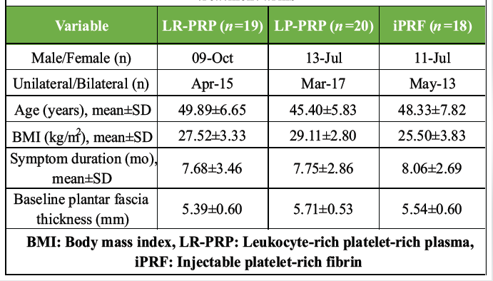
Table 1: Baseline demographic and clinical characteristics across the three treatment arms
Pain outcome (VAS)
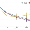
In all three arms, there was an overall decrease in mean VAS scores from injection to 6 months, reflecting significant pain relief in each of the biologic groups (Fig. 1 and Table 2).
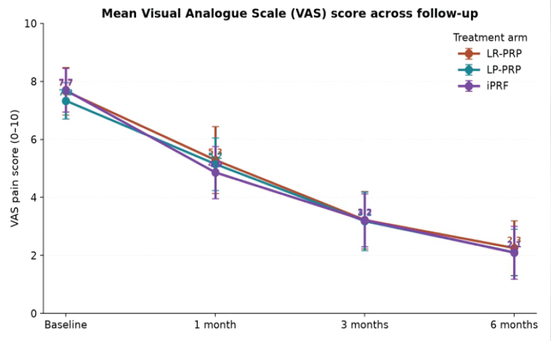
Figure 1: Mean Visual Analog Scale pain scores from baseline to 6 months in the leukocyte-rich platelet-rich plasma, leukocyte-poor PRP, and injectable platelet-rich fibrin arms.
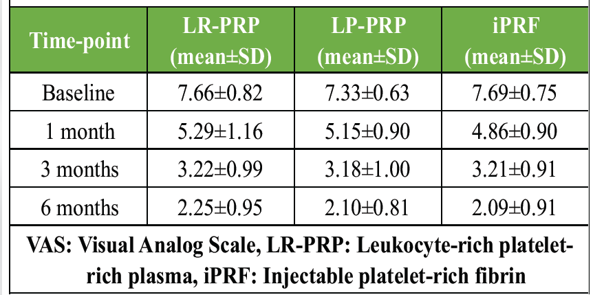
Table 2: Mean VAS pain scores from baseline to 6 months
In Group 1 (LR-PRP), mean VAS declined from 7.66 ± 0.82 at baseline to 5.29 ± 1.16 at 1 month, 3.22 ± 0.99 at 3 months, and 2.25 ± 0.95 at 6 months, representing a 70.6% reduction from baseline. In Group 2 (LP-PRP), the corresponding values were 7.33 ± 0.63, 5.15 ± 0.90, 3.18 ± 1.00, and 2.10 ± 0.81, with a 71.4% reduction at 6 months. In Group 3 (iPRF), the mean VAS decreased from 7.69 ± 0.75 to 4.86 ± 0.90, 3.21 ± 0.91 and 2.09 ± 0.91 across the same time points, equating to a 72.8% reduction. The mean VAS at 1 month was not significantly different between groups (one-way ANOVA, P > 0.05) and was numerically lowest in the iPRF arm. However, at 3 and 6 months, differences between groups were not significant (one-way ANOVA, P > 0.05).
Functional outcome (AOFAS)
There was a dramatic increase in the AOFAS hindfoot score in all arms, which reflects the decrease in pain (Table 3 and Fig. 2).
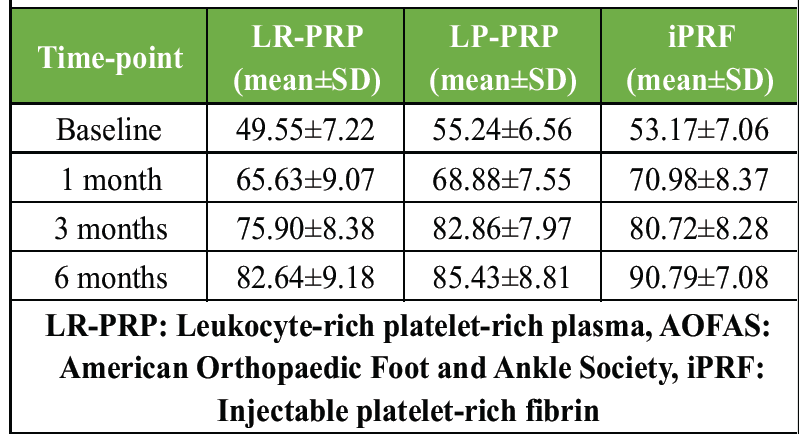
Table 3: Mean AOFAS hindfoot scores from baseline to 6 months
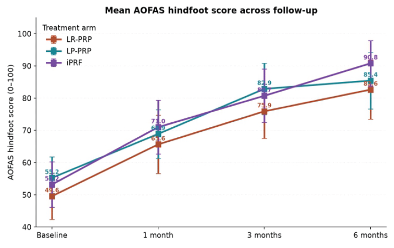
Figure 2: Mean American Orthopaedic Foot and Ankle Society hindfoot scores from baseline to 6 months in the three arms.
In the LR-PRP arm, AOFAS rose from 49.55 ± 7.22 at baseline to 65.63 ± 9.07, 75.90 ± 8.38, and 82.64 ± 9.18 at 1, 3, and 6 months. In the LP-PRP arm, the corresponding scores were 55.24 ± 6.56, 68.88 ± 7.55, 82.86 ± 7.97, and 85.43 ± 8.81. Across the four time points, the greatest improvement occurred in the functional rating of the iPRF arm, with mean scores of respectively, the highest mean functional rating in the study. The change from baseline at 6 months (ΔAOFAS) was 33.08 in LR-PRP, 30.19 in LP-PRP, and 37.62 in iPRF; iPRF was found to have a small but consistent functional benefit over both types of PRP.
Distribution of 6-month outcomes
The interquartile range was smallest in the iPRF arm for AOFAS, suggesting a more uniform functional recovery (Fig. 3). The median VAS was similar in all groups (2.0–2.3), and the median AOFAS was highest in the iPRF arm (92.0) compared to LP-PRP (86.2) and LR-PRP (83.4).
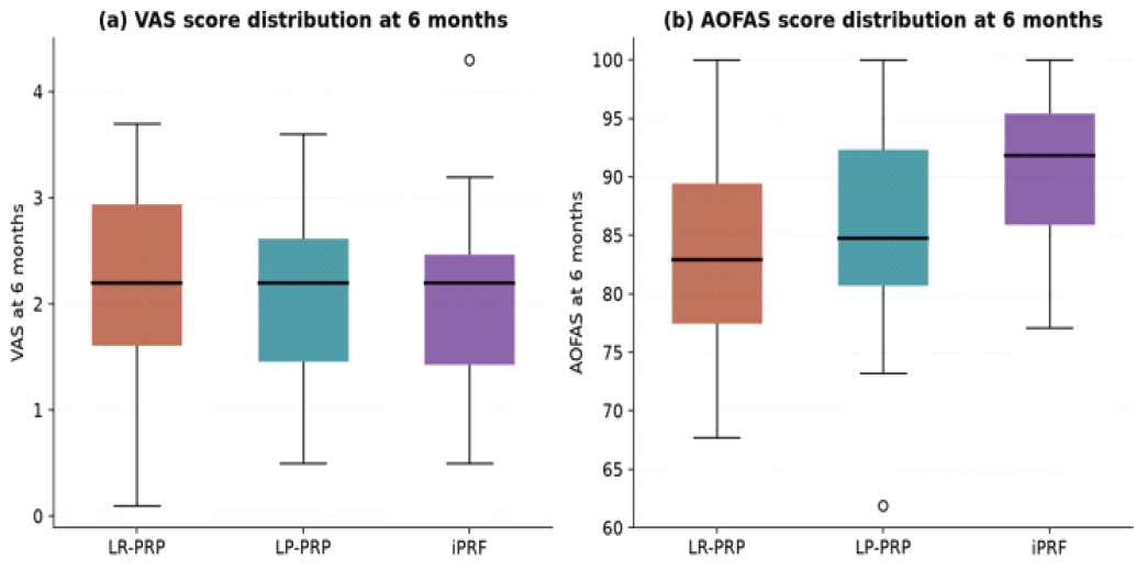
Figure 3: Distribution of 6-month outcomes across treatment arms: (a) Visual Analog Scale pain score and (b) American Orthopaedic Foot and Ankle Society hindfoot score.
Mean change from baseline
The mean reduction in VAS from baseline to 6 months was 5.41 in LR-PRP, 5.23 in LP-PRP and 5.60 in iPRF, exceeding the published minimum clinically important difference of 2 points for VAS in chronic musculoskeletal pain (Fig. 4a). The mean gain in AOFAS was 33.08, 30.19, and 37.62 points, respectively, comfortably exceeding the AOFAS minimum clinically important difference of 7.9–11 points reported in foot and ankle literature (Fig. 4b).
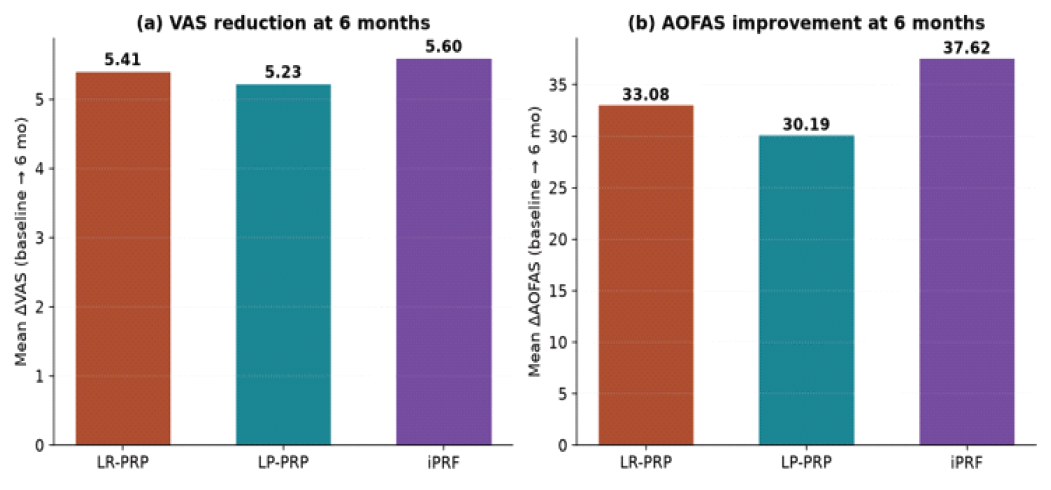
Figure 4: Mean change from baseline at 6 months: (a) Visual Analog Scale reduction and (b) American Orthopaedic Foot and Ankle Society improvement across the three arms.
Ultrasonographic plantar fascia thickness
Mean ultrasonographic plantar fascia thickness decreased from 5.39 ± 0.60 mm to 4.12 ± 0.76 mm in LR-PRP, 5.71 ± 0.53 mm to 4.15 ± 0.75 mm in LP-PRP, and 5.54 ± 0.60 mm to 4.17 ± 0.74 mm in iPRF over 6 months (Fig. 5). The mean reduction (Δ) was 1.27 mm, 1.56 mm and 1.37 mm, respectively, all of which were statistically significant within groups (Paired t-test, P < 0.001) but not between groups (one-way ANOVA, P = 0.21).
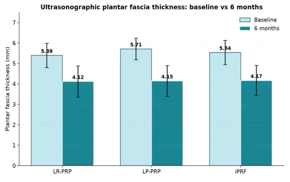
Figure 5: Ultrasonographic plantar fascia thickness at baseline and at 6 months across the three arms.
Adverse events
There were no major complications, including deep infection, plantar fascia rupture, or thromboembolic complications in any arm in the study period. Mild pain <48 h after injection was documented in 4 patients with LR-PRP (21.1%), 3 patients with LP-PRP (15.0%), and 2 with iPRF (11.1%), and mild swelling <72 h in 2, 1, and 1 patients, respectively. The minor event rate in the iPRF arm was lower, as would be expected with a lower volume preparation, which is free from any anticoagulants, and consistent with the safety reports published in the dental and maxillofacial literature.
4th one added
A prospective comparative study suggests that LR-PRP, LP-PRP, and iPRF are all effective and safe biological autologous injections for the treatment of chronic PF, but with a slight edge to the iPRF in terms of results (AOFAS functional scores at 6 months) [15,16]. This is one of the first head-to-head comparisons of these three modern platelet concentrates in PF, where we found a clinically relevant void in the recent systematic reviews conducted [17].
The effect size of pain relief achieved (VAS reduction 5.2–5.6 points, mean) is consistent with the long-term results of PRP according to Qiao et al. in their meta-analyses of randomized controlled trials, which showed that PRP was more effective than corticosteroid injections at 6 and 12 months [18]. Our mean AOFAS scores in the three arms of 82–91 after 6 months are similar to or slightly better than those of Niemiec et al. [19]. These data corroborate the current concept of autologous platelet concentrates as a sustainable regenerative therapeutic approach, in which repeated steroid injections were replaced [20].
The biological basis of iPRF use in PF is its low-speed centrifugation that maintains a dense autologous fibrin matrix that retains platelets, leukocytes, monocytes, and a wide spectrum of growth factors [21,22]. In comparison with PRP, iPRF contains the same growth factors (PDGF, VEGF, TGF-β, and bone morphogenetic proteins) but is released over 10-14 days instead of minutes to hours, providing a prolonged local stimulator to encourage fibroblast proliferation, neoangiogenesis, and collagen organization [23]. This beneficial effect of iPRF on regeneration is supported by pre-clinical studies on other joints, such as the Achilles, the rotator cuff, and, more recently, by clinical data in TMJD and knee OA [24,25].
The results of this study agree with recent double-blind randomized trials conducted by Romandini et al. and Martino et al. in knee OA, which did not show any clinically significant difference between LR-PRP and LP-PRP after 6 months [26]. In vitro evidence has suggested that leukocyte-rich preparations have a greater potential to stimulate the release of pro-inflammatory cytokines [27]. Still, there is no evidence in contemporary clinical studies that this has a measurable impact on pain or function in vivo [28]. This minor-event profile, which shows a slightly higher incidence of transient post-injection pain at LR-PRP, is in line with this body of literature [29].
This study has some strengths: It was prospective, three different biologically distinct platelet concentrates were compared head-to-head at a single center, the patient-reported outcomes were validated, and the fascia thickness was analyzed through a validated ultrasonographic measurement, in addition to the patient-reported outcome [30]. The strengths of the present study are as follows: the study was prospective; three different PC types were compared head-to-head at the same center; there was complete follow-up for 6 months; and an objective structural endpoint (ultrasonographic fascia thickness) was assessed along with the validated patient-reported outcome [31]. A total of a peppering injection technique with ultrasound-guided surface marking, withholding of NSAIDs for 2 weeks, and a post-procedural rehabilitation protocol, which was standardized, minimizes procedural heterogeneity.
Some of the limitations are the small sample size common to a trial, the single-center design, the lack of a corticosteroid or placebo control arm, and follow-up of only 6 months, which does not allow for evaluation of recurrence or durability beyond this time [32]. The assessor was blinded to the ultrasonographic measurement, but full double-blinding of both patient and operator was not possible due to the various centrifugation protocols. Future studies should include a larger, multicentric database, follow-up at 12 and 24 months, and biomarker characterization (platelet count, leukocyte differential, and growth factor profile) to relate dose to clinical response [33]. Cost-effectiveness analysis of iPRF, PRP variants, and corticosteroid injection within the Indian public-hospital setting would also be beneficial.
In a translational sense, our data suggest that in cases of chronic PF unresponsive to standard conservative treatment, APs can be used as a first-line interventional treatment; in particular, iPRF is a very appealing option because of its simple preparation process using a single spin, the absence of a requirement for any anticoagulants, and the favorable functional outcome [34]. The results are in line with the general paradigm in orthobiologics: kinetics and matrix architecture, not the number of platelets, are the key to clinical effectiveness [35].
The aim of the ultrasonographic data is very unique and is often not included in PRP studies for PF. At 6 months, the mean plantar fascia thickness decreased from 5.39 to 4.12 mm in LR-PRP, from 5.71 to 4.15 mm in LP-PRP, and from 5.54 to 4.17 mm in iPRF, in parallel with the improvements in symptoms and function seen in each arm. This sonographic remodeling is similar to the sonographic reversal of fasciosis, which was described in previous cohorts of patients who had been injected with PRP, with reduction in myxoid degeneration, organized collagen bundles, and resolution of perifascial edema. In this condition, the combination of all three arms toward near-normal plantar fascia thickness implies that in this condition, regardless of the leukocytes or the density of the fibrin matrix, the autologous platelet concentrates act through a common pathway in the tissue-remodeling process downstream.
The small advantage of iPRF in functional recovery is biologically plausible, mechanistically. This low-speed centrifugation protocol, which was optimized by Choukroun and further developed by Ghanaati, yields a 3D autologous fibrin scaffold, rich in platelets, monocytes, and lymphocytes, and without the need for bovine thrombin or calcium chloride to activate the fibrin. In vitro release studies indicate that PDGF-BB, VEGF, TGF-β1, and bone morphogenetic protein-2 are released continuously from the iPRF clot over 2 weeks as opposed to the “burst release” that is observed within minutes to hours from traditional PRP [36]. This pharmacokinetic profile may be more representative of the slow turnover of degenerative plantar fascia tissue that requires a sustained mitogenic and angiogenic stimulus for collagen architecture remodeling. Furthermore, the iPRF matrix produces anti-inflammatory cytokines from the resident monocytes, which may reverse the chronic low-grade inflammation that is characteristic of fasciosis.
iPRF is especially appealing from a health-systems point of view in resource-constrained environments, such as tertiary public institutions in India. Preparation involves just one brief centrifugation without using an anticoagulant, no exogenous activator or any proprietary kit, and the cost is less than commercial PRP systems per dose. iPRF can be more easily adopted into district orthopedic practice by the same centrifugal unit used for routine PRP. Further, the absence of long-term side effects of the use of steroids makes iPRF a cost-effective means of regenerating the large burden of PF seen in Indian outpatient orthopedic clinics.
Finally, our results highlight the urgency for reporting the platelet concentrate composition in studies of PF in a standardized fashion. The introduction of the dose, efficiency, purity, and activation of platelets, activation, white cell classification systems, and the quantification of the release of growth factors would help to answer the question of whether the observed clinical differences reflect true biologic variations or just a difference in preparation of the different types of growth factors [37]. These compositional parameters should be included in future multicentric trials, and outcomes should be followed for 12 and 24 months as well as stratified by symptom duration, BMI, and ultrasonographic severity to determine which subgroups benefit most from iPRF compared with PRP variants. Further strengthening of the evidence base would be achieved by pragmatic comparative-effectiveness studies set in a real-world orthopedics clinic, associated with the use of patient-reported outcome registries and economic modeling, which could help inform the rational position of iPRF, LR-PRP and LP-PRP within the treatment algorithm for chronic PF.
Single intralesional injection of LR-PRP, LP-PRP, and iPRF significantly and clinically improved pain (VAS), function (AOFAS), and plantar fascia thickness at the 6-month follow-up in this prospective comparative study with chronic PF who failed conservative therapy. No major adverse events were reported in any of the three preparations across arms. Further, adequately powered multicentric randomized controlled trials with longer follow-up and biomarker profiling are necessary to establish whether the small functional advantage of iPRF observed in this study is truly superior in routine practice and is cost-effective.
1. Autologous platelet concentrates (LR-PRP, LP-PRP, and iPRF) are safe and effective options for patients with chronic plantar fasciitis unresponsive to conservative therapy.
2. iPRF offers practical advantages – simple preparation without anticoagulants, sustained growth-factor release, and a consistent functional benefit compared to PRP variants.
3. Future multicentric, long-term trials with standardized biologic profiling are essential to validate comparative effectiveness, cost-efficiency, and subgroup benefits, ensuring robust integration into routine clinical practice.
References
- 1. Buchanan BK, Sina RE, Kushner D. Plantar fasciitis. In: StatPearls. Treasure Island, FL: StatPearls Publishing; 2026. [Google Scholar] [PubMed]
- 2. Lemont H, Ammirati KM, Usen N. Plantar fasciitis: A degenerative process (fasciosis) without inflammation. J Am Podiatr Med Assoc 2003;93:234-7. [Google Scholar] [PubMed]
- 3. Gustafsson T, Ryman Augustsson S. Case report: Focused shockwave therapy (fESWT) in thumb carpometacarpal joint osteoarthritis: A single case study. Front Rehabil Sci 2025;6:1716305. [Google Scholar] [PubMed]
- 4. Testa G, Vescio A, Perez S, Consoli A, Costarella L, Sessa G, et al. Extracorporeal shockwave therapy treatment in upper limb diseases: A systematic review. J Clin Med 2020;9:453. [Google Scholar] [PubMed]
- 5. Thora A, Tiwari A, Bhatnagar N, Mohindra M. Efficacy of platelet rich plasma in comparison to steroid for the management of chronic plantar fasciitis. Int J Res Orthop 2018;4:371. [Google Scholar] [PubMed]
- 6. Qafesha RM, Ishreiteh HA, Nassourah AL, Tawil OI, Mashaly D. Efficacy and safety of dextrose prolotherapy versus corticosteroid injections in plantar fasciitis: A systematic review and meta-analysis. J Foot Ankle Res 2026;19:e70135. [Google Scholar] [PubMed]
- 7. Wu WS, Chen LR, Chen KH. Platelet-rich plasma (PRP): Molecular mechanisms, actions and clinical applications in human body. Int J Mol Sci 2025;26:10804. [Google Scholar] [PubMed]
- 8. Fei X, Lang L, Lingjiao H, Wei C, Zhou X. Platelet-rich plasma has better mid-term clinical results than traditional steroid injection for plantar fasciitis: A systematic review and meta-analysis. Orthop Traumatol Surg Res 2021;107:103007. [Google Scholar] [PubMed]
- 9. Martín-Vega M, Gómez-Carrión Á, Zaragoza-García I, Ortu˜no-Soriano I, Marcelino-Argemi A, Sanz-Wozniak P, et al. Leukocyte-rich versus leukocyte-poor platelet-rich plasma for Osteoarthritis: A systematic review. Regen Ther 2026;31:101078. [Google Scholar] [PubMed]
- 10. Costa FR, de Souza SA, Martins RA, Costa BR, Pires L, de Macedo AP, et al.: The role of injectable platelet-rich fibrin in orthopedics: Where do we stand? Curr Issues Mol Biol 2025;47:239. [Google Scholar] [PubMed]
- 11. Dashore S, Chouhan K, Nanda S, Sharma A. Platelet-rich fibrin, preparation and use in dermatology. Indian Dermatol Online J 2021;12:S55-65. [Google Scholar] [PubMed]
- 12. Gowda BH, Krishna R, Srinivasan S, Astekar M. Comparing the release of growth factors when partially demineralized tooth matrix is mixed with platelet rich fibrin and injectable platelet rich fibrin: An in-vitro observational study. J Oral Maxillofac Pathol 2024;28:415-21. [Google Scholar] [PubMed]
- 13. Niemczyk W, Niemczyk S, Odrzywolska O, Doroz P, Hochuł D, Zawadzka K. Application of i-PRF in dentistry. Wiad Lek 2024;77:2348-52. [Google Scholar] [PubMed]
- 14. Yadav DA, Meena DI, Singh DA, Gugale DS. Comparative efficacy of autologous platelet-rich plasma versus corticosteroid injections in chronic plantar fasciitis: A prospective randomized study. Int J Med Pharm Res 2026;7:880-5. [Google Scholar] [PubMed]
- 15. Sathyendra KG, Dnyandeo Solankey R, Singh M, Venkatesh Kumar S, Singh G, Gupta MM. Comparative study of local injections of autologous platelet rich plasma versus corticosteroid in management of chronic plantar fasciitis. J Clin Orthop Trauma 2023;43:102225. [Google Scholar] [PubMed]
- 16. Choudhary R, Agrawal AC, Garg AK, Inamdar AP, Verma S. Long-term clinical results of autologous Platelet Rich Plasma (PRP) in recalcitrant plantar fasciitis. 2025. [Google Scholar] [PubMed]
- 17. Vahdatpour B, Kianimehr L, Ahrar MH. Autologous platelet-rich plasma compared with whole blood for the treatment of chronic plantar fasciitis; a comparative clinical trial. Adv Biomed Res 2016;5:84. [Google Scholar] [PubMed]
- 18. Qiao X, Yan L, Feng Y, Li X, Zhang K, Lv Z, et al. Efficacy and safety of corticosteroids, hyaluronic acid, and PRP and combination therapy for knee osteoarthritis: A systematic review and network meta-analysis. BMC Musculoskelet Disord 2023;24:926. [Google Scholar] [PubMed]
- 19. Niemiec P, Szyluk K, Jarosz A, Iwanicki T, Balcerzyk A. Effectiveness of platelet-rich plasma for lateral epicondylitis: A systematic review and meta-analysis based on achievement of minimal clinically important difference. Orthop J Sports Med 2022;10:23259671221086920. [Google Scholar] [PubMed]
- 20. Egle K, Salma I, Dubnika A. From blood to regenerative tissue: How autologous platelet-rich fibrin can be combined with other materials to ensure controlled drug and growth factor release. Int J Mol Sci 2021;22:11553. [Google Scholar] [PubMed]
- 21. Pavlovic V, Ciric M, Jovanovic V, Trandafilovic M, Stojanovic P. Platelet-rich fibrin: Basics of biological actions and protocol modifications. Open Med (Wars) 2021;16:446-54. [Google Scholar] [PubMed]
- 22. Jia K, You J, Zhu Y, Li M, Chen S, Ren S, et al. Platelet-rich fibrin as an autologous biomaterial for bone regeneration: Mechanisms, applications, optimization. Front Bioeng Biotechnol 2024;12:1286035. [Google Scholar] [PubMed]
- 23. Karcı B, Savas HB. Comparison of growth factor levels in injectable platelet-rich fibrin obtained from healthy individuals and patients with chronic periodontitis: A pilot study. BMC Oral Health 2024;24:527. [Google Scholar] [PubMed]
- 24. Bai MY, Vy VP, Tang SL, Hung TN, Wang CW, Liang JY, et al. Current progress of platelet-rich derivatives in cartilage and joint repairs. Int J Mol Sci 2023;24:12608. [Google Scholar] [PubMed]
- 25. Narayanaswamy R, Patro BP, Jeyaraman N, Gangadaran P, Rajendran RL, Nallakumarasamy A, et al. Evolution and clinical advances of platelet-rich fibrin in musculoskeletal regeneration. Bioengineering (Basel) 2023;10:58. [Google Scholar] [PubMed]
- 26. Romandini I, Boffa A, Di Martino A, Andriolo L, Cenacchi A, Sangiorgi E, et al. Leukocytes do not influence the safety and efficacy of platelet-rich plasma injections for the treatment of knee osteoarthritis: A double-blind randomized controlled trial. Am J Sports Med 2024;52:3212-22. [Google Scholar] [PubMed]
- 27. Dejnek M, Moreira H, Płaczkowska S, Barg E, Reichert P, Królikowska A. Leukocyte-rich platelet-rich plasma as an effective source of molecules that modulate local immune and inflammatory cell responses. Oxid Med Cell Longev 2022;2022:8059622. [Google Scholar] [PubMed]
- 28. Gregory NS, Girard TE, Ram A, Casey AB, Malenka RC, Tawfik VL, et al. No evidence of immediate or persistent analgesic effect from a single dose of psilocybin in three mouse models of pain. Nat Commun 2026;17:1916. [Google Scholar] [PubMed]
- 29. Glinkowski WM, Gut G, Śladowski D. Platelet-rich plasma for knee osteoarthritis: A comprehensive narrative review of the mechanisms, preparation protocols, and clinical evidence. J Clin Med 2025;14:3983. [Google Scholar] [PubMed]
- 30. Chattrathikul V, Pinthonglor P, Supanchart C, Sangin S. A comparative study of three liquid platelet concentrates on human primary osteoblast activity: An in vitro study. J Appl Oral Sci 2025;33:e20240575. [Google Scholar] [PubMed]
- 31. Hurley RW, Adams MC, Barad M, Bhaskar A, Bhatia A, Chadwick A, et al. Consensus practice guidelines on interventions for cervical spine (facet) joint pain from a multispecialty international working group. Reg Anesth Pain Med 2022;47:3-59. [Google Scholar] [PubMed]
- 32. Wang M, Ma H, Shi Y, Ni H, Qin C, Ji C. Single-arm clinical trials: Design, ethics, principles. BMJ Support Palliat Care 2024;15:e004984. [Google Scholar] [PubMed]
- 33. Bahgat MM, El-Helw NR, Abdel-Hamid AM, Fata MM, Ashour MS, Atteya AM. Synovial fluid level of myeloperoxidase enzyme following ultrasound-guided regenerative injections in patients with irreducible temporomandibular disc displacement: A double-blinded parallel randomized clinical trial. BMC Oral Health 2026;26:1312. [Google Scholar] [PubMed]
- 34. Armağan C, Karaduman ZO, Arıcan M, Turhan Y, Kaban İ, Uludağ V. Innovative approaches in the treatment of chronic plantar fasciitis: Comparison of pulsed radiofrequency ablation and surgical intervention. Int Orthop 2024;48:2719-26. [Google Scholar] [PubMed]
- 35. Navani A, Jeyaraman M, Jeyaraman N, Ramasubramanian S, Nallakumarasamy A, Azzini G, et al. Precision medicine in orthobiologics: A paradigm shift in regenerative therapies. Bioengineering 2025;12:908. [Google Scholar] [PubMed]
- 36. Abd El Raouf M, Wang X, Miusi S, Chai J, Mohamed AbdEl-Aal AB, Nefissa Helmy MM, et al. Injectable-platelet rich fibrin using the low speed centrifugation concept improves cartilage regeneration when compared to platelet-rich plasma. Platelets 2019;30:213-21. [Google Scholar] [PubMed]
- 37. Berdini M, Clementi G, Torcianti M, Del Bianco D, Cantori I, Procaccini R, et al. Standardization of platelet-rich plasma preparation in orthopedics: A review of the literature and proposal for a reproducible protocol. LabMed 2026;3:8. [Google Scholar] [PubMed]





Related Articles in Journal of Orthopaedic Case Reports
 August 1, 2026 Comparative Outcome Analysis of Leukocyte-Rich Platelet-rich Plasma, Leukocyte-Poor Platelet-rich Plasma, Injectable Platelet-rich Fibrin and Corticosteroid Injections in Periarthritis of the Shoulder: A Double-Blinded, Four-Arm Randomized Controlled Trial
 February 1, 2025 The Use of Platelet-Rich Plasma in the Treatment of Open Foot Injuries: A Case Series and Literature Review
 August 1, 2026 A Case Report – K Wire as Guide in Endoscopic Plantar Fascia Release for Beginners
 August 1, 2026 Platelet-Rich Plasma Augmentation of Osteosynthesis in Femoral Neck Fractures: A Prospective Comparative Study Demonstrating Accelerated Union and Improved Function