

When varus malalignment is ≥5°, successful treatment of posterior medial meniscal root tears requires concomitant high tibial osteotomy along with arthroscopic repair to reduce failure rates and prevent accelerated osteoarthritis progression.
Dr. Madhan Jeyaraman, Department of Orthopaedics, ACS Medical College and Hospital, Dr. MGR Educational and Research Institute, Chennai, Tamil Nadu, India. E-mail: madhanjeyaraman@gmail.com
Abstract
Introduction: Posterior medial meniscal root tears (PMMRTs) are biomechanically similar to meniscectomy and often occur in middle-aged women with low-energy trauma. The literature reports consistently high failure rates, meniscal extrusion, and early progression of osteoarthritis following isolated arthroscopic repair in varus-malaligned knees, while isolated arthroscopic repair is reported to have good outcomes in well-aligned knees. The need for concomitant high tibial osteotomy (HTO) to protect root repair in varus knees is an ongoing controversial topic with emerging high-quality evidence.
Methods: The PubMed/MEDLINE, Embase, Cochrane Library, and Scopus databases were searched from January 2019 to June 2026 for articles to be included in the narrative review, with additional landmark pre-2019 articles included. The searched terms used were: Medical Subject Headings: “Meniscal root tear” AND “high tibial osteotomy” AND “meniscal extrusion” AND “varus malalignment” AND “posterior medial meniscus” AND “knee osteoarthritis”. Original articles, systematic reviews, meta-analyses, and consensus statements were included, but preprints, opinion pieces, and predatory journal sources were not.
Results: PMMRTs accounted for 2.2–9.8% of all knee magnetic resonance imaging (MRI) examinations, and approximately 90% occurred in the posterior horn of the medial meniscus, with a majority of these occurring in overweight women aged 50–60 years. Hoop stress was eliminated with detachment of the roots, and the resulting tibiofemoral contact pressure was comparable to that of total meniscectomy. Isolated repair was associated with an 88.5% rate of MRI failure and a 2.37-fold increased risk of progression of knee osteoarthritis in varus-aligned knees compared to neutral alignment knees. HTO combined with meniscal root repair (HTO-MRR) resulted in significantly improved Hospital for Special Surgery and International Knee Documentation Committee scores compared with HTO alone (n = 725). HTO-MRR has been found to heal between 13% and 93%, whereas isolated repair was 0–50%. A criterion of ≥5° of correctable varus was the most commonly reported one for concomitant realignment.
Conclusion: Concomitant HTO with arthroscopic root repair in varus (≥5°) patients, especially those <65 years, with Kellgren–Lawrence ≤2, is the most suitable joint preserving option. Long-term randomized controlled trials or registry studies are required to establish the upper limits of benefit and to establish subgroups that might benefit from isolated repair with centralization augmentation.
Keywords: Meniscal root tear, high tibial osteotomy, meniscal extrusion, varus malalignment, posterior medial meniscus, knee osteoarthritis.

Posterior medial meniscal root tears (PMMRTs) are tears or avulsions of the meniscal root attachment that occur within 9 mm of the PMMRT [1]. They are now known to play an important role and are a major cause of fast joint changes in the knee. The incidence in patients undergoing magnetic resonance imaging (MRI) of the knee is 2.2–9.8%, and epidemiological series do confirm that there is a marked demographic bias for middle-aged, overweight females after a low-energy trauma. An analysis of 96 patients with degenerative meniscal root tears found a mean age of 56.6 years, a 72% female preponderance, and a mean body mass index (BMI) of 28.9 kg/m2. Because of the distribution of compressive and tensile forces, the posterior horn of the medial meniscus experiences about 90% of all root pathology [2].
The impacts of a PMMRT are far-reaching. The loss of the posterior root attachment results in the entire medial meniscus being lost from its capacity to generate hoop stress and increases the contact pressures between the tibia and the femur to levels that are statistically similar to total meniscectomy [3]. This leads to meniscal extrusion (displacement ≥3 mm beyond the medial tibial plateau margin on non-weight-bearing MRI), which causes peak contact stresses and sets in motion an accelerating cycle of chondral degeneration [4]. If left untreated, the majority of patients with PMMRTs will develop radiographic osteoarthritis (OA) and partial meniscectomy for root tears will result in the conversion to total knee arthroplasty (TKA) in more than 70% of patients at 10-year follow-up [5].
In most appropriate candidates, the preferred surgical approach is arthroscopic root repair, which is most frequently transtibial pullout (TPO) repair. At more than 60-month follow-up, significantly fewer patients who underwent repair had OA progression and fewer needed a TKA, compared with those who had meniscectomy (66% had OA progression in meniscectomy patients vs. 22% in repaired patients) [6]. However, there is a major debate regarding the effect of the alignment of the coronal plane on repair success. An increase in varus malalignment by about 5%/degree of varus leads to an increase in loading on the medial compartment, and finite-element modeling of the biomechanics has demonstrated that varus increases contact pressures on the repair construct [7]. In a cohort study in 2026, a varus knee (≥5°) treated with isolated PMMRT had an 88.5% MRI confirmed failure rate compared to 24.4% in knees that were normally aligned, giving a 2.37-fold increased odds of progression of OA [8].
There is, however, a great deal of controversy over this. Retrospective studies have suggested that high tibial osteotomy (HTO) without concurrent root repair will allow spontaneous healing in up to 50% of the untreated roots, via compartmental offloading [9,10]. In contrast, a 2025 systematic review of 506 knees and a 2025 meta-analysis of 725 individuals show objective benefits of combined HTO plus meniscal root repair (HTO-MRR) over HTO alone with respect to healing rate, joint space width, and patient-reported outcome measures (PROMs) [11,12]. One major knowledge gap is the lack of a published randomized controlled trial (RCT) that prospectively compares isolated repair with HTO alone and combined HTO-MRR with standardized, long-term outcome reporting for varus-aligned patients with PMMRT [11,12].
The purpose of this narrative review is to summarize the current evidence regarding the anatomy, biomechanics, diagnosis, and clinical considerations for the management of PMMRT, to determine evidence-based thresholds for concomitant osteotomy, and to present a clinical decision algorithm that practicing surgeons may use.
A narrative literature review was conducted in the databases, PubMed/MEDLINE, Embase, Cochrane Library and the Scopus database, as well as in the date range. The major search period was from January 2019 to June 2026. Anatomical and biomechanical foundations, classification systems and landmark articles before 2019 were added when relevant.
Search strings
Boolean operators (medical subject headings and free-text keywords) used to search: (“meniscal root tear” OR “posterior medial meniscus root tear” OR “medial meniscus posterior root”) AND (“high tibial osteotomy” OR “realignment osteotomy” OR “varus malalignment”) AND (“outcomes” OR “repair” OR “extrusion” OR “osteoarthritis”). Secondary searches were conducted for the following: “Transtibial pullout repair,” “suture anchor meniscal root,” “meniscal extrusion MRI,” “LaPrade classification meniscal root,” “meniscal centralization suture,” “MMPRT failure varus,” “HTO meniscal repair second look arthroscopy,” “meniscal allograft transplantation varus,” and “knee OA root tear long term.
Inclusion criteria
Peer-reviewed original articles, systematic reviews, meta-analyses, consensus guidelines and clinical trial reports published in PubMed-indexed journals were included. To provide the context for the translation arguments, only large-animal and cadaveric biomechanical studies were also included. Only English-language publications were considered.
Exclusion criteria
Preprints, conference abstracts (no peer-reviewed full text), opinion pieces (without accompanying data), predatory journal sources, and case reports (fewer than five patients for the clinical synthesis) were excluded from the study.
Scale for the assessment of narrative review articles (SANRA) adherence
This review was written in adherence to the SANRA [13]. Throughout the six SANRA domains, namely importance justification, concrete aims, literature search description, referencing, scientific reasoning, and appropriate data presentation, are covered. Evidence was differentiated by study design and evidence by level at the point of citation. RCTs, prospective cohorts, registry analyses, meta-analyses and consensus statements published in the years 2019–2026 were given the highest weight; biomechanical studies support the translational arguments only.
Anatomy and biomechanics of the PMMRT
The posterior root of the medial meniscus attaches to the tibial eminence about 9.6 mm posterior to the attachment of the anterior cruciate ligament and directly anterior to the posterior cruciate ligament (PCL) tibial footprint, with the size of its footprint being about 30 mm2, on average [14]. Root radially oriented tie fibers interdigitate mechanically with circumferential (hoop) stresses within the meniscal body and convert an axial compressive load applied to the tibia and femur into hoop stresses. This hoop stress mechanism redistributes load over the tibial plateau and protects the articular cartilage from focally high contact pressures. The meniscal body is also supported by the meniscotibial ligament (MTL), and there is emerging evidence that an insufficiency of the MTL can precede complete root avulsion and lead to an extrusion and overload of the root process [4,15].
A PMMRT completely removed this hoop stress mechanism. In the landmark cadaveric study by Allaire et al., [3]. Contact pressure peaks at 30° and 60° of knee flexion; repair of the roots does not restore native mechanics completely [16]. 78–81% of patients with PMMRTs and is an independent risk factor for the prevalence of knee OA (3.86-fold increase) [4,17].
Epidemiology and demographical data, LaPrade classification
The PMMRTs are 2.2–9.8% of the total number of MRIs performed on the knee. In a study of 96 patients that occurred in 2025, 72% were female, the mean age was 56.6 years, the mean BMI was 28.9 kg/m2, and 92% of the localization was at the posterior horn of the medial meniscus. The most common mechanism is low-energy trauma, such as deep knee flexion or squatting, on a degeneratively compromised root attachment. Up to 20% of cases have a subchondral insufficiency fracture of the medial tibial plateau that is significantly associated with tear gap width and grade. In 15–60% of surgical series, associated varus malalignment is the most common and controllable risk factor for repair failure [2].
Root tears can be classified into five types according to the LaPrade classification: Type 1 (partial stable, 7%), Type 2 (complete radial, 67.6% – subdivided into 2A 0–3 mm, 2B 3–6 mm and 2C 6–9 mm from the enthesis), Type 3 (bucket-handle with root detachment, 5.6%), Type 4 (complex oblique, 9.9%), and Type 5 (bony avulsion, 9.9%) [1]. Surgical targets are the canonical type 2A–2C tears. The clinical signs are tenderness at the joint line, pain over the inner knee, and walking on the ball of the foot [18].
Diagnosis
MRI is the diagnostic gold standard, and the sensitivity of MRI for complete tears is 77–88%, and the specificity is 90–95% using 3-Tesla imaging. The ghost sign (absent meniscal signal on sagittal images at the PCL level), the truncation sign (abrupt termination of the posterior horn on coronal sequences), a radial linear cleft or fluid gap at the root attachment, and meniscal extrusion ≥3 mm on coronal T2-weighted images are all pathognomonic signs [19,20]. Another study that used arthroscopy to assess the findings in 2025 confirmed that the most reproducible finding was the ghost sign (in this case, with the cleft sign also adding specificity) [20]. Often occurs along with subchondral bone marrow edema. The key diagnostic signs to diagnose PMMRT are presented in Table 1.
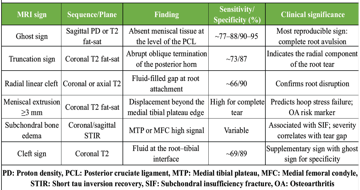
Table 1: Key MRI diagnostic signs in posterior medial meniscal root tear
Additional imaging is required for surgery. Posteroanterior radiographs, taken in 45° flexion (Rosenberg view), are used to measure the joint space width and the Kellgren–Lawrence (KL) grade. Weight-bearing hip–knee–ankle (HKA) alignment radiographs are taken to measure mechanical axis deviation and direct how much correction is needed for osteotomy.
Surgical techniques
The current gold standard is TPO repair, in which the suture is passed through a posteromedial portal in the torn root and then pulled through a transtibial tunnel that is created from the anteromedial tibia to the anatomic root footprint. A cortical button or tie-over post is used to secure sutures. Two-suture and modified Mason–Allen sutures have been shown to offer better load-to-failure properties than single simple sutures [22,23]. Tunnel placement is highly critical, and placement deviations of only 5 mm from the natural footprint make the repair biomechanically similar to an unrepaired root [4].
Suture anchor repair uses knotless adjustable suture anchors to attach to the posterior root attachment through the posterior working portal. A biomechanical cadaveric study was performed in 2025, which showed that suture anchor repair had significantly lower absolute and relative meniscal extrusion after 500 and 1000 loading cycles compared to transtibial fixation (P = 0.012 and P = 0.001, respectively) and lower mean medial compartment contact pressures after 1000 cycles (P = 0.028). The benefits are said to be due to the direct anchorage at the root footprint and decreased cyclic suture-tunnel laxity [24].
All inside repair (e.g., a FasT-Fix suture passer) does not involve bone tunneling but offers less primary fixation strength: 87% survival at 5 years in 43 patients with the modified Mason–Allen configuration [25].
The extruded meniscal body is reattached to the periphery of the tibial plateau with all-inside suture anchors placed posteromedially, known as the meniscal centralization suture technique [4]. A 2025 RCT found that extrusion increased from baseline in 96.2% of all cases (centralized and non-centralized), despite the over 50% reduction in the increase of extrusion at 6 months [26].
Concomitant HTO is performed as a single-stage procedure: Arthroscopic root repair first, followed by medial opening-wedge osteotomy aimed to achieve a 62–65% weight-bearing line of the tibial plateau, which is the maximum amount of offloading achievable for the medial compartment [12].
Why malalignment matters
Perfect root repair is not enough to correct the normal mechanics of the medial compartment if varus malalignment is present. A biomechanical analysis using finite elements in varus knee alignment showed that repair achieved higher peak contact pressure than varus knee alignment, suggesting that the repaired construct is still mechanically overloaded despite anatomic reconstruction. Varus is also associated with greater forces on the repair construct peripherally, which explains the higher failure rates in the clinic [7,27,28].
The presence of persistent post-operative meniscal extrusion is independently associated with clinical failure and TKA conversion. Although 98% of patients healed in the roots, Krych et al. reported a mean increase in extrusion from 1.9 to 2.6 mm at 6-month MRI following transtibial repair [4], and the 2025 RCT showed extrusion increased in 96.2% of patients at 6 months [26]. Chung et al. found that a varus alignment at the time of surgery and an extrusion at the time of surgery >0.7 mm were the greatest independent risk factors for clinical failure and eventual conversion to TKA at long-term follow-up [29].
Evidence for isolated repair versus repair + HTO
The results strongly support combined HTO-MRR over isolated repair in varus-aligned knees. Table 2 summarizes some of the key studies on the subject.
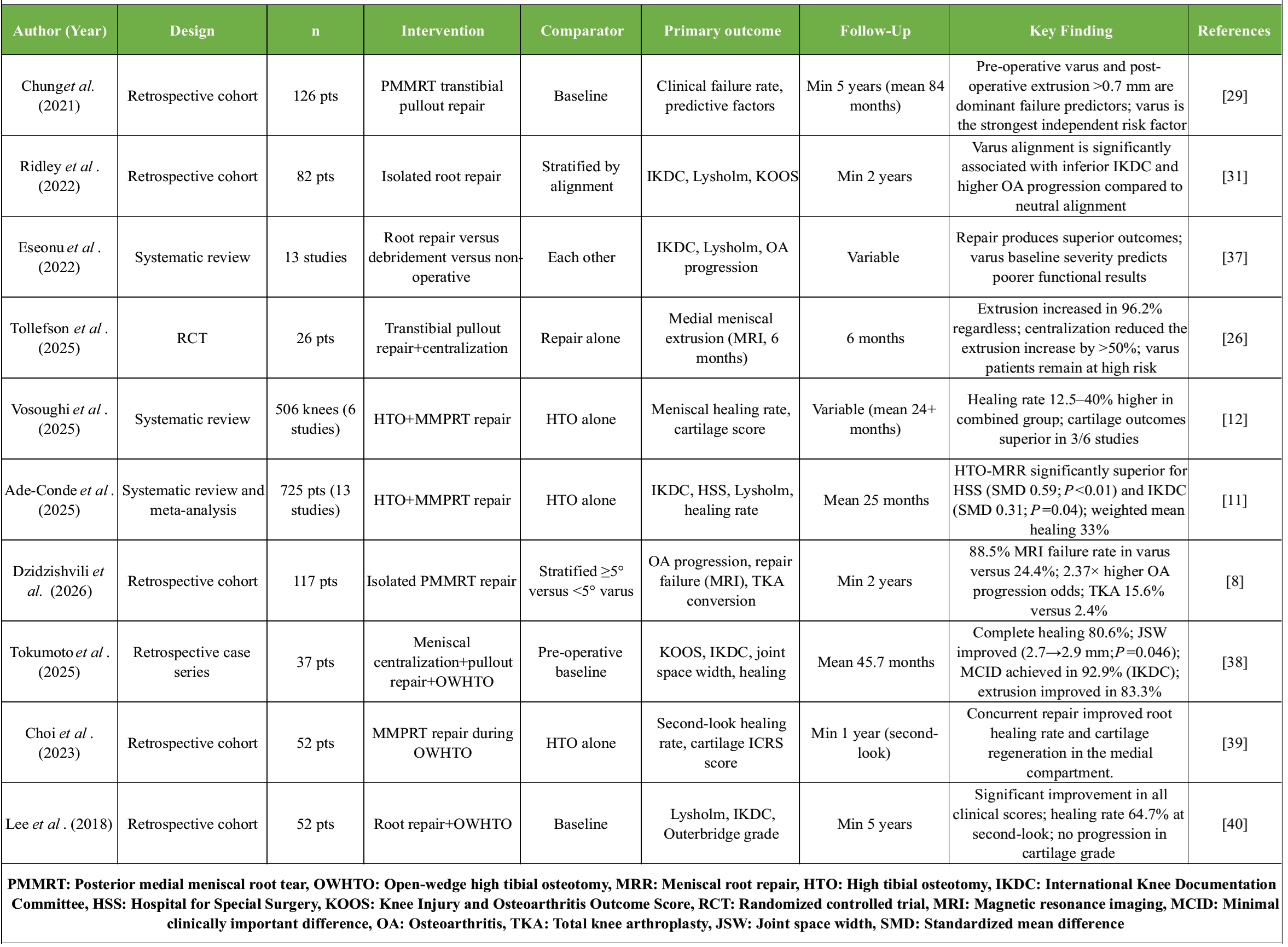
Table 2: Summary of pivotal studies on posterior medial meniscal root tear repair and/or high tibial osteotomy
The rates of OA progression were 32.5% and 53.3% in normally aligned and varus-aligned knees, respectively (P = 0.02). The MRI confirmed repair failure rate was 24.4% in normally aligned knees and 88.5% in varus-aligned knees (P < 0.001). Multivariable logistic regression ranged from a 1.13 to a 4.99 odds ratio for progression of OA with varus alignment (2.37, 95% confidence interval [CI] 1.13–4.99). Conversion to TKA was 2.4% (neutral) versus 15.6% (varus), P = 0.005 [8].
The 2025 systematic review of 506 knees (six comparative studies; HTO vs. HTO + MRR) showed that the healing rate of the whole meniscus was 12.5–40% higher in the combined group than in the HTO group alone (0–15% healing) [12]. In three out of six studies, cartilage outcomes were better. The 2025 meta-analysis of 725 patients (13 studies, 67% female, mean age 54 years, mean follow-up 25 months) confirmed statistically significant superiority of HTO-MRR over HTO alone for both Hospital for Special Surgery score (standardized mean difference [SMD] 0.59; 95% CI 0.34–0.84; P < 0.01) and International Knee Documentation Committee score (SMD 0.31; 95% CI 0.01–0.61; P = 0.04). Studies reported 13–93% healing of the meniscus following the HTO-MRR (weighted mean 33%) [11].
When root repair is not performed at the same time as HTO, studies have reported that untreated PMMRTs heal spontaneously in 40–50% of cases at second-look arthroscopy, which is thought to occur due to reduced contact pressure increasing vascularity and the biological environment [9,10]. Objective outcomes such as healing rate and cartilage score, however, have always been favorable for the combined treatment; moreover, younger and active patients show remarkably good functional outcomes without any surgical cartilage repair procedure (2025 meta-analysis). The key difference in mechanism is that anatomic hoop stress capacity is not restored after a root tear, even if it is spontaneously healed with fibrous tissue, which is the reason that HTO has a relative offloading effect only [11].
Activity demand, age, BMI, KL grade and mechanical axis deviation were chosen as the indicators for HTO. The indicators chosen for HTO were mechanical axis deviation, KL grade, BMI, age, and activity demand [30,31]. A comparative summary of the outcomes is presented in Table 3.
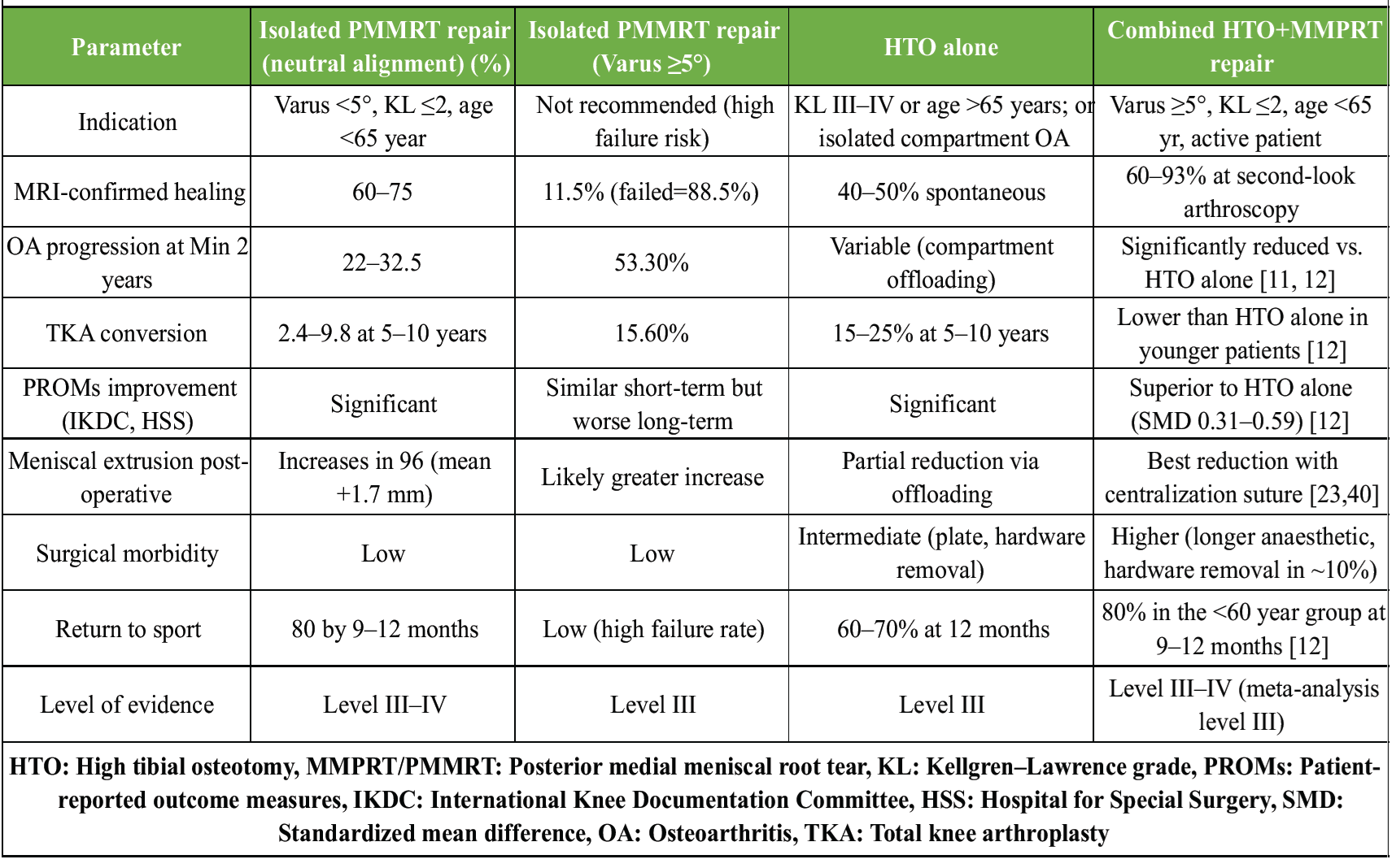
Table 3: Comparative summary: Isolated repair versus HTO alone versus combined HTO+meniscal root repair
Criteria for concomitant HTO with PMMRT repair
Mechanical axis deviation: ≥5° corrected varus on full-length HKA radiographs is most accepted as the threshold. The medial compartment carries an estimated 5% higher load with the increase of varus deviation, and below 5°, the technical reproducibility of correction is limited, and there is no evidence for benefit. The mechanical axis correction is not exactly toward neutral, but rather slightly toward valgus (weight-bearing line at 62–65% of the tibial plateau width) [7]. The decision threshold for concomitant HTO with PMMRT repair is given in Table 4.
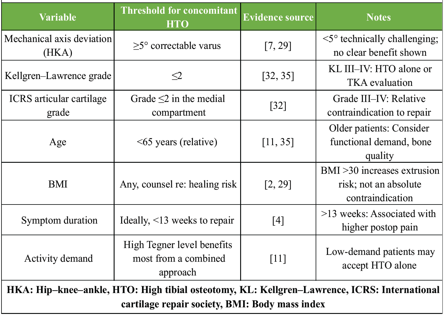
Table 4: Key surgical decision thresholds for concomitant HTO in PMMRT management
KL grading: Repair is recommended for KL grade ≤2 (mild or no radiographic OA). For Grade III and IV, KL is a relative contraindication for repair as joint cartilage degeneration is too severe to be amenable to meniscal function restoration, and more suited to HTO or TKA. Articular cartilage changes in the Grade III–IV range in the MEDIAL compartment are another relative contraindication [32].
In younger patients (<65 years), the combined approach is best indicated. An elevated BMI (>30 kg/m2) will also decrease the rate of healing, but also increase medial loading, and therefore, patients should be counseled. BMI itself does not preclude the combination procedure [2].
Benefit according to activity demand and timing: Young, active patients with a high Tegner activity score benefit most [11]. Repair within 13 weeks of symptom onset is associated with better outcomes, and longer delays have been associated with greater progression of extrusion and pain after repair [4].
Decision algorithm, return to sport timing, long-term OA progression data and future directions
The algorithm for making decisions is presented in the form of a practical evidence-based algorithm in Fig. 1. Briefly: (1) Confirm PMMRT by MRI (ghost sign, truncation sign, extrusion ≥3 mm); (2) Obtain full-length HKA alignment radiograph and Rosenberg-view radiograph; (3) If varus <5° and KL ≤2, isolated arthroscopic pullout repair, ± centralization suture; (4) If varus ≥5° and KL ≤2 and age <65 years, single-stage HTO + arthroscopic root repair, ± centralization suture; (5) If KL III–IV, defer to HTO alone (non-biological compartmental offloading) or TKA assessment [32,33].
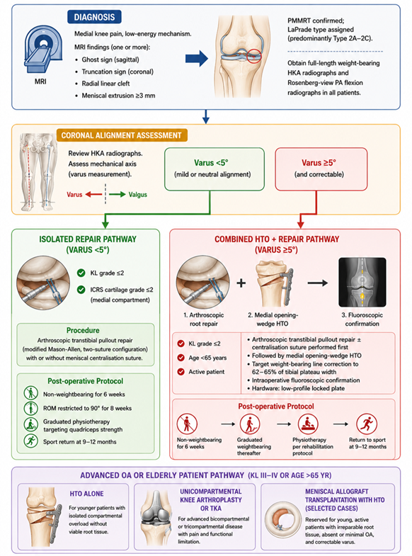
Figure 1: Clinical decision algorithm for arthroscopic repair of posterior medial meniscal root tear with or without concomitant high tibial osteotomy.
Return to sport and rehabilitation: 6 weeks of strict non-weight bearing, progressive weight bearing at 6 weeks, with 8 weeks of 90° restriction, progressive physiotherapy at 12 weeks, and sport-specific training beginning at 6 months after combined HTO-MRR [25,32]. In series using the combined approach, return to recreational sport is reported in 80% of patients younger than 60 years of age [11]. In 2023, a study was conducted that further supported this direct relationship between post-operative quadriceps strength and reduced meniscal extrusion progression, and suggested physiotherapy intensity as one of the modifiable determinants for this outcome [29].
Long-term OA progression data: The landmark study by Chung et al. into long-term outcomes of the same cohort, at a minimum 10-year follow-up, demonstrated that the biggest independent failure predictors overall were varus alignment and post-operative extrusion, with 5-year Kaplan–Meier survival of 92% after TPO repair in neutrally aligned knees [29,34]. On the other hand, meniscectomy for root tears has a conversion rate to TKA >70% at 10 years, suggesting that repair is always the best option in the right candidates [5].
Advances: The next step in the future is the combination of meniscal allograft transplantation (MAT) with HTO for patients younger than those who have irreparably damaged root tissue [7]. There is no level 1 evidence for the use of scaffold-augmented repair with platelet-rich plasma injection. There is a need for registry-based prospective studies and platform RCTs to compare isolated repair, centralization, HTO-MRR and MAT + HTO [11].
The article collates the converging evidence that isolated repair of PMMRT in varus-aligned knees of ≥5° is associated with unacceptably high failure rates and is no longer a standard of care technique in this subgroup. The 2026 American Journal of Sports Medicine cohort with MRI confirmed failure at 88.5% and odds of OA progression 2.37 times higher in varus knees, supports the lack of anatomic accuracy as a mechanical factor, but rather varus as an actual threat to repair integrity [8] (in line with the findings of the finite element modeling, which indicated that varus increased contact pressure at the construct regardless of anatomic accuracy) [7,31].
Compared to previous reviews: Previous reviews (up to 2022) tended to recommend repair for all PMMRTs with KL ≤2, irrespective of alignment. The most recent syntheses, the 2025 systematic review and meta-analysis, showed that HTO-MRR is the objectively best treatment for meniscal healing and PROMs over HTO alone, and that HTO-MRR is the objectively best treatment compared to other interventions and HTO alone in varus-aligned patients [11,12].
There is still conflict on three issues. In the first place, the issue of whether spontaneous root healing after HTO alone is clinically sufficient has not been standardized: Although the healing of untreated roots after HTO has been documented in 40–50% of cases, the nature of healing (fibrous vs. bony) and the biomechanical adequacy are not. PROMs data are conflicting at short-to-mid-term follow-up [9,10]. Second, it is unclear whether centralization sutures are an effective alternative to or an adjunct to HTO in the treatment of moderate varus (3–4°): The 2025 RCT showed the extrusion increase was significantly reduced with centralization sutures compared to the isolated repair of the distal femur, but there was still some post-operative extrusion in almost all patients [26,35]. Third, the ideal valgus overcorrection target continues to be controversial, with suggestions of the medial tibial spine being the goal of a proposed weight-bearing line of 62–65% of tibial plateau width [7].
The amount of literature that is limited is significant. No published RCTs are comparing isolated PMMRT repair with a three-arm design and long-term follow-up with HTO alone and HTO-MRR. Most comparative studies are retrospective, non-randomized, have heterogeneous osteotomy correction, variable suture configurations and inconsistent use of centralization sutures. Follow-up periods are typically <3–4 years, and cohorts of 10 + years are needed for the hard endpoint of conversion to TKA. There is no standardization on MRI-based healing grading, which hinders quantitative synthesis across studies. In multivariate analyses, the accompanying subchondral insufficiency fracture, quality of cartilage, BMI, and timing of surgery are rarely taken into account [35,36,37].
The growing agreement is to obtain HKA films in all patients with a confirmed PMMRT, to treat varus deformity ≥5° concurrently with HTO as part of the surgical plan, to aim for cartilage preservation ages and KL grades, and for standardization of post-operative management, which emphasizes 6-week non-weight-bearing and intensive physiotherapy. Surgeons working in resource-poor areas should be mindful that both techniques require specialized instruments, that both have longer lengths of surgery and that hardware complications (tibial plate prominence, hardware removal) account for 10–15% of secondary procedures in HTO series [25,30,38].
PMMRT tears represent a pivotal challenge in knee preservation, particularly when compounded by varus malalignment. Current evidence underscores the importance of addressing both biomechanical instability and meniscal integrity to optimize outcomes. While isolated repair may suffice in well-aligned knees, the presence of varus deformity necessitates a more comprehensive strategy that integrates alignment correction with root restoration. For younger individuals with early OA, this paradigm offers the most promising avenue to delay disease progression and reduce the likelihood of arthroplasty. Future prospective studies will be essential to validate long-term benefits and refine patient selection criteria, ensuring durable outcomes in this complex clinical scenario.
correct
1. Isolated arthroscopic repair of posterior medial meniscal root tears in varus-aligned knees (≥5°) has unacceptably high failure rates (up to 88.5%) and accelerates osteoarthritis progression.
2. Concomitant high tibial osteotomy (HTO) with root repair significantly improves meniscal healing, cartilage preservation, and patient-reported outcomes compared to HTO alone or isolated repair.
3. The best candidates for combined HTO-root repair are patients under 65 years with Kellgren–Lawrence grade ≤2 and correctable varus ≥5°, making this the most effective joint-preserving strategy.
References
- 1. LaPrade CM, James EW, Cram TR, Feagin JA, Engebretsen L, LaPrade RF. Meniscal root tears: A classification system based on tear morphology. Am J Sports Med 2015;43:363-9. [Google Scholar] [PubMed]
- 2. Guimaraes JB, Manatrakul R, Joseph GB, Feeley B, Lansdown DA, Chen JV, et al. Degenerative medial and lateral menisci root tears: Demographics, clinical presentation, imaging features, and associated findings. Skeletal Radiol 2025;54:255-66. [Google Scholar] [PubMed]
- 3. Allaire R, Muriuki M, Gilbertson L, Harner CD. Biomechanical consequences of a tear of the posterior root of the medial meniscus. Similar to total meniscectomy. J Bone Joint Surg Am 2008;90:1922-31. [Google Scholar] [PubMed]
- 4. Ehrke TR, Keel TJ, Guerin GE, Wilebski BJ, Tollefson LV, LaPrade RF. Meniscus extrusion after a medial meniscus root repair: Where we are and how can we solve it? Sci Prog 2026;109:368504261426669. [Google Scholar] [PubMed]
- 5. Lamba A, Regan C, Levy BA, Stuart MJ, Krych AJ, Hevesi M. Long-term outcomes of partial meniscectomy for degenerative medial meniscus posterior root tears. Orthop J Sports Med 2024;12:23259671241266593. [Google Scholar] [PubMed]
- 6. Mordecai SC, Al-Hadithy N, Ware HE, Gupte CM. Treatment of meniscal tears: An evidence based approach. World J Orthop 2014;5:233-41. [Google Scholar] [PubMed]
- 7. Daniel AV, Weingart AJ, Barrera LE, Carbone AD, Kannan AS, Levy BA. The role of alignment in treating meniscus pathology. Curr Rev Musculoskelet Med 2025;18:149-59. [Google Scholar] [PubMed]
- 8. Dzidzishvili L, Casanova F, López-Torres II, Moews LD, Perez Lloveras GO, Thamrongskulsiri N, et al. Medial meniscus posterior root repair in knees with ≥5° of varus alignment is associated with greater osteoarthritis progression compared with <5° varus alignment. Am J Sports Med 2026;54:1605-17. [Google Scholar] [PubMed]
- 9. Lee OS, Lee SH, Lee YS. Comparison of the radiologic, arthroscopic, and clinical outcomes between repaired versus unrepaired medial meniscus posterior horn root tear during open wedge high tibial osteotomy. J Knee Surg 2021;34:57-66. [Google Scholar] [PubMed]
- 10. Dong C, Kong L, Niu Y, Dong J. Repairing the medial meniscus posterior root during open-wedge high tibial osteotomy does not significantly affect clinical outcomes. Front Surg 2025;12:1672154. [Google Scholar] [PubMed]
- 11. Ade-Conde AM, Cruickshank M, Bouchard MD, Vivekanantha P, Meena A, Malik SS, et al. Combined high tibial osteotomy and root repair improves patient-reported outcomes in medial meniscus posterior root tears: A systematic review and meta-analysis. J ISAKOS 2026;16:101035. [Google Scholar] [PubMed]
- 12. Vosoughi F, Vahedi P, Nakhjiri MT, Keyhani S, Soleymanha M, LaPrade R, et al. High tibial osteotomy and concurrent medial meniscus root repair provides improved objective outcomes compared to high tibial osteotomy alone for knee osteoarthritis: A systematic review. Knee Surg Sports Traumatol Arthrosc 2025;33:3361-74. [Google Scholar] [PubMed]
- 13. Baethge C, Goldbeck-Wood S, Mertens S. SANRA-a scale for the quality assessment of narrative review articles. Res Integr Peer Rev 2019;4:5. [Google Scholar] [PubMed]
- 14. Johannsen AM, Civitarese DM, Padalecki JR, Goldsmith MT, Wijdicks CA, LaPrade RF. Qualitative and quantitative anatomic analysis of the posterior root attachments of the medial and lateral menisci. Am J Sports Med 2012;40:2342-7. [Google Scholar] [PubMed]
- 15. LaPrade RF, Ho CP, James E, Crespo B, LaPrade CM, Matheny LM. Diagnostic accuracy of 3.0 T magnetic resonance imaging for the detection of meniscus posterior root pathology. Knee Surg Sports Traumatol Arthrosc 2015;23:152-7. [Google Scholar] [PubMed]
- 16. Ahn JH, Lee YS, Yoo JC, Chang MJ, Park SJ, Pae YR. Results of arthroscopic all-inside repair for lateral meniscus root tear in patients undergoing concomitant anterior cruciate ligament reconstruction. Arthroscopy 2010;26:67-75. [Google Scholar] [PubMed]
- 17. Li L, Yang X, Yang L, Zhang K, Shi J, Zhu L, et al. Biomechanical analysis of the effect of medial meniscus degenerative and traumatic lesions on the knee joint. Am J Transl Res 2019;11:542-56. [Google Scholar] [PubMed]
- 18. Thaunat M, Fayard JM, Guimaraes TM, Jan N, Murphy CG, Sonnery-Cottet B. Classification and surgical repair of ramp lesions of the medial meniscus. Arthrosc Tech 2016;5:e871-5. [Google Scholar] [PubMed]
- 19. Gannon NP, Wise KL, Macalena JA. Arthroscopic transosseous repair of a medial meniscal posterior-root tear. JBJS Essent Surg Tech 2021;11:e20.00031. [Google Scholar] [PubMed]
- 20. Bi AS. Editorial commentary: Thickened meniscus, extrusion, truncation, ghost sign, cleft sign, and bony edema indicate medial meniscus root tear on magnetic resonance imaging. Arthroscopy 2025;41:3369-70. [Google Scholar] [PubMed]
- 21. Lakkireddy M, Barman M, Maley DK, Eppakayala S, Satapathy D, Yalamanchili RK, et al. A comparative analysis of the standing weight-bearing anteroposterior view versus the rosenberg view radiographs and their correlation with oxford knee scores in grading osteoarthritis of the knee joint. Cureus 2025;17:e92105. [Google Scholar] [PubMed]
- 22. LaPrade CM, Foad A, Smith SD, Turnbull TL, Dornan GJ, Engebretsen L, et al. Biomechanical consequences of a nonanatomic posterior medial meniscal root repair. Am J Sports Med 2015;43:912-20. [Google Scholar] [PubMed]
- 23. Ro KH, Kim JH, Heo JW, Lee DH. Clinical and radiological outcomes of meniscal repair versus partial meniscectomy for medial meniscus root tears: A systematic review and meta-analysis. Orthop J Sports Med 2020;8:2325967120962078. [Google Scholar] [PubMed]
- 24. Cinque ME, Hinz M, Sidrak J, Hollenbeck JF, Buchalter WH, Kanakamedala A, et al. Biomechanical comparison of transtibial pull-out fixation versus suture anchor fixation for repair of medial meniscus posterior root tears. Am J Sports Med 2025;53:2128-35. [Google Scholar] [PubMed]
- 25. Okazaki Y, Sugiu K, Kamatsuki Y, Tamura M, Kawada K, Hasegawa T, et al. Clinical outcomes following medial meniscus posterior root repairs: A minimum of 5-year follow-up study. J Exp Orthop 2025;12:e70262. [Google Scholar] [PubMed]
- 26. Tollefson LV, Kajabi AW, Hedayati E, Knutsen K, Takahashi T, Ellermann J, et al. Medial meniscal extrusion increased on 6-month magnetic resonance imaging, despite successful posterior root repair with or without a centralization suture: A randomized controlled clinical trial. Am J Sports Med 2025;53:2808-16. [Google Scholar] [PubMed]
- 27. Berk AN, Cregar WM, Wang S, Habet NA, Ifarraguerri AM, Trofa DP, et al. The effect of lower limb alignment on tibiofemoral joint contact biomechanics after medial meniscus posterior root repair: A finite-element analysis. J Am Acad Orthop Surg 2024;32:e558-67. [Google Scholar] [PubMed]
- 28. Deichsel A, Peez C, Raschke MJ, Richards RG, Gueorguiev B, Zderic I, et al. Arthroscopic centralization of the medial meniscus reduces load on a posterior root repair under dynamic varus loading: A biomechanical investigation. Am J Sports Med 2024;52:3030-8. [Google Scholar] [PubMed]
- 29. Chung KS, Ha JK, Ra HJ, Kim JG. Preoperative varus alignment and postoperative meniscus extrusion are the main long-term predictive factors of clinical failure of meniscal root repair. Knee Surg Sports Traumatol Arthrosc 2021;29:4122-30. [Google Scholar] [PubMed]
- 30. Lee DW, Lee SH, Kim JG. Outcomes of medial meniscal posterior root repair during proximal tibial osteotomy: Is root repair beneficial? Arthroscopy 2020;36:2466-75. [Google Scholar] [PubMed]
- 31. Ridley TJ, Ruzbarsky JJ, Dornan GJ, Woolson TE, Poulton RT, LaPrade RF, et al. Minimum 2-year clinical outcomes of medial meniscus root tears in relation to coronal alignment. Am J Sports Med 2022;50:1254-60. [Google Scholar] [PubMed]
- 32. Feucht MJ, Izadpanah K, Lacheta L, Südkamp NP, Imhoff AB, Forkel P. Arthroscopic transtibial pullout repair for posterior meniscus root tears. Oper Orthopadie Traumatol 2019;31:248-60. [Google Scholar] [PubMed]
- 33. Suruga M, Iriuchishima T, Kozu T, Yahagi Y, Lee H, Nakanishi K. Magnetic resonance imaging measurements of maximum thickness of medial meniscus exceeding 10 mm are strongly associated with medial meniscus posterior root tear. Arthroscopy 2025;41:3360-8. [Google Scholar] [PubMed]
- 34. Chung KS, Noh JM, Ha JK, Ra HJ, Park SB, Kim HK, et al. Survivorship analysis and clinical outcomes of transtibial pullout repair for medial meniscus posterior root tears: A 5- to 10-year follow-up study. Arthroscopy 2018;34:530-5. [Google Scholar] [PubMed]
- 35. Perry AK, Lavoie-Gagne O, Knapik DM, Maheshwer B, Hodakowski A, Gursoy S, et al. Examining the efficacy of medial meniscus posterior root repair: A meta-analysis and systematic review of biomechanical and clinical outcomes. Am J Sports Med 2023;51:1914-26. [Google Scholar] [PubMed]
- 36. Krych AJ, Song BM, Nauert RF 3rd, Cook CS, Levy BA, Camp CL, et al. Prospective consecutive clinical outcomes after transtibial root repair for posterior meniscal root tears: A multicenter study. Orthop J Sports Med 2022;10:23259671221079794. [Google Scholar] [PubMed]
- 37. Eseonu KC, Neale J, Lyons A, Kluzek S. Are outcomes of acute meniscus root tear repair better than debridement or nonoperative management? A systematic review. Am J Sports Med 2022;50:3130-9. [Google Scholar] [PubMed]
- 38. Tokumoto Y, Nakagawa Y, Nakamura T, Ozeki N, Hoshino T, Sekiya I, et al. Combined medial meniscus repair, centralization, and high tibial osteotomy are associated with improved clinical, radiological, and arthroscopic outcomes in patients with posterior root tears and varus alignment. Arthroscopy 2025;41:5217-26. [Google Scholar] [PubMed]
- 39. Choi YS, Chang MJ, Lee JH, Lee JH, D’Lima DD, Kim TW, et al. Repair of medial meniscus posterior root tear is effective for root healing and cartilage regeneration in opening wedge high tibial osteotomy. Knee Surg Sports Traumatol Arthrosc 2023;31:5799-811. [Google Scholar] [PubMed]
- 40. Lee HI, Park D, Cho J. Clinical and radiological results with second-look arthroscopic findings after open wedge high tibial osteotomy without arthroscopic procedures for medial meniscal root tears. Knee Surg Relat Res 2018;30:34-41. [Google Scholar] [PubMed]






Related Articles in Journal of Orthopaedic Case Reports
 July 1, 2026 All-inside Circumferential Fiber Augmentation of Medial Meniscus Combined with Medial Open-wedge High Tibial Osteotomy
 January 1, 2025 Tibial Nerve injury in Medial Open Wedge-High Tibial Osteotomy – A Rare Complication with 14-Year Follow-Up
 September 1, 2026 Diagnostic Accuracy of Magnetic Resonance Imaging Signs for Detecting Meniscal Root Tears: A Prospective Comparison with Arthroscopic Findings
 August 1, 2026 The Effect of Intraarticular Platelet-Rich Plasma Injection in Knee Osteoarthritis