


Introduction
Developmental malformation instead of a veracious neoplasm is a more apt entity to define osteochondroma and its oddity nature in the scapula. It is a benign growth within the periosteum covered with hyaline cartilage that usually has the tendency to grow away from the epiphyseal plate. Accounting for 35–46% prevalence of all benign tumor family, osteochondromas constitute a major tumor burden to the society. Scapular osteochondroma tends to develop usually in the second decade of life (10–20 years), with men are more likely than women to develop osteochondroma before the age of 30 [1,2]. Metaphyseal region of long bones, especially the tibia, the humerus, and the distal femur combinedly share the region of involvement, with 90% of all exostoses have an outgrowth that is radiologically appreciable [2,3]. Only 3–4.6% of all cases of osteochondroma occur in flat bones like the scapula, and 14.4% of scapular tumors are identified as osteochondroma [4,5]. Although various cases have been reported talking for the location of the mass on the ventral surface of the scapula, the rate of incidence for the dorsal surface location is still unanswered [6,7].
Case Report
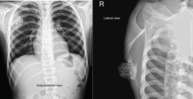
A 8-year male patient presented to the outpatient department of the tertiary care center with the chief complaints of an abnormal painless hard swelling over the right side of the upper back since last 5 years, which was gradually progressive in nature. It was associated with discomfort in lying supine position and inability to take turn toward right side. On examination, swelling measured approximately 4 × 3 cm in size. Having bony hard consistency on palpation of the dorsal aspect of the medial border of the scapula, it was non-tender and fixed to the underlying scapula with normal pinchable overlying skin without any neurovascular deficit of the upper limb. There was no other abnormal swelling elicited, and right shoulder range of motion was within normal limits. On further ruling out other differentials, there was no history of any fever, trauma, weight loss or similar past episodes. Roentgenographic examination revealed bony outgrowth from the dorsomedial aspect of the right scapula with both cortical and cancellous components, which were corresponding to the components of the parent bone, that is, the scapula (Fig. 1).
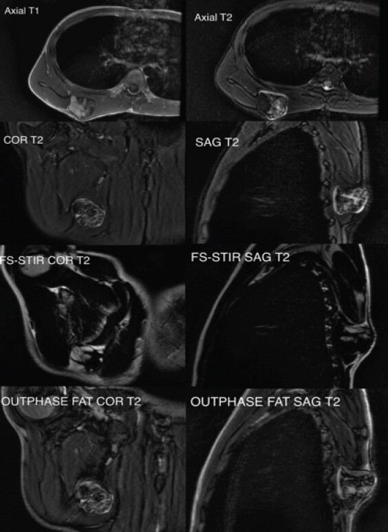
Radiographs of the both forearms and knee joints were performed to rule out other exostotic bony lesions and bowing of ulna commonly found in osteochondroma and a rare entity known as Trevor’s disease (dysplasia epiphysealis hemimelica) associated with osteochondroma mainly in the epiphysis of the knee joint. Computed tomography scan revealed a pedunculated stalked mushroom-like mass arising from the dorsal surface at the medial border of the scapula. Magnetic resonance imaging (MRI) sequences delineated visible cartilaginous cap blended with surrounding fatty marrow and edema (Fig. 2). To clearly delineate the cartilaginous cap measurement and to differentiate from edematous medullary fat, fat-suppressed short tau inversion recovery images obtained, which showed high signal intensity (cartilage) against the dark background (macroscopic medullary fat) and the thickness of the cartilaginous cap to be 14 mm. Out-of-phase (Opposed phase-FS) fat-suppressed images also obtained to differentiate between benign fatty marrow infiltration (microscopic fat with signal drop out) from near total tumor replacement (hyper-intense signal), which does not delineated signal drop out.
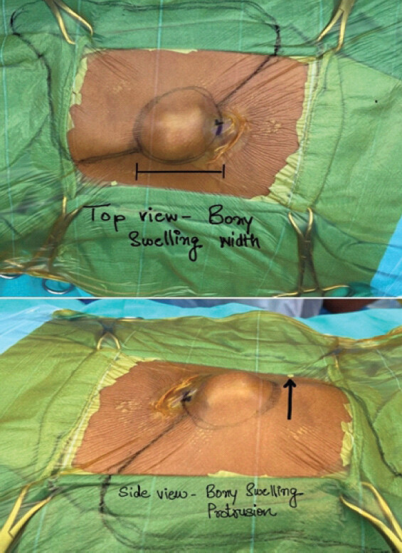
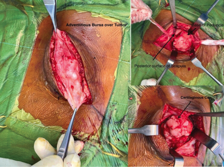
Neither any pathological fracture nor any chest wall abnormality elicited. Blood investigations were within normal limits. Based on the above findings, a provisional diagnosis of osteochondroma of the right dorsal scapula was made, and the patient was planned for excisional biopsy. In en block excision of the tumor, an incision given directly over the globular swelling in the prone position (Fig. 3), and after retracting the overlying soft tissue and bursa, the whole of the tumor became exposed.
Peduncle of the tumor identified and was resected without any post-resection void in the scapular body; thus, there was not any need of fixation (Fig. 4).
Excised specimen was sent for histopathological examination, which confirmed the diagnosis as osteochondroma. On gross examination, a globular bony tissue measured 3.8 × 3.2 × 4 cm was obtained, with the cut surface showed brownish white color (Fig. 5).
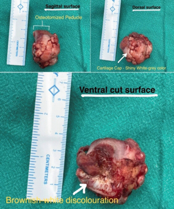
Cartilaginous cap measured 14 mm in thickness. Histopathological examination showed a bone tumor with three distinct layers comprising of fibrous perichondrium, cartilaginous cap, and bony trabeculae, with their junction showed endochondral ossification (Fig. 6).
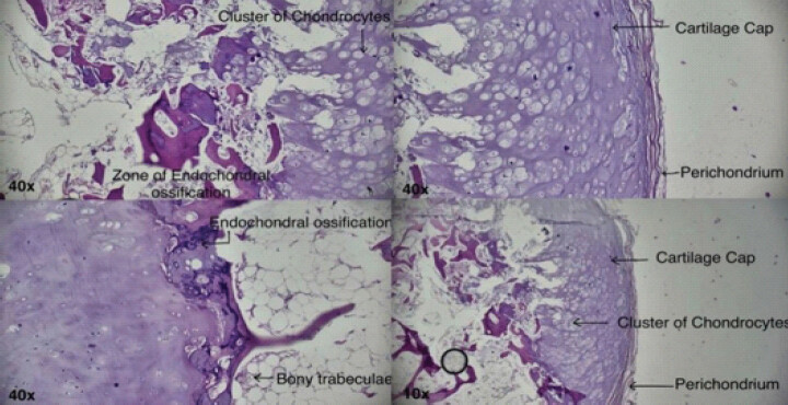
There were no signs of malignant transformation. Post-operative period was uneventful and shoulder range of motion was within normal functional limits. After 1 month, 3 months, 6 months and 1 year of sequential follow-up, there was no evidence of recurrence (Fig. 7).
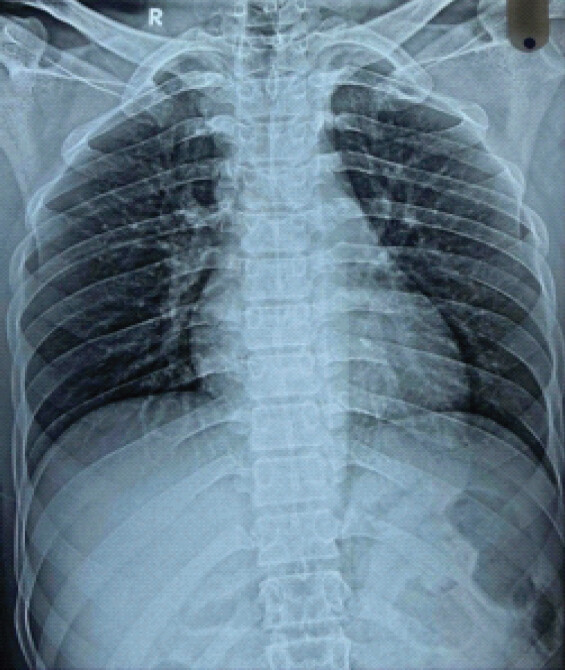
Discussion
It is quite infrequent for the scapula to have an osteochondromatous growth arising from it. With a prevalence of 14.4% of all benign tumors of the scapula, osteochondroma is considered to be a very rare tumor of flat bones [5]. Although predominantly located on the ventral surface of the omoplate (63%) [8], the dorsal scapular osteochondromas droves the patient to seek medical treatment much earlier, as presented in our case scenario due to inability of the patient to sleep in the supine position amid swelling at the upper back region and cosmetic reasons, which is the reason of interest nowadays. On the contrary, the overlying anatomical tissues are immediately compressed by ventral scapular osteochondromas, which directly causes symptoms, or indirectly by reactive bursitis, which causes snapping scapula syndrome, pseudo-winging of scapula and limits range of motion [8] with which the patient usually presents late. Likewise, lateral scapular location may result in subacromial impingement syndrome [8]. Beauchamp-Chalifour and Pelet in 2018 demonstrated a patient with trapezius weakness due to impingement of spinal accessory and suprascapular nerves amid the mass effect of dorsal superomedial angle of scapula osteochondroma [9]. Therefore, it is of immense importance to have a proper history from the patient about the symptoms to know about the correct region of involvement and the underlined pathology behind the manifestation. Location of the lesion on the scapula can aid for the differential diagnosis. The scapula was classified into two zones by the musculoskeletal tumor society: The S1 zone consists of “blade-spine” part of the scapula, and the S2 zone consists of the “glenoid-acromial complex.” Although severe benign tumors like aneurysmal bone cysts and giant cell tumors are more likely to reside in the S2 zone, osteochondromas are frequently seen in the S1 zone, as presented in our case, which is more prone to malignancies [9] as represented in the diagram (Fig. 8).
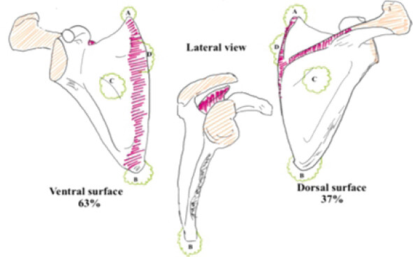
According to their morphology, osteochondromas can range in size from 1 cm to 20 cm and have cartilage cap that are typically <2 cm thick [8]. A cartilage cap thicker than 2 cm typically signifies the development of malignancy [10]. Malignant transformation of a scapular osteochondroma is an unusual complication, estimated to happen in <1–2% for solitary, sporadic lesions. Risk of malignancy increases to about 10% in patients of multiple hereditary exostosis [11]. Although rare, for osteochondroma to occur, the scapula can be aptly labeled as “location of tension” because the ilium, the scapula, and the pubic rami are the most common sites associated with malignant changes, which we have tried to delineate by the diagrammatical representation in Fig. 8 [12]. The cartilage has the appearance of a chaotic growth plate that is undergoing endochondral ossification till the time of physeal closure, continued growth in adulthood should strongly raise the suspicion of malignant transformation into chondrosarcoma [5,9]. As such, there are no differentiating features for dorsal and ventral osteochondromas as far as roentgenography is concerned, except for diagnosing malignant transformation, where MRI plays a pivotal role. “Nora lesion” can mimic an osteochondroma; however, it is quite a rare entity and can be differentiated on the basis of MRI, which characteristically shows only the outer cortical contact with the parent bone and no continuation of the medullary canal, unlike osteochondroma [13]. The surgical approach and the challenges associated with the location of the tumor mass are also a topic of discussion. Considering ventral surface osteochondroma, the subscapularis muscle usually breached due to impingement of the growing mass between the tumor and rib cage. Therefore, dissection of the subscapularis plays a pivotal role in tumor exposure. Anatomically, while taking the medial parascapular approach, there are three muscles which come across while exposing the ventral tumor mass, that is, the Trapezius, the rhomboid major and the latissimus dorsi. Along with the medial border of the scapula, all these muscles form what we call as the “Triangle of Auscultation.” In 2019, Prakash et al. reported a case series using the same approach mentioned above for ventral scapular osteochondromas excision [8]. There are other approaches also being reported on reviewing literature, as mentioned in Table 1.
Different approaches and muscle division undertaken to expose tumor
| References | Year | Techniques | Muscular plane |
|---|---|---|---|
| Fageir et al. [14] | 2009 | Medial border of scapula approach | Trans trapezius and detachment of rhomboid major |
| Perez et al. [15] | 2011 | Mini-Thoracoscopic approach | Trans serratus anterior, rhomboid major and trapezius |
| Kwon and Kelly et al. [16] | 2012 | Medial border of scapula approach | Trans trapezius and detachment of rhomboid major |
| Tungdim et al. [17] | 2017 | Medial border of scapula approach | Trans trapezius and rhomboid major |
| Steven’s et al. [18] | 2018 | Sparing of trapezium with incision at medial scapular border | Rhomboid major reflection from tip of scapula |
Approach for excision of dorsal scapular osteochondroma is relatively easy with less soft tissue insult as compared to that for ventral scapular osteochondroma. The idea is to respect as much anatomy as possible while going from skin toward the tumor peduncular stalk so that there occurs minimal damage to musculature and neurovascular bundle, which aid to quicker post-operative recovery and minimal complications.
On reviewing the literature, McWilliams in 1914 reported the first case of scapular osteochondroma with adventitious bursitis [28]. Since 1914, 19 cases of ventral scapular osteochondroma along with the large encapsulated bursa have been reported [29]. Similarly, less than 50 cases of snapping scapula syndrome secondary to ventral scapular osteochondroma have been documented [30]. Dorsal scapular osteochondromas are considered exceedingly rare; only 14 cases were published till date, enough to describe the oddity of the location (Table 2). Locations of the reported dorsal scapular lesions in accordance with the image in Fig. 8 were five at the inferior angle of the scapula (35.7%), three at the superomedial margin (21.4%), five at the medial-central body (35.7%), and one at the lateral border of the scapula. Only two cases out of fourteen (14.2%) lying in the unusual age group, that is, in the first decade of life, as our patient.
Reported cases of dorsal scapular osteochondroma in best of our search since 1914
| Author (year) | Age (year)/gender | Presentation | Side/location | Tumor size (cm) | Treatment | Follow-up in months |
|---|---|---|---|---|---|---|
| Yadkikar and Yadkikar (2013) [19] | 11/F | Progressive pain and difficulty in sleeping supine | Left, Dorsal centromedial scapular region | 3 × 2.5 | Excisional biopsy | 12 |
| Jadhav et al. (2016) [7] | 12/M | Problem in lying supine in bed | Right, Dorsal inferior scapular angle | 4 × 3 | En block excision | 12 |
| Nekkanti et al. (2018) [5] | 19/M | Upper back pain and swelling increased over time | Left, Dorsal medial border of body | 3 × 3 | Complete excision | – |
| Beauchamp-Chalifour and Pelet (2018) [9] | 25/M | Pain and winging of scapula | Right, Dorsal superomedial angle of scapula | 3 × 3 | Complete excision | 36 |
| Bektas and Ozmanevra (2019) [6] | 15/F | Left side upper back lump, painful left shoulder movement, difficulty in sleeping supine | Left, Dorsal superomedial scapula | 6 × 4 | Complete excision with the help of osteotomy | 12 |
| Shahid et al. (2021) [20] | 23/M | Painless scapular mass | Left, Dorsal medial aspect of body | 1.5 × 1.8 | Observe | – |
| Altwaijri et al. (2022) [21] | 2/F | Worsening painful swelling | Left, Dorsal medial aspect of body | 3 × 2.5 | Complete excision | 6 |
| Das et al. (2023) [22] | 10/M | Painless swelling, discomfort in lying supine | Right, Dorsal inferior angle of scapula | 4.8 × 4 | En-block excision | 12 |
| Chun et al. (2023) [3] | 8/M | Mid back pain, winging of scapula, scoliosis | Left, Dorsal inferior angle of scapula | 2.3 × 1.3 | Chiropractor did scapular manipulation, observe | 6 |
| Qureshi et al. (2023) [23] | 25/F | Non-specific pain at shoulder, neck, hard lump at upper back | Right, Dorsal at superomedial border | – | – | – |
| Jangir et al. (2024) [24] | 21/M | Upper back swelling with discomfort | Right, dorsal infero- medial border of scapula | 5.5 × 6 | En-block excision | 3 |
| Raja and Rao (2024) [25] | 12/F | Painful swelling of scapula | Left, Dorsal medial aspect of body | 4 × 4 | Extraperiosteal resection | 1.5 |
| Khan and Chand (2024) [26] | 12/M | Progressive painful swelling, difficulty in lying supine | Left, Dorsal lateral border of scapula | 2 × 2 | Excisional biopsy | 12 |
| Kalekar and Kumar et al. (2025) [27] | 15/F | Left shoulder pain, decrease range of motion, swelling | Left, Dorsal inferior scapular blade | 3.5 × 2 | En-block excision | – |
Conclusion
Symptoms caused by osteochondromas can vary depending on where they are located. Although osteochondroma is uncommon in the scapula, it should be remembered that it is the most typical benign tumor of the scapula. Depending on the extent of the mass, osteochondroma’s dorsal involvement may also result in clinical symptoms due to its mass effect, even though it is more common in the ventral region and causes a clinical snapping scapula.
Clinical Message
A meticulously rehearsed sequence of history taking, examination, appropriate imaging, and excisional biopsy is the prerequisite for the diagnosis of osteochondroma at a location as unusual as the dorsal scapula. En bloc excision is the choice of procedure for favorable outcome. Regular follow-up is necessary amid its notorious nature, although rare, to transformed into malignancy, especially for the scapular lesions.
Conflict of Interest:
Nil
Source of Support:
Nil
Consent:
The authors confirm that informed consent was obtained from the patient for publication of this article
References
- Oliveira MA, Alfaro Y, Kotzias Neto A, Korman MC. Subscapular osteochondroma as a differential diagnosis of winged scapula. Rev Bras Ortop (Sao Paulo) 2019;54:241–6. Google Scholar | PubMed
- Ngongang FO, Fodjeu G, Fon AC, Fonkoue L, Guifo ML, Bitang A, Mafok LJ. Surgical treatment of rare case of scapula osteochondroma in a resource limited setting: A case report. Int J Surg Case Rep 2019;61:130–4. Google Scholar | PubMed
- Chun DI, Cho JH, Choi IH, Yi Y, Kim JY, Kim JH. Osteochondroma of ventral scapula associated with chest pain due to rib cage compression: A case report. Medicine (Baltimore) 2018;97:e0510. Google Scholar | PubMed
- Mozaffarian K, Farahani MJ, Vosoughi AR. Bilateral sandwiched scapulae: A rare presentation of hereditary multiple exostoses. J Clin Orthop Trauma 2016;7 Suppl 1:5–7. Google Scholar | PubMed
- Nekkanti S, Moogali A, Meka A, Nair M. An unusual presentation of osteochondroma on the dorsal surface of the scapula: A review of two patients. J Orthop Case Rep 2018;8:38–41. Google Scholar | PubMed
- Bektas YE, Ozmanevra R. An unusual location of osteochondroma: Dorsal scapula. Cureus 2019;11:e6464. Google Scholar | PubMed
- Jadhav PU, Banshelkikar SN, Seth BA, Goregaonkar AB. Osteochondromas at unusual sites- case series with review of literature. J Orthop Case Rep 2016;6:52–4. Google Scholar | PubMed
- Prakash S, Kalra P, Khan Y, Dhal A. Ventral scapular osteochondroma excision through 'triangle of auscultation': A case series. J Orthop Surg (Hong Kong) 2020;28:2309499019892848. doi: 10.1177/2309499019892848. Google Scholar | PubMed | CrossRef
- Beauchamp-Chalifour P, Pelet S. Osteochondroma of the scapula with accessory nerve (XI) compression. Case Rep Orthop 2018;2018:7018109. Google Scholar | PubMed
- Tomo H, Ito Y, Aono M, Takaoka K. Chest wall deformity associated with osteochondroma of the scapula: A case report and review of the literature. J Shoulder Elbow Surg 2005;14:103–6. Google Scholar | PubMed
- Fukunaga S, Futani H, Yoshiya S. Endoscopically assisted resection of a scapular osteochondroma causing snapping scapula syndrome. World J Surg Oncol 2007;5:37. Google Scholar | PubMed
- Enneking W, Dunham W, Gebhardt M, Malawar M, Pritchard D. A system for the classification of skeletal resections. Chir Organi Mov 1990;75 Suppl 1:217–40. Google Scholar | PubMed
- Gruber G, Giessauf C, Leithner A, Zacherl M, Clar H, Bodo K. Bizarre parosteal osteochondromatous proliferation (Nora lesion): A report of 3 cases and a review of the literature. Can J Surg 2008;51:486–9. Google Scholar | PubMed
- Fageir MM, Edwards MR, Addison AK. The surgical management of osteochondroma on the ventral surface of the scapula. J Pediatr Orthop B 2009;18:304–7. Google Scholar | PubMed
- Pérez D, Cano JR, Caballero J, López L. Minimally-invasive resection of a scapular osteochondroma. Interact Cardiovasc Thorac Surg 2011;13:468–70. Google Scholar | PubMed
- Kwon OS, Kelly JI. Delayed presentation of osteochondroma on the ventral surface of the scapula. Int J Shoulder Surg 2012;6:61–3. Google Scholar | PubMed
- Tungdim PH, Singh II, Mukherjee S, Pertin T. Excision of solitary osteochondroma on the ventral aspect of left scapula presenting as pseudowinging in a 4-year-old boy: A rare case report. J Orthop Case Rep 2017;7:36–40. Google Scholar | PubMed
- Stevens J, Stirling P, Patton J. Trapezius-sparing approach to osteochondromas on the ventral surface of the scapula. Tech Shoulder Elb Surg 2018;19:124–8. Google Scholar | PubMed
- Yadkikar SV, Yadkikar VS. Osteochondroma on dorsal surface of the scapula in 11 years old child-a case report. Int J Med Res Amp Health Sci 2013;2:305–8. Google Scholar | PubMed
- Shahid O, Shahid M, Shaik L, Masud M, Ranjha S. Rare case of osteochondroma on the dorsal aspect of the scapula. Cureus 2021;13:e17051. Google Scholar | PubMed
- Altwaijri NA, Fakeeha J, Alshugair I. Osteochondroma of the scapula: A case report and literature review. Cureus 2022;14:e30558. Google Scholar | PubMed
- Das R, Arya S, Krishna A, Ghosh S, Mukartihal R, Keezhadath S. Osteochondroma of dorsal scapula: A case report and review of literature. J Orthop Case Rep 2023;13:104–9. Google Scholar | PubMed
- Qureshi I, Hamed HS, Nathani P, Al Sadairi FM, Bughio S. Solitary osteochondroma of scapula: A common entity at an uncommon location. J Med Res Surg 2023;4:115–7. Google Scholar | PubMed
- Jangir R, Kumar M, Berwal A, Muzahid M, Tailor RR, Dulhanani M. Dorsal scapular osteochondroma in a young male: A case report and literature review. J Orthop Case Rep 2024;14:10–5. Google Scholar | PubMed
- Raja KB, Rao KL. Osteochondroma of the scapula in a 12-year-old girl: An unusual case report. J Orthop Traumatol Rehabil 2024;16:200–3. Google Scholar | PubMed
- Khan DJ, Chand S. Osteochondroma dorsal scapula - rare site of a common tumor. J Orthop Case Rep 2024;14:95–8. Google Scholar | PubMed
- Kalekar DR, Kumar DP. Uncommon presentation of dorsal scapular osteochondroma in a young patient: A case report with literature review. Int J Med Pharm Res 2025;6:1040–3. Google Scholar | PubMed
- Moya D, Salamone H, Vaccarelli A, Grand DM, Alfano F, Gómez D. Acute exostosis bursata: A rare complication of scapular osteo-chondromas-case report and literature review. Rev Asoc Argent Orthop Traumatol 2025;90:361–8. Google Scholar | PubMed
- Ogawa K, Inokuchi W. Solitary osteochondroma of the ventral scapula associated with large bursa formation and pseudowinging of the scapula: A case report and literature review. Case Rep Orthop 2018;2018:5145642. Google Scholar | PubMed
- Clarke DO, Crichlow A, Christmas M, Vaughan K, Mullings S, Neil I. The unusual osteochondroma: A case of snapping scapula syndrome and review of the literature. Orthop Traumatol Surg Res 2017;103:1295–8. Google Scholar | PubMed
© 2026 Journal of Orthopaedic Case Reports - Published by Indian Orthopaedic Research Group
About the Authors
-

Ashish Garg
-

Prabodh Kantiwal
-

Deepak Kumar
-

Ghulam Mustafa Najar
Related Articles in Journal of Orthopaedic Case Reports
 August 1, 2026 Surgical Excision of a Rare Ventral Scapular Osteochondroma using the Prone “Chicken-Wing” Position: A Case Report
 August 1, 2026 Vanishing Osteochondromas of the Distal Femur and Proximal Humerus: A Two-Case Report
 July 1, 2026 Posterior Scapular Osteochondroma in a Pediatric Patient: An Uncommon Presentation of a Common Benign Tumor
 July 1, 2026 Posterior Femoral Neck Exostosis in a Young Adult: Open Resection and Prophylactic Dynamic Hip Screw Fixation – A Case Report