


Introduction
Clavicular fractures are relatively common and are often treated conservatively or with surgery, depending on the fracture characteristics and patient demographics. These fractures are commonly accompanied by chest trauma, but pneumothorax is a rare complication and tends to be more common in patients with rib fractures or high-energy trauma [1,2].
Subcutaneous emphysema is defined as the presence of air pockets in the subcutaneous tissue planes, and is commonly associated with pneumothorax, pneumomediastinum, or tracheobronchial injury. Occasionally, the air may dissect extensively along fascial planes to involve the neck, chest wall, back and even the scrotum, leading to extensive surgical emphysema and pneumoscrotum. This air tracking can result in respiratory distress and death due to compromise of the airway and chest wall mobility [3,4,5].
Pneumoscrotum can be defined as the presence of air inside the scrotal sac. It is often caused by air migration from the chest or abdomen through the fascial planes and inguinal canal to the scrotum, and is usually associated with pneumomediastinum and widespread subcutaneous emphysema [3,5].
We present a rare case of clavicle fracture associated with bilateral pneumothorax, extensive subcutaneous emphysema, and pneumoscrotum in the absence of rib fractures, with recurrence due to persistent air leak requiring repeated intercostal drainage and ventilatory support.
Case Report
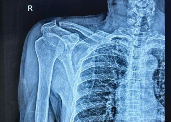
A 74-year-old male presented with a history of blunt injury to the chest and right shoulder. On initial assessment, radiological evaluation revealed a fracture of the middle third of the right clavicle along with a fracture involving the body and spine of the scapula (Fig. 1). No rib fractures were identified on initial imaging (Fig. 2).
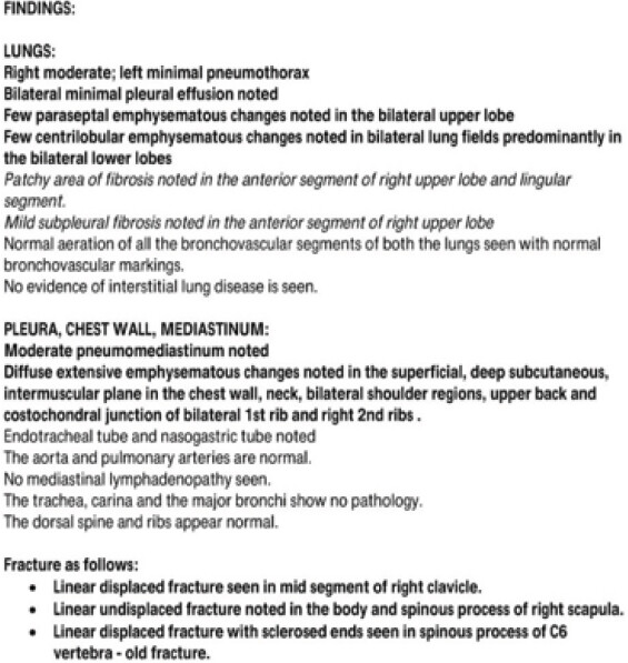
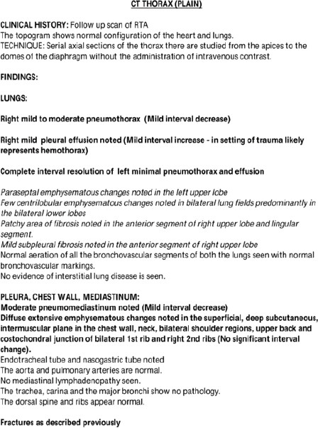
During the hospital stay, the patient developed progressive swelling over the neck and chest wall accompanied by respiratory distress and oxygen desaturation, for which he was intubated, and ventilatory support was initiated. A high-resolution computed tomography (CT) scan of the thorax was performed, which revealed bilateral pneumothorax, more on the right side, along with extensive subcutaneous emphysema involving the superficial and deep tissue planes of the chest wall, neck, bilateral shoulder region, and upper back. No rib fractures were seen. The image findings were consistent with bilateral pneumothorax and massive surgical emphysema (Fig. 2, 3, 4, 5, 6).
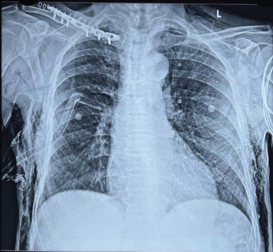
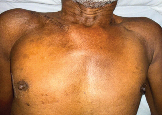
A right intercostal drain (ICD) was placed and monitored with serial chest radiograph imaging and drain output. Progressively, the patient exhibited clinical improvement, and the subcutaneous emphysema reduced.
Under general anesthesia, the patient underwent open reduction and internal fixation of the right clavicle with plate and screws. The intraoperative period was uneventful, and the patient was monitored in the intensive care unit postoperatively.
After clinical and radiological improvement, the ICD was removed. However, on the following day, the patient developed recurrent swelling over the neck and chest wall associated with respiratory distress and desaturation, and he was intubated again. A repeat CT scan of the thorax showed persistent extensive subcutaneous emphysema with a reduction in the size of the pneumothorax compared to the previous scan. In view of recurrent symptoms, a right-sided ICD was reinserted (Fig. 3 and 4).
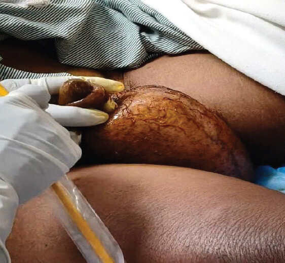
During this period, the patient also developed significant scrotal swelling, which was diagnosed as pneumoscrotum secondary to extensive subcutaneous emphysema. The swelling gradually subsided following intercostal drainage and supportive management (Fig. 5).
Management was undertaken through coordinated multidisciplinary care involving the pulmonology, cardiothoracic surgery, anesthesia, and intensive care teams. Their recommendations guided respiratory stabilization, ICD management, perioperative care, and subsequent treatment following recurrence. Bronchoscopy was considered by the pulmonology team only in the event of further clinical deterioration to evaluate for a possible occult tracheobronchial injury. As the patient’s respiratory status improved with intercostal drainage and supportive treatment, bronchoscopic evaluation was not required.
The patient was subsequently monitored with serial chest radiographs and ICD output. The ICD was removed after confirming adequate lung expansion and clinical improvement. The patient improved symptomatically, was shifted to the ward, and sutures were removed on post-operative day 15. The patient was discharged in stable condition and was followed for 1 year and showed no complications.
Discussion
Although clavicle fractures are common, the occurrence of bilateral pneumothorax and extensive subcutaneous emphysema without associated rib fractures is rare. Pneumothorax in clavicle fractures may occur due to injury to the lung apex by a displaced fracture fragment or due to underlying lung parenchymal injury following blunt trauma [2].
| Time from injury | Clinical event |
|---|---|
| Day 0 | Blunt chest trauma resulting in right midshaft clavicle fracture and scapular fracture; admitted to hospital. |
| Day 1 | Developed progressive neck and chest wall swelling with respiratory distress and oxygen desaturation, requiring endotracheal intubation and mechanical ventilation. |
| Day 1 | HRCT thorax revealed bilateral pneumothorax with extensive subcutaneous emphysema without rib fractures. |
| Day 1 | Right-sided intercostal drain (ICD) inserted. |
| Days 2–4 | Gradual clinical improvement with reduction in subcutaneous emphysema. |
| Day 5 | Open reduction and internal fixation of the right clavicle using plate and screws. |
| Day 9 | ICD removed following clinical and radiological improvement. |
| Day 10 | Recurrent subcutaneous emphysema with respiratory distress and oxygen desaturation, requiring re-intubation. |
| Day 10 | Repeat CT thorax demonstrated persistent extensive subcutaneous emphysema with a reduced pneumothorax compared with the initial scan. |
| Day 10 | Right-sided ICD reinserted for suspected persistent air leak. |
| Subsequent days | Pneumoscrotum developed and resolved with continued intercostal drainage and supportive management. |
| Before discharge | Clinical and radiological recovery achieved; ICD removed after confirmation of lung expansion. |
| 1-year follow-up | Patient remained asymptomatic with no recurrence or complications. |
HRCT: High-resolution computed tomography
The likely mechanism in this case is explained by the Macklin effect, in which blunt chest trauma leads to alveolar rupture, allowing air to track along the bronchovascular sheaths into the mediastinum, resulting in pneumomediastinum. From the mediastinum, air can dissect into the subcutaneous tissues of the neck, chest wall, and abdominal wall, leading to extensive subcutaneous emphysema. The air may further extend through the inguinal canal into the scrotum, resulting in pneumoscrotum [1,6,7,8,9,10].
Subcutaneous emphysema may be associated with respiratory distress, secondary to airway compression and chest wall rigidity. Life-threatening subcutaneous emphysema may warrant airway support and intercostal drainage, even with minimal pneumothorax [7,8].
Return of the subcutaneous emphysema after ICD removal suggests an air leak, which could be from lung parenchymal damage or apical pleural trauma caused by the clavicle fracture. In these cases, prolonged intercostal drainage and close monitoring are required before the drain can be removed [2,8].
Pneumoscrotum is a rare complication and is usually seen in association with pneumomediastinum and subcutaneous emphysema. This condition should be recognized as it is usually treated conservatively by addressing the underlying cause, and surgery is rarely needed [3,4,5].
A definite causal link between the clavicle fracture and bilateral pneumothorax could not be established. However, the temporal sequence of events, absence of associated rib fractures, and radiological findings suggested a trauma-related thoracic air leak as the most plausible explanation. The patient was managed with serial cardiothoracic surgical assessments, repeat chest imaging, and continuous ICD monitoring to evaluate and treat the suspected persistent air leak. Other potential causes of extensive subcutaneous emphysema and pneumoscrotum, such as tracheobronchial, esophageal, or intra-abdominal injuries, were considered but were considered unlikely based on the clinical course and imaging findings.
This case highlights the need to promptly recognize subcutaneous emphysema and pneumomediastinum in patients with clavicle fractures presenting with respiratory symptoms, even when rib fractures are not present. Although this presentation is exceptionally rare, the management described should not be considered universally applicable. Instead, this case highlights the importance of individualized management based on the patient’s clinical status, imaging findings, and multidisciplinary assessment.
Limitations
The exact source of the air leak could not be definitively identified; therefore, the proposed mechanism (alveolar rupture, pleural injury, or lung parenchymal tear with Macklin effect) remains speculative. The contribution of perioperative factors, including general anesthesia, positive-pressure ventilation, and surgical intervention, to the recurrence of subcutaneous emphysema could not be definitively established. Although relevant literature was reviewed, comparison with a larger number of similar published cases may have further strengthened the discussion. The extent of pneumothorax, subcutaneous emphysema, and pneumoscrotum was assessed qualitatively based on clinical and radiological findings, without standardized quantitative measurements.
Conclusion
Bilateral pneumothorax, pneumomediastinum, massive subcutaneous emphysema, and pneumoscrotum in the absence of rib fractures are rare but severe complications of clavicle fractures. If subcutaneous emphysema keeps recurring after removal of the ICD, it indicates an air leak and needs extended drainage and monitoring. Prompt diagnosis and treatment are necessary to avoid respiratory complications and ensure a good outcome.
Clinical Message
Severe thoracic complications, including bilateral pneumothorax and massive subcutaneous emphysema, can rarely be encountered in patients with clavicular fractures even without rib fractures. Recurrence of emphysema on removal of the intercostal drain suggests ongoing air leak. Observation and timing of drain removal are crucial for avoiding recurrence and complications.
Conflict of Interest:
Source of Support:
Nil
Consent:
The authors confirm that informed consent was obtained from the patient for publication of this article
References
- Wintermark M, Schnyder P.The Macklin effect: A frequent etiology for pneumomediastinum in severe blunt chest trauma. Chest 2001;120:543-7. Google Scholar | PubMed
- Geraci G, Pisello F, Sciumè C, Sunseri A, Romeo M, Li Volsi F.Clavicle fracture complicated by pneumothorax. Case report and literature review. G Chir 2007;28:330-3. Google Scholar | PubMed
- Simaioforidis V, Kontos S, Fokitis I, Lefakis G, Koritsiadis S.Subcutaneous emphysema of the scrotum (pneumoscrotum) due to traumatic pneumothorax: A case report. Cases J 2008;1:293. Google Scholar | PubMed
- Raffin E, Tundo G, Schroeck F.Pneumoscrotum with extensive penile and abdominal subcutaneous emphysema: A case report of uncertain etiology. Urol Case Rep 2017;11:50-2. Google Scholar | PubMed
- Heimbach D, Hofmockel G, Wirth M, Frohmüller H.Pneumoscrotum. Case report and review of the literature. Urologe A 1993;32:503-6. Google Scholar | PubMed
- Kumeda H, Saito G.Spontaneous pneumomediastinum diagnosed by the Macklin effect. J Surg Case Rep 2022;2022:rjab634. Google Scholar | PubMed
- Morales-Eslava BA, Suástegui-Mares JE, Chuc-Baeza GA, Sánchez-Arzate A.The Macklin effect: An underestimated cause of pneumomediastinum. Cureus 2024;16:e69328. Google Scholar | PubMed
- Alrahmani AA, Aldarsouni FG, Alwasel LS, AlGhamdi FS, Abdelhamid SW, Twier K.The macklin effect following trauma: A case report of concurrent pneumothorax, pneumoperitoneum, pneumomediastinum, and pneumoretroperitoneum in an intubated young adult. Cureus 2024;16:e74901. Google Scholar | PubMed
- Carzolio-Trujillo HA, Navarro-Tovar F, Padilla-Gómez CI, Hernández-Martínez IA, Herrera-Enríquez J.Blunt chest trauma with pneumomediastinum and pneumoperitoneum secondary to Macklin effect: Case report. Cir Cir 2016;84:409-14. Google Scholar | PubMed
- Pavrey R, Makwana N, Das N.A rare co-occurrence of spontaneous pneumomediastinum, pneumothorax, and pneumoperitoneum: Macklin effect. World J Emerg Med 2024;15:246-8. Google Scholar | PubMed
© 2026 Journal of Orthopaedic Case Reports - Published by Indian Orthopaedic Research Group
About the Authors
-

Dr. K Dharmaraj
-

Dr. Arjun Ganesh
-

Dr. Mohan Choudhary
-

Dr. Kevin Lourdes
-

Dr. Raja Sujith Kumar
Related Articles in Journal of Orthopaedic Case Reports
 June 1, 2026 Delayed Pseudoaneurysm of the Subclavian Artery Following Clavicle Fracture Fixation: A Case Report
 April 1, 2026 Non-infectious Subcutaneous Emphysema of the Upper Extremity: A Unique Mimicker of Necrotizing Fasciitis – A Case Report
 February 1, 2026 Post-Traumatic Subcutaneous Emphysema of the Upper Extremity Associated with an Open Scapular Fracture: A Case Report and Review of Literature
 December 1, 2025 Novel Lag Screw Application In The Reduction Of Neer Type V Distal Clavicle Fracture: A Case Report And Technical Note