


Introduction
Vertebral body ablation and augmentation are widely employed effective percutaneous treatments in the setting of painful vertebral body fractures related to neoplasm. Techniques for vertebral augmentation in the thoracic and lumbar spine are widely reported. However, techniques in the cervical spine, especially involving the axis vertebra (C2), are limited [1]. Previously described techniques are not only technically difficult but also involve serious complication risk [1]. Historical approaches rely on techniques that do not utilize direct imaging guidance or require an open surgical approach. We describe a novel, fully image-guided approach for C2 ablation and vertebroplasty in the setting of a painful osseous metastases and fracture using conventional fluoroscopy and ultrasound.
Case Report
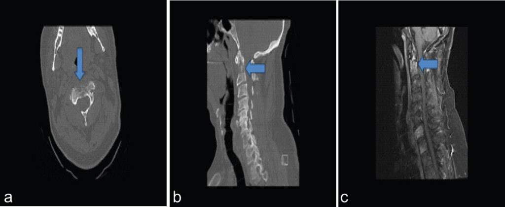
A 52-year-old male with history of renal cell carcinoma presented with acute onset intractable neck pain. Computed tomography (CT) of the cervical spine revealed a destructive lytic lesion in C2, resulting in a non-displaced type II dens fracture. Magnetic resonance imaging with gadolinium administration demonstrated a marrow replacing enhancing lesion within the body of C2 (Fig. 1). The patient’s pain was inadequately controlled despite optimal medical management. Neurosurgery and radiation oncology consultations determined that the patient was not a candidate for surgical intervention or radiation therapy, respectively.
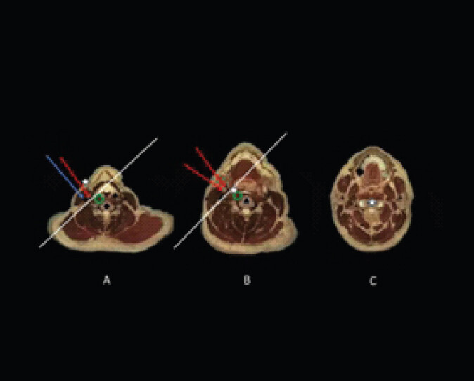
The decision was made to proceed with percutaneous C2 vertebroplasty and radiofrequency ablation of the lesion. A detailed review was conducted using pre-procedure axial imaging and anatomic references for vascular and nonvascular structures in the right neck. An anterolateral trajectory plan was generated to safely reach the C2 vertebral body under real time ultrasound and biplanar fluoroscopic imaging. This skin entry point mapped to approximately the fifth cervical (C5) vertebral body level (Fig. 2a). A plan was made to advance the Trocar medial to the sternocleidomastoid muscle and the common carotid artery (Fig. 2b). Careful consideration was made to remain below the carotid bifurcation to avoid damage to any branches of the external carotid artery. Once confirmed medial and posterior to the vascular structures, the trocar would be advanced to the deep retropharyngeal space (Fig 2c). Using biplanar fluoroscopy, the trocar was guided cranially toward the inferior endplate of C2 through the longus coli muscle toward the level of the C2-C3 disc space while maintaining gentle medial traction (Figs. 2c and Fig. 3a).
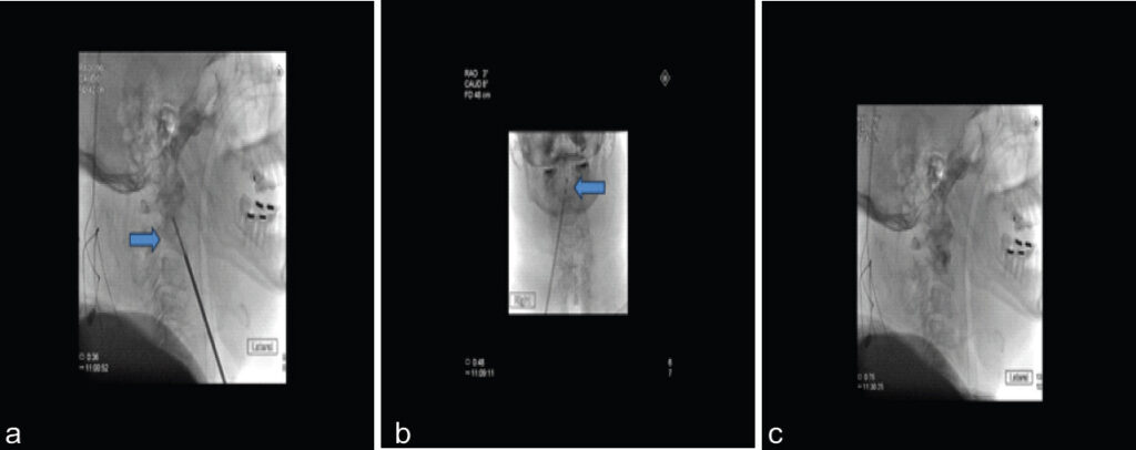
With the trocar in place, in a coaxial fashion, a complementary bone drill was advanced to manually create a tract inside the C2 vertebral body toward the base of the dens (Fig. 3a). A radiofrequency ablation probe was subsequently advanced through the trocar and confirmed as terminating at the base of the dens using fluoroscopy. Following ablation, polymethyl methacrylate was injected under intermittent fluoroscopy (Fig. 3b and 3c).
Discussion
Osteolytic lesions of C2 often present a difficult painful clinical scenario to manage, given their precarious location. Most attempts to intervene carry high risk of serious neurovascular compromise or other complications. Traditional treatments include radiotherapy and open surgery. Percutaneous vertebral augmentation has been used as an adjunctive therapy historically, especially for pain control and stabilization [2]. Vertebroplasty techniques using an open surgical anterior approach have been described as well [1,3]. Other approaches have been described which are technically difficult and associated with high-risk complications. A posterolateral approach is associated with vertebral artery injury [4], a translateral approach with carotid artery injury [5], and a transoral approach with a high risk of infection, including retropharyngeal abscess or meningitis [6]. While some reported approaches rely on specialized equipment (e.g., curved needles), our technique does not require advanced instrumentation [7].
Most of these previously reported complications can be minimized using an image-guided percutaneous anterolateral approach with a combination of ultrasound and fluoroscopic guidance. Herein, we describe a novel technique that is performed under direct real-time imaging guidance using ultrasound to direct the needle medial to the sternocleidomastoid muscle and common carotid artery. Precision is required to traverse the anterior neck and ensure a trajectory below the carotid bifurcation as many small external carotid branches may be violated with a higher approach. Following vertebral body access, the procedure is similar to radiofrequency ablation and cementoplasty at other levels. The risk of injury to vascular structures within the neck such as the carotid arteries and branches of the external carotid artery is minimal due to real-time ultrasound visualization.
The patient described in this case had an osteolytic lesion extending to the base of the dens. There is a paucity of data in the literature for percutaneous vertebroplasty at C2 with most of the data describing interventions involving the C2 vertebral body and not the dens [8]. Nevertheless, studies have shown vertebroplasty of C2 provides rapid pain relief in up to 80% of patients and spine stability in 87% of patients [1, 9]. This medial, caudal-to-cranial approach can be used for percutaneous ablation and stabilization of lesions involving the C2 vertebral body, including those extending into the dens, as in this case. A percutaneous approach using image guidance enables less blood loss, shorter anesthesia time, and lower risk for damage to neurovascular structures in the neck. For appropriately selected patients, percutaneous vertebroplasty offers a favorable benefit-risk profile compared with non-operative medical management, with consistently high reported success rates [10]. The technique described herein may be adopted by operators with experience in vertebral augmentation in non-cervical spine levels at institutions with access to ultrasound and fluoroscopy equipment.
Conclusion
This case describes a novel percutaneous image-guided approach to metastatic lesions in the dens for purposes of safe radiofrequency ablation and cement stabilization – resulting in pain relief for a patient with a paucity of other treatment options. Using an anterolateral approach that combines ultrasound and fluoroscopic guidance enables this procedure to be performed percutaneously by experienced operators with standard equipment. Interventional radiologists and spine surgeons experienced in vertebral augmentation may use this technique if presented with a patient experiencing a lesion and/or fracture of C2. Orthopedic surgeons and neurosurgeons managing patients with cervical spine metastases and/or fractures may also consider this approach as a referral option when surgical intervention is not feasible.
Clinical Message
Patients who present with intractable pain secondary to metastatic involvement and fracture of C2 may be treated percutaneously under image guidance with standard radiofrequency ablation and cementoplasty techniques using an anterolateral caudal-to-cranial specific trajectory described herein.
Conflict of Interest:
Source of Support:
Nil
Consent:
The authors confirm that informed consent was obtained from the patient for publication of this article
References
- Shetty SR, Ganigi PM, Mandanna BK.Open C2 Vertebroplasty: Case report, technique, and review of literature. Asian J Neurosurg 2017;12:710-4. Google Scholar | PubMed
- Pilitsis JG, Rengachary SS.The role of vertebroplasty in metastatic spinal disease. Neurosurg Focus 2001;11:e9. Google Scholar | PubMed
- Dorman JK.Vertebroplasty of the C2 vertebral body and dens using an anterior cervical approach: Technical case report. Neurosurgery 2010;67:E1143-6. discussion E6. Google Scholar | PubMed
- Sun HY, Lee JW, Kim KJ, Yeom JS, Kang HS.Percutaneous intervention of the C2 vertebral body using a CT-guided posterolateral approach. AJR Am J Roentgenol 2009;193:1703-5. Google Scholar | PubMed
- Guo WH, Meng MB, You X, Luo Y, Li J, Qiu M.CT-guided percutaneous vertebroplasty of the upper cervical spine via a translateral approach. Pain Physician 2012;15:E733-41. Google Scholar | PubMed
- Martin JB, Gailloud P, Dietrich PY, Luciani ME, Somon T, Sappino PA.Direct transoral approach to C2 for percutaneous vertebroplasty. Cardiovasc Intervent Radiol 2002;25:517-9. Google Scholar | PubMed
- Swarnkar A, Zain S, Christie O, Mirchia K.Vertebroplasty of C2 pathologic fracture: A unique case report using a curved-needle technique. Cureus 2022;14:e25463. Google Scholar | PubMed
- Wenger M.Vertebroplasty for metastasis. Med Oncol 2003;20:203-9. Google Scholar | PubMed
- Mont'Alverne F, Vallee JN, Cormier E, Guillevin R, Barragan H, Jean B.Percutaneous vertebroplasty for metastatic involvement of the axis. AJNR Am J Neuroradiol 2005;26:1641-5. Google Scholar | PubMed
- Barr JD, Jensen ME, Hirsch JA, McGraw JK, Barr RM, Brook AL.Position statement on percutaneous vertebral augmentation: A consensus statement developed by the Society of Interventional Radiology (SIR), American Association of Neurological Surgeons (AANS) and the Congress of Neurological Surgeons (CNS), American College of Radiology (ACR), American Society of Neuroradiology (ASNR), American Society of Spine Radiology (ASSR), Canadian Interventional Radiology Association (CIRA), and the Society of NeuroInterventional Surgery (SNIS). J Vasc Interv Radiol 2014;25:171-81. Google Scholar | PubMed
© 2026 Journal of Orthopaedic Case Reports - Published by Indian Orthopaedic Research Group
About the Authors
-

Dr. Junman Kim
-

Mr. Frank Prologo
-

Dr. Rameez Rehman
-

Dr. Maxwell E Cooper
-

Neil J Resnick
Related Articles in Journal of Orthopaedic Case Reports
 September 1, 2026 Pediatric Cervical Spine Aneurysmal Bone Cyst with Recurrent Atlantoaxial Rotatory Subluxation Requiring C1–C2 Fusion: A 7-Year Follow-Up Case Report
 August 1, 2026 O-arm Navigation-assisted Excision of Cervical Osteoid Osteoma Adjacent to the Vertebral Artery: Technical Note and Case Report
 June 1, 2026 Efficacy and Safety of Microwave Ablation for Osteoid Osteoma: A Retrospective Analysis of 35 Cases
 June 1, 2026 Clinicoradiological Outcomes after Percutaneous Radiofrequency Ablation of Benign Bone Tumors: A Prospective Observational Study