


Introduction
Osteochondroma is a cartilage-capped bony projection that shows continuity of cortical and medullary bone with the parent bone. It is the most frequent benign bone tumor and typically arises from the metaphyseal region of long bones. Flat bone involvement is less common, but among scapular tumors, osteochondroma remains an important diagnosis because it may produce mechanical symptoms due to the close relationship between the scapula and the thoracic wall [1,2,3,4].
Ventral scapular lesions are particularly challenging. Because they grow toward the thoracic cage, the mass may be poorly visible on routine clinical inspection and sometimes on plain radiographs. Symptoms are usually caused by mass effect and include pain, crepitus, snapping, scapulothoracic bursitis, limitation of shoulder motion, or a static scapular deformity that mimics winging [5,6,7,8,9,10,11,12,13].
True scapular winging is usually dynamic and related to neuromuscular dysfunction, such as long thoracic nerve or spinal accessory nerve involvement. In contrast, pseudo-winging caused by a ventral scapular mass is typically static, may be present at rest, and does not necessarily increase with resisted forward elevation [5,6,7,8]. This distinction is essential because misdiagnosis can delay appropriate imaging and surgical treatment.
The index presentation of this patient was previously reported in an 11-year-old boy with painful pseudo scapula alata caused by a ventral scapular osteochondroma [1]. The present manuscript reports the clinically relevant subsequent recurrence of the same pathology in adolescence, after an initially successful excision, and discusses practical lessons for diagnosis, surgical technique, and follow-up.
Case Report
Index episode
The patient was an 11-year-old boy who initially presented with progressive pain along the medial border of the left scapular region and a static scapular deformity. There was no history of trauma, and neurovascular examination of the left upper limb was normal. Clinical assessment demonstrated pseudo-winging of the scapula rather than a dynamic neuromuscular scapular winging pattern. The clinical timeline of both the index and the recurrent episodes is summarized in Table 1.
Clinical timeline
| Time point | Clinical event | Key findings/Management |
|---|---|---|
| Age 11 years | Initial presentation | Painful static pseudo-winging of the left scapula; normal neurovascular examination. |
| Age 11 years | Diagnostic work- up | Radiographs and computed tomography demonstrated a ventral scapular osteochondroma near the inferior scapular region. |
| Age 11 years | Index surgery | Posterior parascapular excision; histology confirmed benign osteochondroma. |
| Early follow-up | Initial outcome | Resolution of pain and pseudo-winging with restoration of scapulothoracic motion. |
| Age 14 years | Recurrent symptoms | Recurrent pain and static scapular prominence; recurrence suspected clinically. |
| Age 14 years | Repeat imaging | Cross-sectional imaging confirmed recurrent ventral scapular osteochondroma. |
| Age 14 years | Revision surgery | Complete re-excision through posterior parascapular approach. |
| Latest follow-up | Outcome | Pain-free shoulder motion, restored scapular contour, and no clinical pseudo-winging. |
Radiographs showed a bone-forming lesion arising from the ventral aspect of the scapula near the inferior angle. Computed tomography (CT) demonstrated a sessile exostosis measuring approximately 3 × 1.2 cm with a cartilage cap of approximately 4 mm, projecting toward the thoracic wall [1]. Surgical excision was performed through a posterior parascapular, muscle-sparing approach. Histopathology confirmed osteochondroma without malignant transformation. The early post-operative period was uneventful, with disappearance of pseudo-winging, restoration of painless scapulothoracic motion, and no evidence of recurrence during the early follow-up reported in the original publication [1].
Recurrent presentation
At 14 years of age, after an initially favorable course following the first operation, the patient returned with recurrent pain and renewed prominence of the left scapular region. The deformity was again clinically static and compatible with recurrent pseudo-winging (Fig. 1). Shoulder movement was preserved, and there were no neurological complaints or neurovascular abnormalities in the ipsilateral upper limb.
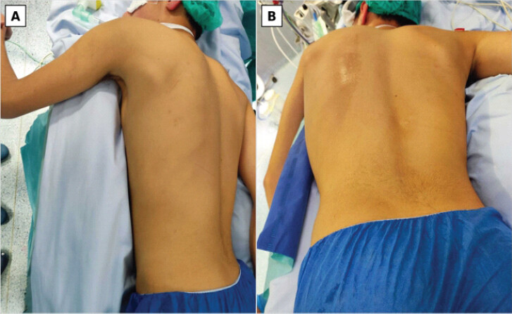
Given the previous history, recurrence of ventral scapular osteochondroma was suspected. Plain radiographs were obtained, followed by cross-sectional imaging, which confirmed a recurrent osteocartilaginous lesion arising from the ventral surface of the scapula. The imaging appearance was consistent with recurrent osteochondroma, and no features suggesting malignant transformation were identified clinically or radiologically.
Treatment
Surgical re-excision was indicated because of pain, recurrent static deformity, and mechanical scapulothoracic conflict. The patient was positioned prone under general anesthesia. A posterior parascapular approach was used, with careful soft-tissue handling and avoidance of unnecessary muscle detachment. The scapula was mobilized to expose the ventral lesion (Fig. 2a). The recurrent osteochondroma was excised flush with its base, aiming to remove the complete cartilage-capped exostosis and any residual cartilaginous tissue.
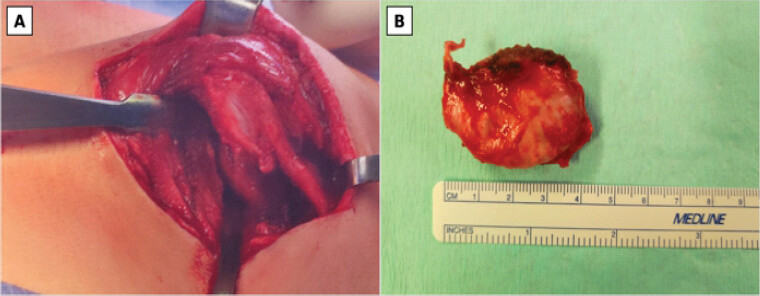
The specimen was sent for histopathological examination (Fig. 2b), which confirmed benign osteochondroma. There was no evidence of malignant transformation. The wound was closed in layers. Postoperatively, the arm was protected initially, followed by progressive rehabilitation with pendular exercises and gradual restoration of shoulder and scapulothoracic motion.
Outcome and follow-up
The post-operative course was uneventful. The patient had progressive resolution of pain and improvement in the scapular contour. At the most recent follow-up, he had painless shoulder mobility, normal scapulothoracic motion, and no clinically evident pseudo-winging. The patient and family were satisfied with the functional and cosmetic outcome. Ongoing surveillance until skeletal maturity was recommended because the recurrence occurred while the patient was still skeletally immature.
Discussion
This case is relevant for three reasons. First, it documents the recurrence of a previously published ventral scapular osteochondroma that had initially produced pseudoscapula alata. Second, it illustrates the importance of distinguishing static pseudo-winging caused by a mechanical mass effect from true dynamic scapular winging caused by neuromuscular dysfunction. Third, it emphasizes that complete surgical resection, including the cartilage cap and base of the lesion, is essential when treating symptomatic osteochondroma in a skeletally immature patient.
The scapula is an uncommon site for osteochondroma, but when these lesions arise from the ventral surface, even a relatively small mass may cause symptoms due to scapulothoracic conflict. Several authors have described patients presenting with pseudo-winging, snapping scapula, pain, or rib irritation from ventral scapular osteochondromas [6,7,8,9,10,11,12,13,14,15,16,17,18]. In the original episode of the present patient, the lesion was located on the ventral side of the scapula and produced static deformity and pain, leading to surgical excision [1].
The diagnostic challenge lies in the clinical resemblance between pseudo-winging and true scapular winging. Dynamic winging is classically associated with neuromuscular pathology, whereas pseudo-winging caused by osteochondroma is often fixed or static. This distinction is more than semantic: management differs substantially. A suspected static deformity should prompt careful palpation, comparison of scapular contour, plain radiographs, and, when needed, CT to identify the exact location, morphology, and relationship of the lesion to the thoracic wall [5,6,7,8,10]. Magnetic resonance imaging may be useful when cartilage cap thickness, bursal formation, soft-tissue involvement, or malignant transformation is a concern [2,3].
Recurrence after osteochondroma excision is unusual when the lesion is completely removed. When recurrence occurs, incomplete removal of the cartilage cap or persistent growth potential before skeletal maturity should be considered. In this patient, recurrence in adolescence suggests that long-term follow-up is advisable after excision of symptomatic lesions in children, even when the early clinical and radiographic outcome is excellent. The recurrent presentation was recognized promptly because the previous diagnosis was known and the clinical deformity was static.
Surgical treatment remains the preferred option for symptomatic scapular osteochondromas causing pain, deformity, pseudo-winging, or mechanical conflict [7,8,9,10,11,12,13,14,15,16,17,18]. Both open and arthroscopic techniques have been described, but open posterior parascapular approaches remain reliable for ventral lesions requiring direct visualization and complete excision. Muscle-sparing exposure is particularly attractive in pediatric and adolescent patients because it may reduce morbidity and accelerate functional recovery. In revision surgery, careful exposure is required to avoid residual cartilage and to protect the surrounding soft tissues.
The main limitation of this report is that it is based on a single patient and relies on clinical follow-up after revision excision. Nevertheless, the case adds value because it represents a rare recurrent presentation of a previously reported ventral scapular osteochondroma and highlights a practical message: apparent cure after excision should not preclude follow-up until skeletal maturity, when the original lesion occurred in childhood.
Conclusion
Recurrent ventral scapular osteochondroma is rare but clinically important. In adolescents with a recurrent static scapular deformity after previous excision, recurrent osteochondroma should be considered. Accurate differentiation between pseudo-winging and true neuromuscular winging, appropriate cross-sectional imaging, complete excision of the lesion including the cartilage cap, and follow-up until skeletal maturity are key elements for successful management.
Clinical Message
A static scapular deformity is not always neurological winging. In a child or adolescent, particularly after previous excision of a ventral scapular osteochondroma, recurrent pain and pseudo-winging should raise suspicion of recurrence. Complete resection and continued follow-up are essential to prevent persistent symptoms and delayed diagnosis.
Conflict of Interest:
Nil
Source of Support:
Nil
Consent
The authors confirm that informed consent was obtained from the patient for publication of this article
How to Cite this Article
Chaves RN, Lopes AL, Neves SM, Teles A, Pinto HL, Sousa RM. Recurrent Ventral Scapular Osteochondroma Presenting with Pseudo-winging in an Adolescent: A Case Report and Follow-up of a Previously Published Case. Journal of Orthopaedic Case Reports 2026 October;16(10): 203-207.
References
- Sousa RM, Sapage R, Branco C, Sousa D, Reis J, Geraldes R. Pseudo scapula alata: A case report of a scapular osteochondroma. Int J Res Orthop 2021;7:144-7. [Google Scholar] | [PubMed]
- Murphey MD, Choi JJ, Kransdorf MJ, Flemming DJ, Gannon FH. Imaging of osteochondroma: Variants and complications with radiologic-pathologic correlation. Radiographics 2000;20:1407-34. [Google Scholar] | [PubMed]
- Kitsoulis P, Galani V, Stefanaki K, Paraskevas G, Karatzias G, Agnantis NJ. Osteochondromas: Review of the clinical, radiological and pathological features. In Vivo 2008;22:633-46. [Google Scholar] | [PubMed]
- Bovee JV. Multiple osteochondromas. Orphanet J Rare Dis 2008;3:3. [Google Scholar] | [PubMed]
- Gooding BW, Geoghegan JM, Wallace WA, Manning PA. Scapular winging. Shoulder Elbow 2014;6:4-11. [Google Scholar] | [PubMed]
- Chillemi C, Franceschini V, Ippolito G. Osteochondroma as a cause of scapular winging in an adolescent: A case report and review of the literature. J Med Case Rep 2013;7:220. [Google Scholar] | [PubMed]
- Flugstad NA, Sanger JR, Hackbarth DA. Pseudo-winging of the scapula caused by scapular osteochondroma: Review of literature and case report. Hand (N Y) 2015;10:353-6. [Google Scholar] | [PubMed]
- Tittal P, Pawar I, Kapoor SK. Pseudo-winging of scapula due to benign lesions of ventral surface of scapula - two unusual causes. J Clin Orthop Trauma 2015;6:30-5. [Google Scholar] | [PubMed]
- Rameez R, Ul-Hassan M, Kotwal HA, Kangoo KA, Nazir A. Painful pseudowinging and snapping of scapula due to subscapular osteochondroma: A case report. J Orthop Case Rep 2016;6:96-9. [Google Scholar] | [PubMed]
- Ogawa K, Inokuchi W. Solitary osteochondroma of the ventral scapula associated with large bursa formation and pseudowinging of the scapula: A case report and literature review. Case Rep Orthop 2018;2018:5145642. [Google Scholar] | [PubMed]
- Alatassi R, Koaban S, Almugebel I, Alshehri A. Scapular osteochondroma with winging: A case report. Int J Surg Case Rep 2018;45:138-42. [Google Scholar] | [PubMed]
- Sivananda P, Rao BK, Kumar PV, Ram GS. Osteochondroma of the ventral scapula causing scapular static winging and secondary rib erosion. J Clin Diagn Res 2014;8:LD03-5. [Google Scholar] | [PubMed]
- Vaishya R, Dhakal S, Vaish A. A solitary osteochondroma of the scapula. BMJ Case Rep 2014;2014:bcr2013202273. [Google Scholar] | [PubMed]
- Ngongang FO, Fodjeu G, Fon AC, Fonkoue L, Guifo ML, Bitang A, Mafok LJ. Surgical treatment of rare case of scapula osteochondroma in a resource limited setting: A case report. Int J Surg Case Rep 2019;61:130-4. [Google Scholar] | [PubMed]
- Clarke DO, Crichlow A, Christmas M, Vaughan K, Mullings S, Neil I. The unusual osteochondroma: A case of snapping scapula syndrome and review of the literature. Orthop Traumatol Surg Res 2017;103:1295-8. [Google Scholar] | [PubMed]
- Pawar E, Modi N, Yadav AK, Mhatre J, Khemkar S, Mishra S. A solitary ventral scapular osteochondroma causing pseudo-winging of scapula: A case report. J Orthop Case Rep 2021;11:82-5. [Google Scholar] | [PubMed]
- Jindal V, Khandekar A, Gupta P, Bansal S. Solitary osteochondroma of the scapula in a young male: A case report. J Orthop Case Rep 2022;12:34-7. [Google Scholar] | [PubMed]
- Faur C, Abu-Awwad A, Patrascu JMJr, Abu-Awwad SA, Tudoran C. Superomedial scapula angle osteochondroma with winging: A case report. J Clin Med 2023;12:5106. Clinical Message. [Google Scholar] | [PubMed]
© 2026 Journal of Orthopaedic Case Reports - Published by Indian Orthopaedic Research Group
About the Authors
-

Rui Nobre Chaves
-

António Lemos Lopes
-

Sara Machado Neves
-

Afonso Teles
-

Henrique Lascasas Pinto
-

Rita Macedo Sousa
Related Articles in Journal of Orthopaedic Case Reports
 February 1, 2026 Symptomatic Ventromedial Scapular Osteochondroma Presenting with Restriction of Shoulder Movements: A Case Report
 October 10, 2023 Unveiling Scapular Pseudo-winging: A Case Report on Ventral Osteochondroma of the Scapula in a 21-year-old
 August 1, 2026 Surgical Excision of a Rare Ventral Scapular Osteochondroma using the Prone “Chicken-Wing” Position: A Case Report
 July 1, 2026 Posterior Scapular Osteochondroma in a Pediatric Patient: An Uncommon Presentation of a Common Benign Tumor