


Introduction
Hip fractures in older people carry high morbidity, and surgical fixation is the standard of care to facilitate early mobilization. Vascular complications, while uncommon, have been reported following intramedullary fixation [1]. Arterial injuries most often involve branches of the profunda femoris artery; injury to the superior gluteal artery (SGA) is exceedingly rare [2]. The SGA exits the pelvis above the piriformis to supply the gluteal musculature, and a handful of cases have documented SGA injury or pseudoaneurysm formation following intertrochanteric or subtrochanteric fracture fixation [1,3,4]. These injuries can cause massive hemorrhage, gluteal compartment syndrome, and hemodynamic shock if not promptly recognized [2,3,4,5].
Anticoagulation adds further complexity to hip fracture management. Our patient was on apixaban for Factor V Leiden thrombophilia. Recent evidence demonstrates that hip fracture surgery without delay for DOAC clearance is safe and not associated with increased blood loss or complications [6]. We present this rare vascular complication of cephalomedullary nailing (CMN) in an anticoagulated patient and review the relevant literature.
The patient was informed that data concerning this case would be submitted for publication and provided consent to do so.
Case Report
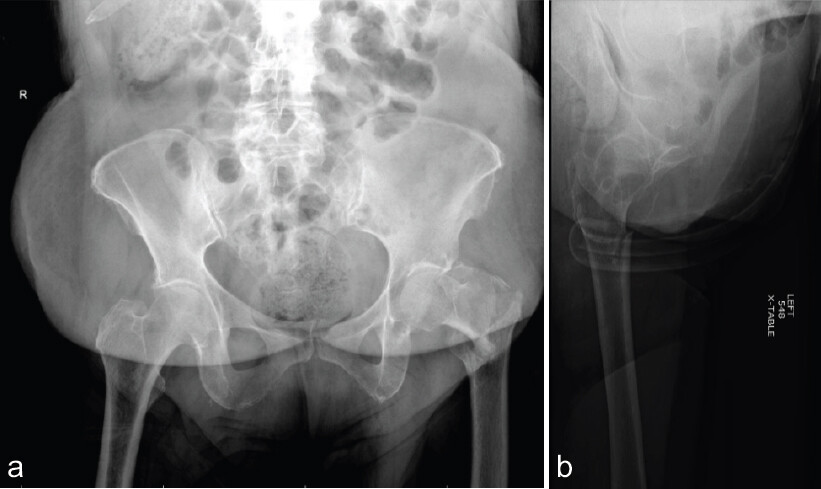
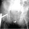
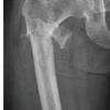
A 75-year-old female with a medical history of hypertension, type 2 diabetes, stage 3 chronic kidney disease, Factor V Leiden thrombophilia (on apixaban for prior deep vein thrombosis), stroke, and giant cell arteritis sustained a low-energy fall. She was found at home approximately 8 h after the fall and was brought to the emergency department. On evaluation, she was alert and oriented with a primary complaint of left hip pain and inability to bear weight. Physical examination revealed left hip tenderness with a positive log-roll and axial load pain. The limb was neurovascularly intact. Initial hemoglobin was 10.4 g/dL. Radiographs of the hip and femur demonstrated a displaced intertrochanteric femur fracture (Fig. 1a and 1b). Given her anticoagulation status and fracture pattern, a decision was made to proceed with surgical fixation once medically optimized.
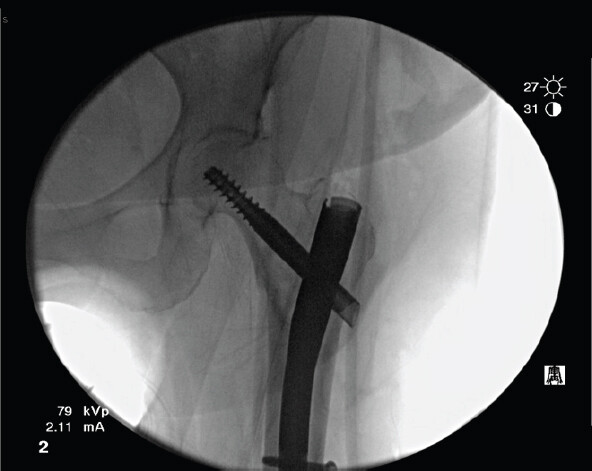
The patient was taken to the operating room the same day for open reduction and internal fixation of the left hip fracture. A 12-mm intermediate-length Trochanteric Fixation Nail Advanced (DePuy Synthes) CMN was placed in antegrade fashion via a proximal femoral trochanteric entry point. The surgery was uncomplicated with approximately 400 mL of blood loss recorded. A vacuum dressing was applied over the incision. There were no arterial injuries noted intraoperatively, and hemostasis appeared adequate upon wound closure (Fig. 2).
In the PACU, 3 h after surgery, the patient was noted to be hypotensive (blood pressure 80/50 mmHg) and tachycardic. The post-operative hemoglobin returned to 6.9 g/dL. She was resuscitated with two units of packed red blood cells (PRBCs), intravenous fluids, started on vasopressor support (phenylephrine), and transferred to the surgical intensive care unit. Immediate focus was placed on determining the source of bleeding. Her surgical dressing remained dry with no external signs of bleeding, but on examination, her left thigh and buttock appeared increasingly firm and swollen. By the next morning (post-operative day 1), despite transfusions, her hemoglobin again fell from 8.1 to 6.8 g/dL, accompanied by rising serum lactate to 5.6 mmol/L, increasing the concern for ongoing internal hemorrhage.
A CT angiogram (CTA) of the abdomen and pelvis with runoff was obtained on post-operative day 1 to localize the bleeding source. The CTA revealed a large hematoma in the left gluteal region, measuring approximately 7.5 × 6 × 9 cm, with active contrast “blush” indicative of active arterial extravasation in the vicinity of the SGA distribution. Given the location of the hematoma lateral to the gluteal musculature, an injury to a branch of the left SGA was suspected. Interventional radiology was consulted urgently.
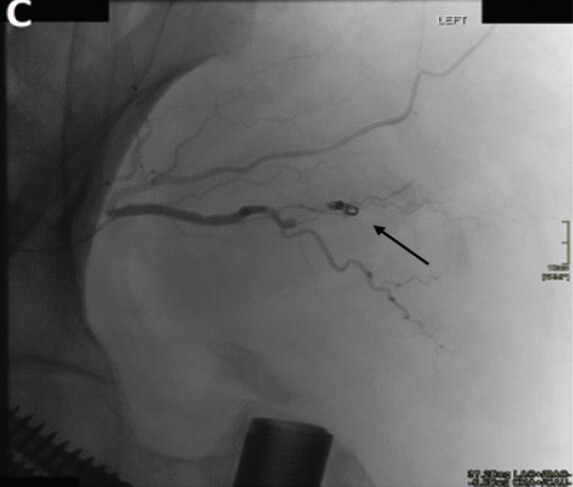
The patient was brought to the angiography suite on post-operative day 1 for urgent pelvic angiography and possible embolization. Angiography of the left internal iliac artery confirmed active arterial bleeding from a branch of the SGA. A selective catheterization of the SGA branch and coil embolization were then performed, achieving hemostasis of the SGA branch. The patient’s hemodynamics stabilized following the procedure (Fig. 3). Post-embolization hemoglobin was 6.9 g/dL, and one additional unit of PRBC was transfused.
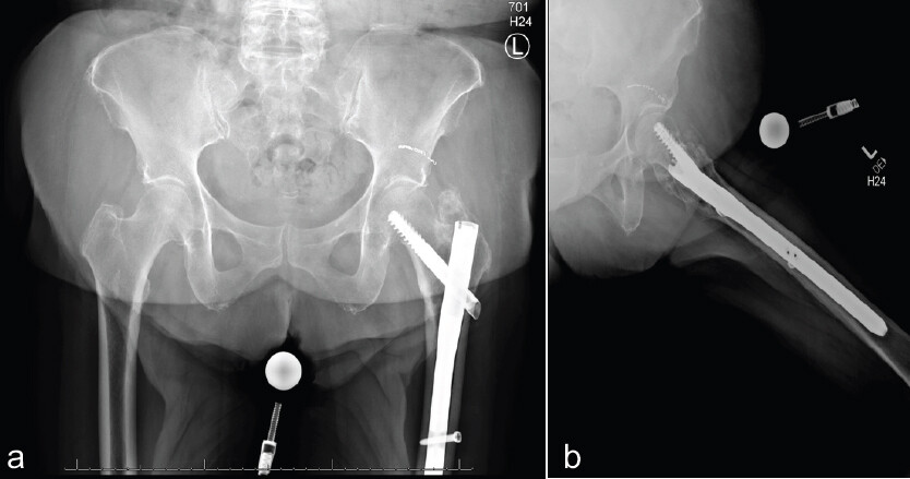
The patient remained in the intensive care unit for continued close monitoring. Over the following 24–48 h, her hemoglobin stabilized at approximately7.9–8.2 g/dL without further transfusion requirements. Vasopressor support was weaned off, and the patient had no signs of compartment syndrome or neurological deficit in the left lower extremity. Given the history of thrombophilia, a low-dose heparin infusion was restarted on post-operative day 4 once bleeding was controlled, with a later transition back to therapeutic apixaban prior to discharge as coordinated with hematology. On post-operative day 11, the patient was discharged to a subacute rehabilitation facility in good condition, having received a total of five units of blood postoperatively. Subsequent follow-up demonstrated appropriate fracture healing progress and no further hemorrhagic complications (Fig. 4a and b).
Discussion
Mechanism of injury to the SGA
Vascular injury during intramedullary nailing of proximal femur fractures is uncommon, and SGA injury is particularly rare [2, 4]. The exact mechanism in our case is uncertain, but several possibilities exist. The guidewire or reamer may have strayed from the ideal trajectory, perforating an SGA branch near the greater sciatic notch; Carroll et al. similarly implicated guidewire trajectory in a deep-branch SGA pseudoaneurysm after CMN [5]. Alternatively, a sharp rather than blunt trochanteric incision may lacerate the vessel, as Ward et al. speculated in their case of SGA injury during subtrochanteric fracture nailing [4]. Even a slightly anterior or medial entry point can endanger the SGA’s deep branch as it runs between the gluteus medius and minimus, a risk highlighted by both anatomical studies and clinical series [7,8,9]. Comminuted fracture fragments may also contribute – sharp edges or displaced lesser trochanteric fragments can injure adjacent vessels, and Fernandes et al. attributed their case of SGA hemorrhage after pertrochanteric nailing to reaming or nail insertion [3].
SGA injuries also occur with pelvic ring fractures and iliosacral screw fixation, sometimes causing gluteal compartment syndrome or delayed hemorrhagic shock [2]. Regardless of mechanism, angiographic embolization is the definitive treatment given the vessel’s deep pelvic location, and published reports consistently demonstrate successful hemostasis with this approach [1,3,4,5,9,10].
Literature review of reported cases
Including the present patient, we identified several reported cases of SGA injury or pseudoaneurysm following intramedullary fixation of proximal femur fractures. Table 1 summarizes the key details of these cases from the literature. Notably, the majority of patients were elderly females with intertrochanteric or subtrochanteric fractures, reflecting the typical osteoporotic hip fracture demographic, though one case involved a young adult with a high-energy femoral shaft fracture. The timing of presentation ranged from acute post-operative hemorrhage (within 24–48 h, as in our case) to delayed presentations several weeks later due to pseudoaneurysm formation. Common clinical signs were an unexplained drop in hemoglobin, large thigh or gluteal hematoma, and hemodynamic instability or anemia not proportional to the surgical loss. In all cases, CT angiography or conventional angiography was used to localize the bleeding, with IR-guided coil embolization of the SGA (or its branch) successfully achieving hemostasis [1,3,4,10].
Published cases of superior gluteal artery (SGA) injury after cephalomedullary nailing or femoral intramedullary fixation for hip fractures
| Source (Year) | Patient & Fracture | Presentation Timing | Management | Outcome and Cause if Given |
|---|---|---|---|---|
| Ward et al. [4] (2013) | 81-year-old female, incomplete subtrochanteric stress (atypical) fracture – prophylactic IM nail | Post-op Day 1: acute anemia, buttock hematoma. | Angiography → Coil embolization of SGA branch. | Recovered; attributed to sharp dissection injury. |
| Ailaney et al. [1] (2019) | 79-year-old female, atypical femoral fracture – IM nail fixation. | Immediate post-op: acute blood loss anemia, extensive thigh ecchymosis. | CTA → SGA extravasation; Angiography → Coil embolization of SGA pseudoaneurysm. | Recovered. |
| Carroll et al. [5] (2020) | 72-year-old female, intertrochanteric hip fracture – CMN fixation. | Post-op Day 0–1: thigh/gluteal swelling, anemia. | Angiography → Coil embolization of deep branch of SGA. | Recovered; guidewire malposition. |
| Fernandes et al. [3] (2020) | 66-year-old female, pathological pertrochanteric fracture – long IM nail. | Post-op Day 5: hemoglobin drop, large gluteal hematoma. | Angiography → Selective SGA branch embolization. | Recovered |
| Nambiar et al. [10] (2022) | 84-year-old female, periprosthetic hip fracture – revision long CMN fixation. | Post-op Day 4: expanding gluteal hematoma, pseudoaneurysm. | Angiography → Coil embolization. | Recovered; first reported during revision fixation. |
| Forin Valvecchi et al. [14] (2022) | 35-year-old male, femoral shaft fracture – IM nail. | Post-op day 14 in rehab: acute hip pain and Hgb drop. | Angiography → Selective embolization of bleeding SGA branch. | Recovered. |
| Başak et al. [9] (2024) | 72-year-old female, intertrochanteric fracture – short CMN fixation. | 3 weeks post-op: gluteal hematoma, anemia. | Ultrasound/CT → Angio → Coil embolization. | Recovered |
Hgb = hemoglobin; IM = intramedullary; CMN = cephalomedullary nail; ex-fix = external fixator; CTA = CT angiography.
Role of anticoagulation and timing of surgery
Our patient’s anticoagulated status (apixaban for Factor V Leiden) likely contributed to the severity of her hemorrhage. It is well known that anticoagulation can increase bleeding risk from any source. In this case, the decision was made not to significantly delay surgery, given the urgent nature of hip fracture fixation in an elderly patient. Current evidence supports this approach: recent studies have found that performing hip fracture surgery within 24 h in patients on DOACs does not significantly increase intraoperative or post-operative bleeding compared to delaying surgery for drug clearance [11,12]. Wang et al. showed that expedited surgery (≤24 h) in DOAC-treated hip fracture patients resulted in lower pre-operative blood loss and no difference in total blood loss or transfusion rate, as well as shorter hospital stays with fewer complications, compared to delayed surgery [11]. Similarly, Brameier et al. and Kolodychuk et al. reported that the use of DOACs is not an automatic reason to postpone hip fracture operations, as timely surgery can be accomplished safely with appropriate precautions [6,13].
Conclusion
SGA injury is a rare but serious complication of intramedullary hip fracture fixation. This case underscores the importance of recognizing disproportionate hemoglobin decline, unexpected hypotension, or gluteal swelling as harbingers of internal arterial injury. Early CTA and prompt embolization are the cornerstones of management and were critical in this patient’s survival. Anticoagulation and advanced age should inform perioperative planning but not delay timely fixation, as outcomes remain favorable with a multidisciplinary approach. Surgeons should maintain awareness of SGA injury risk, use meticulous technique, and engage interventional radiology early when post-operative hemorrhage is suspected.
Clinical Message
Acute superior gluteal artery hemorrhage is a distinct and previously unreported presentation in patients on therapeutic direct oral anticoagulant therapy following CMN. Unlike delayed pseudoaneurysm formation (the more commonly described SGA complication), acute hemorrhage presents within hours of surgery with rapid hemodynamic deterioration and expanding gluteal hematoma, demanding immediate recognition and action. CT angiography is the diagnostic modality of choice, and selective coil embolization by interventional radiology is both limb- and life-saving. Surgeons should employ meticulous technique at the trochanteric entry point, maintain a low threshold for vascular imaging when post-operative bleeding is disproportionate to surgical blood loss, and engage interventional radiology early. This case further demonstrates that DOAC use need not delay timely surgical fixation of fragility hip fractures, but mandates heightened post-operative vigilance for hemorrhagic complications.
Conflict of Interest:
Nil
Source of Support:
Nil
Consent:
The authors confirm that informed consent was obtained from the patient for publication of this article
How to Cite this Article
Pang A, Zamzam M, Pellerito A, Eller E. Superior Gluteal Artery Injury After Cephalomedullary Nailing of a Hip Fracture in a Patient on Anticoagulation: A Case Report and Literature Review. Journal of Orthopaedic Case Reports 2026 October;16(10): 233-237.
References
- Ailaney N, O'Connell R, Giambra L, Golladay G. Superior gluteal artery pseudoaneurysm following intramedullary nailing of an atypical femoral fracture. BMJ Case Rep 2019;12:e231490. [Google Scholar] | [PubMed]
- Gee MJ, Tolat A, Sinha J. Acute gluteal and thigh compartment syndrome following pelvic fracture and superior gluteal artery bleed: A case report and review of the literature. Eur J Trauma Emerg Surg 2007;33:188-91. [Google Scholar] | [PubMed]
- Fernandes PX, Medeiros DM, Teixeira RC, Nunes AM, Seco AS, Caetano AC. Injury to the superior gluteal artery during intramedullary fixation of a proximal femoral fracture: A case report. J Orthop Case Rep 2020;9:27-31. [Google Scholar] | [PubMed]
- Ward JP, Strauss EJ, Tejwani NC. Injury to the superior gluteal artery during intramedullary fixation of an atypical subtrochanteric stress fracture: A case report. Bull Hosp Jt Dis 2013;71:297-300. [Google Scholar] | [PubMed]
- Carroll C, Warren M, Nammour M, Taillac H, Mautner J. Superior gluteal artery bleed after cephalomedullary nail fixation. Ochsner J 2020;20:339-42. [Google Scholar] | [PubMed]
- Brameier DT, Tischler EH, Ottesen TD, McTague MF, Appleton PT, Harris MB. Use of direct oral anticoagulants among patients with hip fracture is not an indication to delay surgical intervention. J Orthop Trauma 2024;38:148-54. [Google Scholar] | [PubMed]
- Vahabi A, Limon YK, Oncel C, Gursoy MK, Aktuglu K. Structures at risk with intrapelvic guide wire migration during cephalomedullary fixation of hip fractures: CT-based analysis. J Orthop Trauma 2026;40:e33-8. [Google Scholar] | [PubMed]
- Farhang K, Desai R, Wilber JH, Cooperman DR, Liu RW. An anatomical study of the entry point in the greater trochanter for intramedullary nailing. Bone Joint J 2014;96-B:1274-81. [Google Scholar] | [PubMed]
- Başak F, Ardalı D, Candan ES, Taşcı M, Söylemez UP, Söylemez MS. Pseudoaneurysm formation in the superior gluteal artery following short femoral nail placement in an intertrochanteric femur fracture: A case report. J Orthop Case Rep 2024;14:153-7. [Google Scholar] | [PubMed]
- Nambiar M, Ng D, Maingard J, Vrazas J. Embolisation of superior gluteal artery pseudoaneurysm, after hip cephalomedullary nail revision. BMJ Case Rep 2022;15:e247322. [Google Scholar] | [PubMed]
- Wang J, Giammanco PA, Nilssen P, Stone J, Breda K, Little M. Expedited hip fracture surgery in patients on direct oral anticoagulants does not increase perioperative blood loss. J Orthop Trauma 2025;39:314-9. [Google Scholar] | [PubMed]
- Schiepers TE, Willems HC, Smeeing DP, Bosma E, Kempen DH, Emmelot-Vonk MH. Impact of early (<24 hours) vs. Delayed (≥24 hours) surgery on hemoglobin decrease in patients with hip fracture on direct oral anticoagulants: A multicenter retrospective cohort study. JBJS Open Access 2026;11:e25.00334. [Google Scholar] | [PubMed]
- Kolodychuk NL, Godshaw B, Nammour M, Starring H, Mautner J. Early hip fracture surgery is safe for patients on direct oral anticoagulants. OTA Int 2023;6:e252. [Google Scholar] | [PubMed]
© 2026 Journal of Orthopaedic Case Reports - Published by Indian Orthopaedic Research Group
About the Authors
-

Aaron Pang
-

Mazen Zamzam
-

Antonio Pellerito
-

Erik Eller
Related Articles in Journal of Orthopaedic Case Reports
 May 1, 2026 Intraoperative Traction for Cephalomedullary Nailing in Amputation Patients
 October 1, 2024 Pseudoaneurysm Formation in the Superior Gluteal Artery Following Short Femoral Nail Placement in an Intertrochanteric Femur Fracture: A Case Report
 August 1, 2026 Radiological Outcomes of Reamed Short Proximal Femoral Nailing in Intertrochanteric Fractures in Patients Aged 50 Years and Above
 August 1, 2026 Evaluation of Orthopedic Outcomes of Proximal Femoral Nailing for Intertrochanteric Fractures and Predictive Value of Pre-operative Glycemic Status, Neutrophil-to-Lymphocyte Ratio and Platelet-to-Lymphocyte Ratio: A Prospective Study