


Introduction
Giant cell tumor (GCT) of bone is a benign but locally aggressive neoplasm and is among the most frequently encountered benign bone tumors in orthopedic practice. The most commonly affected anatomical sites, in descending order of frequency, include the distal femur, proximal tibia, distal radius, and proximal humerus. Involvement of the small bones of the hand and foot is relatively rare, with reported incidences of approximately 2.4% in the hand and 1.2–1.8% in the foot [1]. The occurrence of GCT in the distal fibula is exceptionally uncommon, constituting <1% of all reported cases.
Several surgical options have been proposed for the management of GCT of the lateral malleolus, ranging from extended intralesional curettage with adjuvant therapies to en bloc resection of the distal fibula followed by ankle reconstruction. Reconstructive techniques described in the literature include inter tibio-fibular or tibiotalar arthrodesis, 180° rotation of the proximal fibula [3–8], cortical or strut graft reconstruction of the remaining fibula, fibular allograft reconstruction, repair of the lateral ligamentous complex of the ankle, and utilization of the distal peroneal tendons to re-establish ankle stability.
Case Report
A 26-year-old male patient presented with pain in the left ankle region and difficulty in walking for a period of 6 months. On examination, a vertically ovoid swelling was present on the lateral aspect of the ankle with restriction of ankle movement. Radiograph of the left ankle showed an eccentric, expansile lytic lesion with a soap-bubble appearance, 6.5 cm in size at the epiphyseo-metaphyseal part of the distal fibula. Magnetic resonance imaging showed that the lesion was compressing the peroneal muscles and tendons and the talar bone (Fig. 1, 2, 3).
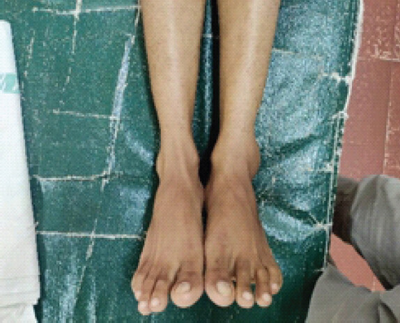
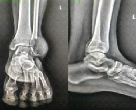
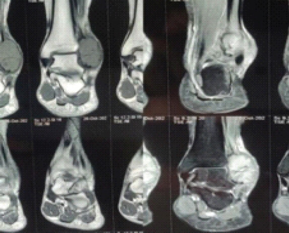
Operative steps
Treatment options were clearly discussed with the patient, and the patient wanted an ankle reconstruction. At his request, surgery was planned to reconstruct the ankle joint using an ipsilateral proximal fibula graft. A direct lateral incision was made over the distal half of the fibula to access the tumor. The superficial peroneal nerve’s cutaneous branch was identified and preserved. After exposing the tumor, we performed a wide surgical resection of the tumor up to 7.5 cm from the lateral malleolus tip, which was sent for biopsy. To reconstruct the ankle, an ipsilateral proximal fibular graft of adequate length was harvested after safeguarding the common peroneal nerve, as shown in Fig. 4, 5, 6.
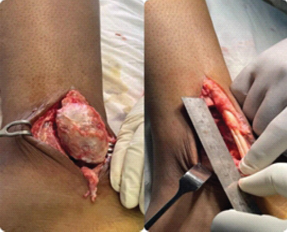
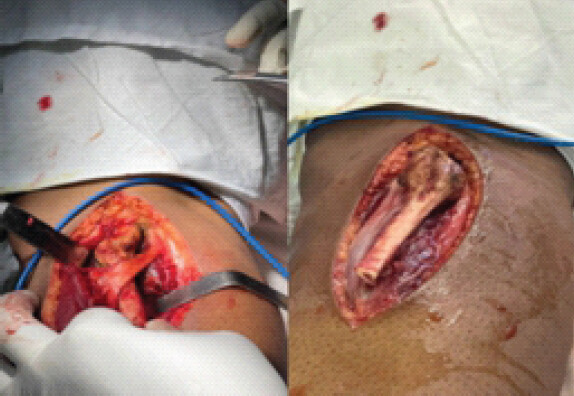
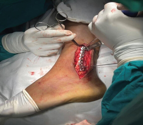
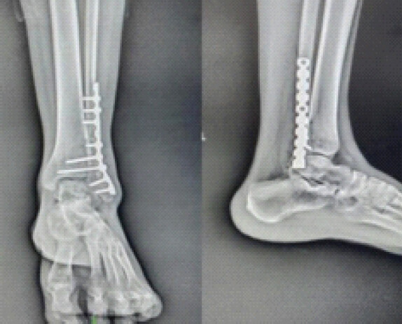
The proximal fibula graft was then prepared and approximated to the proximal part of the distal fibula and temporarily fixed with K-wires. An adequate length reconstruction plate was then used to fix the graft. The fibular collateral ligament and the syndesmotic ligaments were repaired, and a syndesmotic screw was also kept as shown in the post-operative X-ray (Fig. 7).
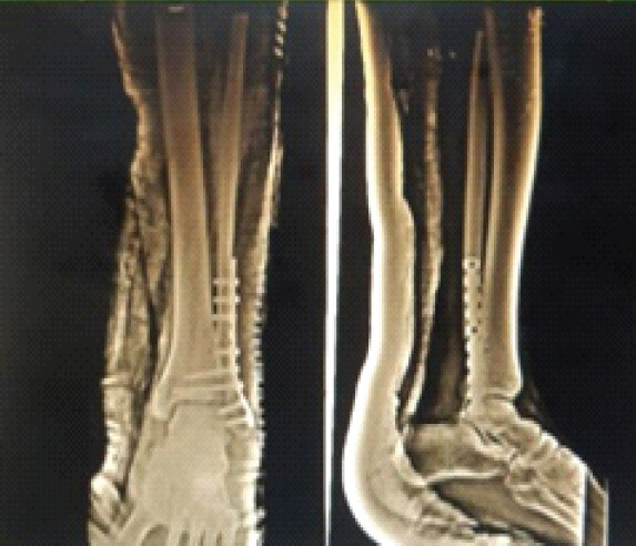
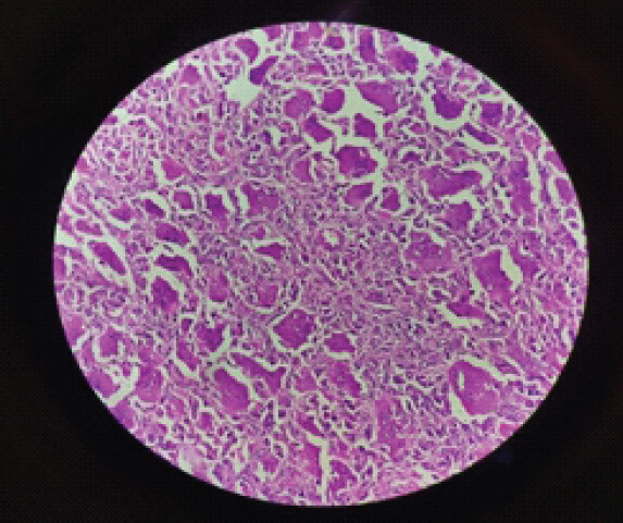
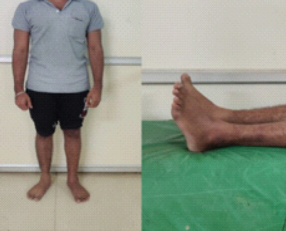
The ankle was immobilized in an above-knee plaster of Paris slab and bandage for 6 weeks, After that the slab was removed, and non-weight-bearing exercises were advised for another 6 weeks. Radiological and Clinical follow-up was carried out regularly (Fig. 9 and 10)..
Discussion
GCT of the lateral malleolus is an uncommon clinical entity [9], and the management of aggressive lesions at this location remains particularly challenging. Owing to its rarity, there are no established treatment guidelines, and the available literature is limited to isolated case reports and small case series. The distal 10 cm of the fibula plays a vital role in maintaining the stability of the ankle mortise. Therefore, reconstruction of the distal fourth of the fibula following tumor excision is essential to preserve ankle stability. Lesions occurring in this region require meticulous planning, especially in young and active individuals, to maintain ankle function and prevent long-term disability.
Furthermore, deformities involving the ankle can significantly impair gait and functional mobility, adversely affecting a patient’s daily activities, social interactions, and occupational performance. Patients with GCT of the lateral malleolus commonly present with pain, swelling, and restricted ankle movements. Previous studies have demonstrated that ankle stability depends not only on the integrity of the distal fibula but also on the preservation of its ligamentous attachments. During weight-bearing, the fibula contributes to deepening the ankle mortise and transmits approximately 7.1% of the body’s weight. Complete excision of the distal fibula without appropriate reconstruction may result in valgus instability of the ankle and subsequent degenerative arthritic changes [5].
Capanna et al. [2] described six different methods of fibular excision and reconstruction based on the location of the distal fibular lesion. These techniques include reconstruction using a proximally harvested fibula rotated through 180°, cortical grafting, and augmentation of ankle stability with the peroneal tendons [10]. Additional reconstructive options aimed at preserving ankle joint congruity include the use of tricortical iliac crest grafts and fibular allografts stabilized with dynamic compression plates. Although each technique offers distinct advantages, they are also associated with specific limitations. Moreover, harvesting the proximal fibula carries the potential risk of injury to the common peroneal nerve and the lateral collateral ligament complex.
Leibner et al. [10] proposed seven principles for distal fibular reconstruction, emphasizing preservation of the native fibula whenever oncologically feasible. In the present case, the patient had a Grade II lesion and expressed a strong desire to retain normal ankle function. Consequently, our treatment strategy focused on complete tumor excision while restoring the integrity of the ankle mortise. Resection of the fibula at a level 6–8 cm proximal to the distal tip can disrupt the syndesmotic ligaments and interosseous membrane, thereby increasing inversion and rotational stresses across the ankle joint. Clinically, this may manifest as deep muscular pain, weakness, and difficulty with running and other strenuous activities. Therefore, reconstruction and stabilization of the syndesmosis are recommended. In our patient, the tumor was excised completely, followed by reconstruction using a 180° rotated proximal fibular graft, syndesmotic repair, and fixation with a reconstruction plate.
Conclusion
GCTs of the lateral malleolus are extremely rare and pose challenges in their management. Thus, we recommend that all aggressive and large GCTs of the lateral malleolus must be excised adequately, and the bony defect should be reconstructed using autogenous bone graft with internal fixation. It is mandatory to preserve the ankle mortise during reconstructive surgery to achieve a good functional outcome.
Clinical Message
Giant cell tumor of the lateral malleolus poses a challenge in management; therefore, proper excision and reconstruction of the ankle mortise are essential for a good functional outcome.
Conflict of Interest:
Nil
Source of Support:
Nil
Consent:
The authors confirm that informed consent was obtained from the patient for publication of this article
How to Cite this Article
Raj NE, Mukharjee GS, Amal PS. Giant Cell Tumor of the Lateral Malleolus Treated by Tumor Excision and Reconstruction using A 180° Rotated Proximal Fibular Autograft – A Rare Case Report. Journal of Orthopaedic Case Reports 2026 October;16(10): 276-280.
References
- Bhowmick K, Boopalan PR. Saving the ankle in distal fibular giant cell tumour - a case report. J Clin Orthop Trauma 2019;10:1054-8. [Google Scholar] | [PubMed]
- Capanna R, Van Horn JR, Biagini R, Ruggieri P, Bettelli G, Campanacci M. Reconstruction after resection of the distal fibula for bone tumor. Acta Orthop Scand 1986;57:290-4. [Google Scholar] | [PubMed]
- Nadkarni S, Punit AS, Nair RV. Giant cell tumour of distal fibula managed by en block resection and reconstruction with ipsilateral proximal fibula. J Orthop Case Reports 2015;5:52-4. [Google Scholar] | [PubMed]
- Balke M, Schremper L, Gebert C, Ahrens H, Streitbuerger A, Koehler G. Giant cell tumor of bone: Treatment and outcome of 214 cases. J Cancer Res Clin Oncol 2008;134:969-78. [Google Scholar] | [PubMed]
- Babhulkar SS, Pande KC, Babhulkar S. Ankle instability after fibular resection. J Bone Joint Surg Br 1995;77:258-61. [Google Scholar] | [PubMed]
- Eger W, Schorle C, Zeiler G. Giant cell tumor of the distal fibula: Fifteen-year result after en bloc resection and fibula reconstruction. Arch Orthop Trauma Surg 2004;124:56-9. [Google Scholar] | [PubMed]
- Yadav SS. Ankle stability after resection of the distal third of the fibula for giant-cell lesions: Report of two cases. Clin Orthop Relat Res 1981;156:105-7. [Google Scholar] | [PubMed]
- Herring CL Jr, Hall RL, Goldner JL. Replacement of the lateral malleolus of the ankle joint with a reversed proximal fibular bone graft. Foot Ankle Int 1997;18:317-23. [Google Scholar] | [PubMed]
- Vaishya R, Kapoor C, Golwala P, Agarwal AK, Vijay V. A rare giant cell tumor of the distal fibula and its management. Cureus 2016;8:e666. [Google Scholar] | [PubMed]
- Leibner E, Ad-El D, Liebergall M, Ofiram E, London E, Peyser A. Lateral malleolar reconstruction after distal fibular resection: A case report. J Bone Joint Surg Am 2005;87:878-82. [Google Scholar] | [PubMed]
© 2026 Journal of Orthopaedic Case Reports - Published by Indian Orthopaedic Research Group
About the Authors
-

N Ebel Raj
-

G S Mukharjee
-

P S Amal
Related Articles in Journal of Orthopaedic Case Reports
 October 1, 2026 Early Recognition of Glomus Tumor of Hand Using Clinical Tests: Experience from an Indian Tertiary Center
 October 1, 2026 Giant Cell Tumor of the Distal Phalanx of the Great Toe: Case Report
 September 1, 2026 “A Giant Shoulder”: Proximal Humerus Giant Cell Tumor Managed With Extended Curettage and Proximal Humerus Internal Locking System Plating with Fibular Strut Graft – A Case Report
 September 1, 2026 Management of Scapular Osteochondroma: An Uncommon Report of Two Cases